铝参与神经原纤维缠结形成的实验研究
赵海花 赖 红 唐忠艳 李兆圣 吕永利
铝参与神经原纤维缠结形成的实验研究
赵海花 赖 红*唐忠艳 李兆圣 吕永利
(中国医科大学人体解剖学教研室,沈阳110001)
目的 探讨铝与神经原纤维缠结(NFTs)形成之间的相关性。方法 选用16只雌性ICR小鼠,分为正常对照组与染铝组(200mg/kg·bw,染铝8个月)。组织荧光双重染色法观察铝与NFTs在小鼠大脑新皮层神经元内的定位。Western blot法半定量检测新皮层内tau蛋白及其磷酸化水平。结果 组织荧光染色表明NFTs阳性荧光表达随铝阳性荧光增强而增强,两者分布呈对应关系;Western blot结果显示长期铝暴露导致tau蛋白水平下降(P<0.01,与对照组相比),但磷酸化水平升高(P<0.01,与对照组相比)。结论 铝参与大脑新皮层神经元内NFTs形成与累积过程,tau蛋白的过度磷酸化可能是其成因之一。
铝;神经元纤维缠绕;tau;桑色素染色;噻嗉红染色
阿尔茨海默病(Alzheimer’s disease,AD)是一种能导致老年期神经生理紊乱的一种神经退行性疾病,临床上表现为痴呆,神经病理学表现以淀粉样斑块(amyloid plaques)及神经原纤维缠结(neurofibrillary tangles,NFTs)为主[1]。NFTs存在于神经元胞浆内,主要由高度磷酸化的微管相关蛋白tau逐级纤聚而成[2],为一种不易被水解的聚合物,且随年龄增长可逐渐累积,其数量与AD认知功能下降程度密切相关[3,4]。铝元素广泛存在于人们日常生活、工作中,能通过食物、空气、水及某些药物等多途径进入人体,蓄积于包括脑在内的多种组织器官中[5,6]。众多研究提示铝与NFTs形成之间的潜在联系,至今仍是争论的焦点之一。本文将通过双重组织荧光染色观察并记录铝与NFTs形成之间的关联,同时采用western blot检测tau蛋白及其磷酸化水平,为明确铝参与NFTs形成提供宝贵的形态学与分子生物学资料。
材料和方法
1.动物模型的建立
16只3月龄雌性ICR小鼠(由中国医科大学实验动物中心提供),平均分为正常对照组与染铝组。染铝组动物每日按200mg/kg·bw的量随每日饮水口服AlCl3溶液,染铝时间为8个月。
2.动物标本的制备
小鼠为12月龄时进行取材,10%水合氯醛(0.05ml/10g·bw,ip)麻醉,经左心室灌注生理盐水,至血液完全被其替代,断头取脑,左侧脑组织参照小鼠脑立体定位图谱[7]取感觉区皮层及运动区皮层。脑组织按1:5比例加入蛋白裂解液,超声粉碎,4℃裂解过夜,次日4℃12000rpm低温离心40min,取上清,每管50μl分装,置于-80℃深冻冰箱内,用于western blot检测;右侧脑组织经4%多聚甲醛中固定约1周后常规石蜡包埋、连续冠状切片(片厚8μm)用于组织荧光双重染色。
3.组织荧光双重染色
Morin[8](3,5,7,2′,4′-pentahydroxyflavone,桑色素,Sigma,M4008-2G,)为一种荧光染料,能与铝离子形成一种荧光复合物,在420nm处激发出绿色荧光。TR[9](Thiazin red,噻嗪红,Chroma 1A416)为一种红色的荧光染料,与NFTs结合后在530nm-560nm处激发红色荧光。
本研究采用Morin联合TR染色法观察染铝小鼠大脑新皮层神经元内铝负载与NFTs形成之间的关联,具体步骤:1)Morin染色:常规石蜡切片脱蜡至水,经0.01MPBS浸洗2次共5min后置于1%盐酸水溶液中预处理10min,双蒸水浸洗2次共5min,切片浸入 Morin染液(含0.2%Morin,85%乙醇,0.5%醋酸)10min,双蒸水浸洗2次共5min,避光;2)TR染色:待 Morin染色结束,切片浸入含0.001%TR水溶液中15min,避光;双蒸水浸洗2次共5min,甘油封片,观察。
4.Western blot检测
样品经10%SDS-PAGE分离;湿转法将蛋白转至PVDF膜上,5%BSA 37℃封闭1h,加入兔抗p tau ser396 1:1000(SAB,#11102),兔抗total tau 1:1000(SAB,#21093)及鼠抗β-actin 1:1000(santa cruz,sc-47778),4℃孵育过夜,TBST 漂洗。加入HRP标记羊抗兔或羊抗鼠二抗,1:5000稀释,37℃孵育1h,TBST漂洗,ECL发光,BIO-RAD凝胶电泳图像分析仪采图,Quantity One软件包分析。实验重复3次。
5.统计学分析
应用SPSS 11.0软件,对两组数据进行均值t检验,结果以均值(M)±标准误(S.E.)表示(¯x±s),P<0.05为有显著性差异。
结 果
1.铝中毒小鼠神经元铝负载与NFTs的关联
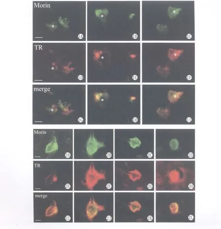
Morin联合TR双重组织荧光染色法观察小鼠大脑新皮质Ⅳ-Ⅴ层神经元铝与NFTs定位情况。图1显示:铝荧光染色较弱时(图1A),神经元几乎未见NFTs荧光阳性反应(图1D)。随着神经元内铝荧光范围的增加(图1B),NFTs染色出现(图1E)。至神经元内铝浓染时(图1C),神经元内也出现与之对应浓染的NFTs(图1F)。图2显示当神经元内铝与NFTs均呈强荧光表达时,神经元形态可出现神经元胞体膨大(图2B,F,J)、萎缩变形(图2C,G,K)与固缩(图2D,H,L)等不同程度的退行性改变。
2.铝中毒对小鼠新皮层tau蛋白及磷酸化水平的影响
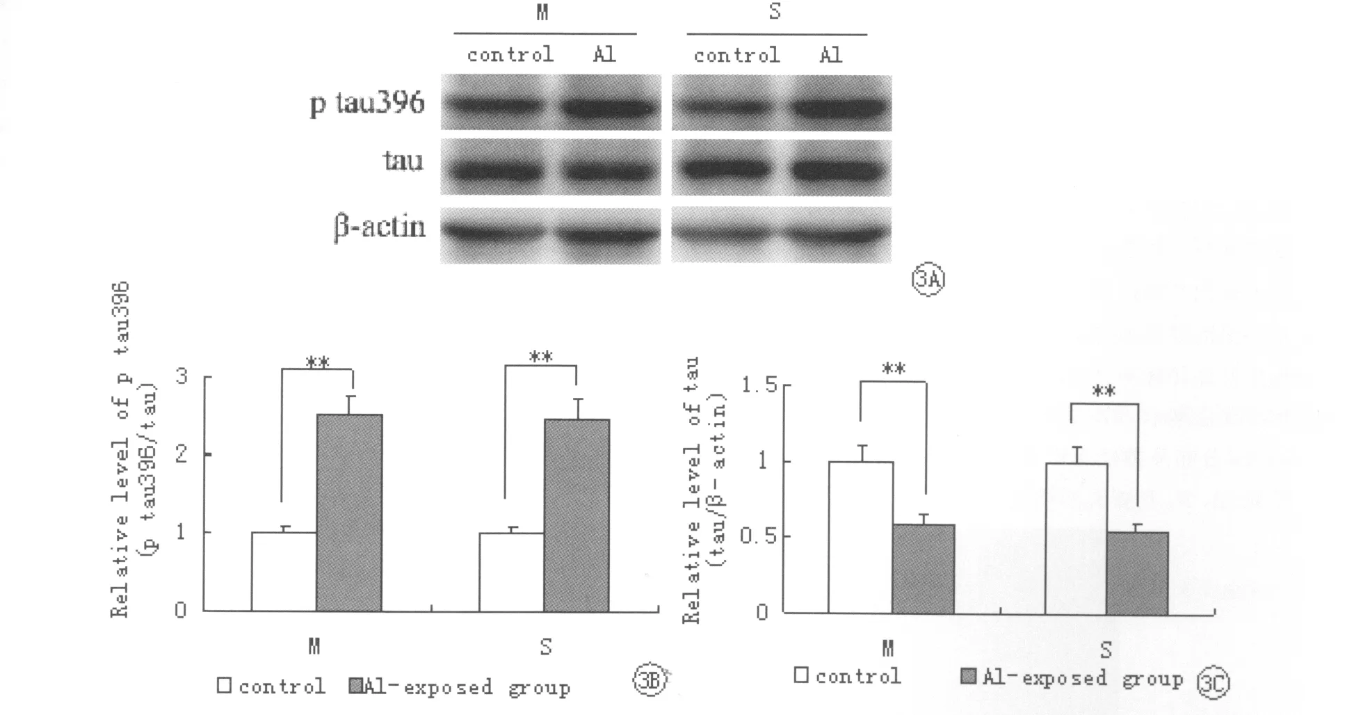
染铝8个月后,对小鼠运动区皮层与感觉区皮层内的tau蛋白及其磷酸化水平进行western blot半定量检测。结果如图3所示:在相对分子量为47kDa处有一清晰的p-tau396和tau条带,以β-actin为内参采用半定量法测IOD(积分光密度)值进行分析。染铝组小鼠两脑区的tau蛋白总量较对照组显著下降(P<0.01),tau蛋白磷酸化水平较对照组显著升高(P<0.01)。
讨 论
铝是否能造成NFTs一直是存在争议的问题,Singer[10]与 Walton[11]等的研究表明铝在兔神经元内能够以Al-NFTs的形式存在于神经元胞浆中;相反,Mizoroki等[12]的研究认为虽然体外实验中铝能引发tau聚合反应,但体内实验结果为阴性。为此,本研究采用Morin与TR联合组织荧光染色对长期铝暴露小鼠皮层神经元进行染色,结果铝在神经元内不同程度的蓄积伴随与之相应的NFTs阳性荧光染色,并且证实了Al-NFTs复合物的存在。同时,我们还发现内含Al-NFTs复合物的神经元形态发生不同程度的改变。前述Mizoroki等的研究对小鼠连续2周腹腔注射Al后未引起tau的异常磷酸化,其原因可能是动物接触铝的时间较短所致。
NFTs主要由高度磷酸化的微管相关蛋白tau逐级纤聚而成。有研究表明[13]通常情况下tau对铝敏感性一般,但当tau磷酸化时,其对铝的敏感性可增加3.5倍。并且铝诱导的tau沉积在金属螯合剂EDTA作用下溶解,而铝诱导的磷酸化tau的沉积却仍保持凝集状态,提示磷酸化作用可增加铝对tau的沉积作用。目前已发现的tau蛋白磷酸化位点高达40多种,其中ser396位点是轻度认知功能障碍向阿尔茨海默病过度的可靠预报位点之一,也与NFTs形成密切相关[14]。本研究发现长期铝暴露虽然减少了小鼠新皮层运动区与感觉区tau蛋白的水平,但是却能显著增加小鼠运动及感觉区皮层内tau蛋白在ser396位点上的磷酸化程度,tau蛋白过度磷酸化也增加了形成NFTs的机率,也与组织荧光染色结果相符。因此,我们得出长期铝暴露诱导tau蛋白过度磷酸化作用可能是NFTs形成的基础之一。

图3 Western blot检测两组小鼠运动区皮层(M)与感觉区皮层(S)tau蛋白及其磷酸化水平。Fig.3 The relative level of tau and p tau396in motor(M)and sensory(S)cortex of each group detected by western blot.A:The bands of p tau396,tau andβ-actin.B:The relative level of p tau396.C:The relative level of tau.** P<0.01vs control group.
[1]Gupta VB,Anitha S,Hegde ML,et al.Aluminum in Alzheimer’s disease:are we still at a crossroad?Cell Mol Life Sci,2005,62(2):143-158
[2]Schneider A,Mandelkow E.Tau-based treatment strategies in neurodegenerative disease.Neurotherapeutics,2008,5(3):443-457
[3]Thal DR,Holzer M,Rub U,et al.Alzheimer-related tau-pathology in the perforant path target zone and in the hippocampal stratum oriens and radiatum correlates with onset and degree of dementia.Exp Neurol,2000,163(1):98-110
[4]Sawa GM,Wharton SB,Ince PG,et al.Age,neuropathology,and dementia.N Engl J Med,2009,360(22):2302-2309
[5]Yumoto S,Nagai H,Kobavashi K,et al.26Al incorporation into the brain of sucking rats through maternal milk.J Inorg Biochem,2003,97(1):155-160
[6]Priest ND.The biological behaviour and bioavailability of aluminium in man,with special reference to studies employing aluminium-26as a tracer:review and study update.J Environ Monit,2004,6(5):375-403
[7]George P,Keith BJF.The mouse brain in stereotaxic coordinates.2nded.San Diego:Academic Press;2001.
[8]Shaw CA,Petrik MS.Aluminum hydroxide injections lead to motor deficits and motor neuron degeneration.J Inorg Biochem,2009,103(11):1555-1562
[9]Galván M,David JP,Delacourte A,et al.Sequence of neurofibrillary changes in aging and Alzheimer’s disease:A confocal study with hosphor-tau antibody,AD2.J Alzheimers Dis,2001,3(4):417-425
[10]Singer SM,Chambers CB,Newfry GA,et al.Tau in aluminum-induced neurofibrillary tangles.Neurotoxicology,1997,18(1):63-76
[11]Walton JR.Evidence for participation of aluminum in neurofibrillary tangle formation and growth in Alzheimer’s disease.J Alzheimers Dis,2010,22(1):65-72
[12]Mizoroki T,Meshitsuka S,Maeda S,et al.Aluminum induces tau aggregation in vitro but not in vivo.J Alzheimers Dis,2007,11(4):419-427
[13]Li W,Ma KK,Sun W,et al.Phosphorylation sensiti-zes microtubule-associated protein tau to Al(3+)-induced aggregation.Neurochem Res,1998,23(12):1467-1476
[14]万章,王春梅 .Tau蛋白过度磷酸化在阿尔茨海默病发病机制中的作用 .医学研究生学报,2010,23(5):539-542
图 版 说 明
EXPLANATION OF FIGURES
图1 Morin法联合TR法组织荧光染色显示染铝小鼠大脑皮层神经元铝负载与NFTs的关联。A,D,G:神经元出现铝负载但NFTs不明显(*);B,E,H:随神经元内铝浓度增加,NFTs出现(*);C,F,I:铝浓度进一步升高伴随更多的NFTs(*)。标尺=10μm。
图2 Morin法联合TR法组织荧光染色显示染铝神经元NFTs分布及神经元形态变化。A,E,I:正常形态神经元;B,F,J:膨大的神经元;C,G,K:萎缩变形的神经元;D,H,L:固缩的神经元。标尺=10μm。
Fig.1 Morin and TR staining showed the relationship between Al loading and NFTs in cortical neurons of.Alexposed mice.A,D,G:Al-positive staining began to appear in neurons,but NFTs was not obvious(*).B,E,H:With Al-positive staining in neurons increasing,NFTs occurred(*).C,F,I:Al loading further increased with more NFTs (* ).Bar =10μm.
Fig.2 Morin and TR staining showed the distribution of NFTs in neurons and the neuronal morphology.A,E,I:The normal form of the neuron.B,F,J:Neurons with an enlargement of cell body.C,G,K:Shrinking deformation of cell body.D,H,L:Pyknotic neuron.Bar=10μm.

Experimental study on the relationship between aluminum exposure and neurofibrillary tangle formation
Zhao Haihua,Lai Hong,Tang Zhongyan,Li Zhaosheng,LüYongli
(Department of Anatomy,Basic Medical College,China Medical University,Shenyang110001,China)
ObjectiveTo investigate the relationship between the neurofibrillary tangle(NFT)formation and the aluminum exposure.Methods16female ICR mice were divided into 2groups:control and aluminum-exposed group(200mg/kg·bw in drinking water,for 8months).Double histofluorescent staining was used to show the distribution of aluminum and NFTs in cortical neurons of mice.Western blot was used to detect the relative level of tau and phospho-tau 396in the neocortex of each group.Results Histofluorescence showed that the degree of NFT positive staining was associated with increasing aluminum loading in neurons.Western blot showed that long term aluminum exposure induced increased tau hyperphosphorylation(P<0.01,vs control group)and decreased tau (P<0.01,vs control group)level.ConclusionAluminum plays a role in the NFT formation by increasing the tau hyperphosphorylation level.
Aluminum;Neurofibrillary tangles;Tau;Morin staining;Thiazin red staining
R 322.81
A
10.3870/zgzzhx.2012.01.006
2011-07-23
2011-11-01
辽宁省科学技术计划项目(2010225034)
赵海花,女(1978年),汉族,讲师。
*通讯作者(To whom correspondence should be addressed)

