儿童肱骨髁上骨折闭合复位两种克氏针固定方法效果观察
吴敏 夏群英 易申德 蔡军 杜香平 熊斌 吴欣乐

[摘要] 目的 觀察内外交叉与单纯外侧交叉两种克氏针固定方法治疗儿童肱骨髁上骨折的临床效果。 方法 回顾性分析自2016年1月至2019年12月在江西省儿童医院小儿骨科诊断肱骨髁上骨折并行手术闭合复位+克氏针固定的患儿共218例。根据患儿采用的克氏针固定方法分为内外交叉组(桡侧两根+尺侧一根)和单纯外侧组(单纯桡侧三根)。观察两组术后有无骨感染、Volkmann挛缩、肘内翻、针尾激惹及神经损伤等,并对结果进行统计学分析。 结果 所有病例无骨感染、Volkmann挛缩及肘内翻畸形等发生。内外交叉组术后发生针尾激惹内外交叉组14例,单纯外侧组6例,组间比较差异无统计学意义(P>0.05)。内外交叉组出现2例(1.32%)医源性尺神经损伤,单纯外侧组未出现,组间比较差异有统计学意义(P<0.05)。随访时骨折全部愈合,末次随访肘关节功能评价两组优良率比较,差异无统计学意义(P>0.05)。 结论 内外交叉克氏针与单纯外侧交叉克氏针治疗小儿肱骨髁上骨折稳定可靠,临床治疗效果无显著差异,但内外交叉克氏针固定有损伤尺神经的风险。
[关键词] 儿童;肱骨髁上骨折;克氏针;闭合复位
[中图分类号] R269.8 [文献标识码] B [文章编号] 1673-9701(2022)04-0075-04
[Abstract] Objective To observe the clinical effects of two Kirschner wire fixation methods (i.e. internal and external cross and simple lateral cross) in the treatment of supracondylar humerus fracture in children. Methods A total of 218 children who were diagnosed with supracondylar humerus fracture in the department of orthopedics, Jiangxi Provincial Children′s Hospital from January 2016 to December 2019 and received surgical closed reduction combined with Kirschner wire fixation were retrospectively analyzed. According to the Kirschner wire fixation method used for patients, they were divided into the internal and external cross group (two wires on the radial side + one wire on the ulnar side) and the simple lateral cross group (three wires on the radial side only). The two groups were observed for bone infection, Volkmann contracture, cubitus varus, Kirschner wire tail irritation and nerve injury after surgery, and the results were statistically analyzed. Results There was no bone infection, Volkmann contracture, and cubitus varus deformity in all cases. Kirschner wire tail irritation occurred in 14 cases in the internal and external cross group and 6 cases in the simple lateral cross group, without statistically significant difference between the two groups (P>0.05). There were 2 cases (1.32%) of iatrogenic ulnar nerve injury in the internal and external cross group, but no case in the simple lateral cross group, with statistically significant difference between the two groups(P<0.05). All fractures healed at follow-up, and there was no statistically significant difference in terms of the excellent rate in elbow joint function evaluation between the two groups at the last follow-up (P>0.05). Conclusion The methods of Internally and externally crossed Kirschner wires and simple laterally crossed Kirschner wires are stable and reliable in the treatment of supracondylar humerus fractures in children, and there is no statistically significant difference in terms of clinical treatment effect. However, the method of internally and externally crossed Kirschner wires may damage the ulnar nerve.
[Key words] Children; Supracondylar humerus fracture; Kirschner wire; Closed reduction
肱骨髁上骨折为小儿最常见的骨折之一,占肘关节损伤的50%~70%,多发生在5~8岁的儿童,男女发生率无明显差异[1-2]。骨折严重时容易引起前臂缺血,导致Volkmann挛缩,从而造成终身残疾。肘内翻为其最常见的晚期并发症,需要截骨矫形手术。根据Gartland分型,Ⅰ型石膏固定保守治疗,Ⅱ、Ⅲ型需要手术治疗[3]。目前主流的手术方法为在全麻或臂丛麻醉下行闭合复位+克氏针内固定[4-5]。但克氏针固定方法颇有争议,争议焦点是内外交叉穿针还是单纯外侧穿针。临床上大多数医生均根据个人经验选择不同的克氏针固定方法。本研究通过回顾性收集分析江西省儿童医院2016年1月至2019年12月行肱骨髁上骨折闭合复位克氏针固定患儿资料并进行随访,比较两种克氏针固定方法之间的治疗效果,现报道如下。
1 资料与方法
1.1一般资料
回顾性分析自2016年1月至2019年12月在江西省儿童医院小儿骨科诊断肱骨髁上骨折并行手术闭合复位+克氏针固定的患儿共218例。根据患儿采用的克氏针固定方法分为内外交叉组(桡侧两根+尺侧一根)和单纯外侧组(单纯桡侧三根)。其中,内外交叉组151例,男96例,女55例,年龄1~11岁,平均(5.87±3.14)岁;单纯外侧组67例,男47例,女20例,年龄2~13岁,平均(4.67±2.66)岁。两组患儿一般资料比较,差异无统计学意义(P>0.05),具有可比性。本研究已经医院医学伦理委员会批准(伦理审查批件号:JXSETYY-YXKY-20200064)。
納入标准[6]:①诊断为肱骨髁上骨折;②受伤时间在14 d内的新鲜骨折;③骨折行闭合复位克氏针内固定;④获得患儿家属知情同意并签字。排除标准[6]:①术前合并神经血管损伤者;②开放性骨折或合并其他部位的多发性骨折者;③合并其他脏器损伤者;④有内分泌代谢异常、肿瘤、感染、血液等疾病者;⑤既往有同部位骨折史者。
1.2方法
全麻成功后,将患儿患肢置于C臂机上呈外展位。患儿平卧位,助手握持肱骨近端,另一助手握持前臂近端。通过缓慢持续牵引完全伸直位肘关节,对于完全移位的严重骨折,因伸直位牵引会使肱二头肌腱及肱肌等肘前结构处于紧张状态,更锁紧了向前移位的骨折近端,同时也使骨折近端、下方的软组织受到更严重的挤压,因而复位困难。可以适当屈肘40°~50°持续纵向牵引。复位时先纠正侧方(尺偏或桡偏)移位,恢复冠状面的力线之后,于牵引下固定近端,使前臂旋前或旋后,矫正远端的旋转畸形。助手缓慢屈肘,同时术者双手四指向后牵拉近端,双拇指同时向前推远端,纠正骨折远端向后的移位。复位成功后维持极度屈肘位固定,用C型臂透视确认骨折对位对线情况。①内外交叉组。由肱骨外髁经皮平行或分散钻入两根直径1.6 mm克氏针,然后,用左手拇指沿内上髁向下方滑动至尺神经沟处,以拇指保护尺神经后再由内上髁顶点进针钻入一根克氏针,所有克氏针均穿透对侧骨皮质,避免克氏针交叉点在骨折线上。②单纯外侧组。由肱骨外髁经皮交叉钻入三根直径1.6 mm克氏针,斜向近端穿透对侧皮质,克氏针尽量呈最大扇形分布,避免克氏针交叉点在骨折线上;剪短针尾,碘伏纱布包绕,无菌敷料覆盖。术后肘关节屈曲70°~90°,长臂玻璃纤维石膏托固定,吊带功能位悬吊于胸前。术后1个月复查X线片,可见骨痂形成或骨折线模糊则门诊拆除石膏及拔除克氏针,并开始向家属讲授轻柔的主、被动功能锻炼,针道伤口每3天换药直至愈合。
1.3 观察指标及评价标准
观察两组术后有无骨感染、Volkmann挛缩、肘内翻、针尾激惹及神经损伤等并记录。术后肘关节功能恢复参照Flynn临床功能美观评定标准进行评价[6],分为优、良、可、差4个等级。与健侧相比,提携角和伸屈功能丢失0°~5°为优,6°~10°为良,11°~15°为可,>15°为差。计算两组关节功能的优良率,优良率=(优+良)例数/总例数×100%,并进行统计学分析。
1.4统计学方法
采用SPSS 17.0统计学软件进行数据分析,计量资料以均数±标准差(x±s)表示,组间比较采用t检验;计数资料以[n(%)]表示,组间比较采用χ2检验,P<0.05为差异有统计学意义。
2 结果
2.1 基本情况
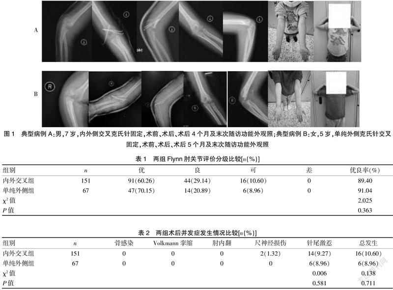
所有病例均获得随访,时间为2~13个月。所有患儿在术后1个月门诊拆除石膏及拔除克氏针,无一例术后发生骨折再移位。在随访期间内所有骨折全部愈合。两组各典型病例见图1。
2.2 两组Flynn肘关节评价分级比较
两组Flynn术后肘关节功能评价在优良率方面比较,差异无统计学意义(P=0.363>0.05)。见表1。
2.3 两组术后并发症发生情况比较
所有病例无骨感染、Volkmann挛缩及肘内翻畸形发生。内外交叉组出现14例(9.27%)针尾激惹,单纯外侧组出现6例(8.96%),拔除克氏针后经过门诊换药后全部愈合。两组并发症总发生率比较,差异无统计学意义(P=0.581>0.05)。内外交叉组术后发生2例(1.32%)医源性尺神经损伤症状,1例表现为手尺侧及小指皮肤感觉麻木,另外1例表现为手尺侧皮肤感觉麻木且分并指受限。术后立即给予拔除尺侧克氏针,重新石膏固定,同时给予口服维生素B6及甲钴胺片,分别于术后第4、7个月神经症状完全恢复。单纯外侧组未出现尺神经损伤病例。见表2。
3 讨论
肱骨髁上骨折主要发生在儿童生长的第1个十年内,与此年龄段内肘关节韧带松弛、过伸位时关节结构的相互关系及髁上区域的骨性结构特点有关。随着儿童骨性结构的发育成熟,该类损伤在青少年中并不常见。移位型的骨折依靠麻醉下闭合复位克氏针内固定大部分能得到满意的疗效,但由于骨折本身的复杂性,关于克氏针的数量、置针的方法仍存在争议。Sankar等[7]回顾279例手术治疗的患儿发现,8例骨折术后有复位的丢失,其中单侧2枚克氏针固定7例,交叉克氏针固定1例;而3枚克氏针固定的37例中未出现一例复位丢失。Bauer等[8]认为,术中应做旋转应力测试,如果存在不稳定,则应在外侧置针后再在内侧小切口切开置入另外一根针。Abzug等[9]认为,Gartland Ⅲ型肱骨髁上骨折至少需要3枚克氏针固定。目前国内小儿骨科医师大部分使用三根克氏针固定,但单纯外侧交叉克氏针还是内外交叉克氏针固定仍没有一致的意见。生物力学实验证明[10-11],单纯桡侧交叉克氏针和内外交叉克氏针在应力稳定性方面是相同的,但内外交叉克氏针的抗旋转力要大于前者,尤其是在内侧柱粉碎时。据此,采用内外交叉克氏针固定似乎有着更好的稳定性,所以目前大部分学者均为内外交叉克氏针固定。本研究收集到的大部分病例亦是使用内外交叉固定方法。
然而,meta分析表明,内外交叉克氏针对尺神经有损伤的风险[12-13]。内外交叉克氏针固定的内侧进针点位于肱骨内上髁,尺神经沿此旁的滑车沟走行,进针时需要依靠术者经验触摸内上髁定位进针。当骨折线由外上斜向内下时,内侧置钉存在一定困难。特别是当骨折后软组织肿胀明显,内上髁触摸不清的情况下,会给穿针造成更大的困难。此时如果反复的尺侧穿针会导致神经损伤的风险进一步上升。国外报道[13],闭合复位内外交叉克氏针固定存在1.4%~15.6%尺神经损伤的概率,与医生的经验水平密切相关,而国内因医患关系原因报道较为少见。为了避免尺神经损伤,术中神经监测、有限内侧切开[14],甚至B超引导下穿针[15]等各种方法时有报道。然而,就算有限切开仍无法避免神经损伤出现的概率,而且切开复位又会带来远期的相关并发症[16-17]。已有研究发现,内外交叉克氏针对尺神经和内上髁的刺激明显,会引起尺神经直接损伤和周围软组织卡压的间接损伤,从而导致神经活动减弱[18]。目前有一些研究[19-20]仅是对这些尺神经损伤进行短期的随访,但是缺乏长期的随访及预后研究。本研究中内外交叉组出现2例(1.32%)医源性的尺神经损伤,虽然术后立即给予拔除尺侧克氏针,且患者分别于术后第4、7个月神经症状完全恢复。但是,拔除后剩余的两根克氏针固定不仅存在术后骨折再移位的风险,而且在当前医疗环境下,还容易增加不必要的医患纠纷。
另外,生物力学研究亦有其局限性,依靠人工复合骨制造的简单横行骨折模型很难模拟临床真实的情况。骨折周围的神经血管、肌肉、韧带等附着的软组织生物力学、骨折线的形态以及石膏外固定的作用也未能纳入实验研究中。Wang等[21]的生物力学研究发现,当骨折线由外下向内上时,单纯外侧交叉克氏针提供的旋转稳定性明显要高于内外交叉克氏针。Ji等[22]认为,外侧穿针可以更容易钻入高于干骺交界线的位置,针的分布更为分散,能提供更高的稳定性。Gopinathan等[23]还推荐了一种外侧分散置针的方法,将骨折线分为四等分,分别经其中三个等分置针可以与内侧交叉克氏针固定在生物力学上无明显差异。一项多中心队列研究[24]发现,内外交叉克氏针医源性神经损伤风险明显高于单纯外侧组。而单纯外侧和内外交叉克氏针固定在影像学和疾病预后方面比较,均无明显统计学差异。本研究结果显示,内外交叉组出现2例(1.32%)医源性的尺神经损伤,而单纯外侧交叉克氏针组未出现。两组在术后关节功能恢复、并发症发生率比较无明显差异。
綜上所述,内外交叉克氏针与单纯外侧交叉克氏针治疗小儿肱骨髁上骨折稳定可靠,临床治疗效果无显著差异,但内外交叉克氏针固定有损伤尺神经的风险。
[参考文献]
[1] Baidoo PK,Kumah AR,Segbefia M,et al. Treatment and outcomes of pediatric supracondylar humeral fractures in Korle Bu Teaching Hospital[J].OTA International:The Open Access Journal of Orthopaedic Trauma,2021,4(2):e124.
[2] 文玉伟,王强,宋宝健,等.闭合复位与桡侧交叉克氏针治疗儿童Gartland Ⅲ型肱骨髁上骨折[J].中华小儿外科杂志,2020,41(9):824-830.
[3] 吕云亮,杨蕊,杨超.经皮克氏针固定儿童不稳定性肱骨髁上骨折[J].中国矫形外科杂志,2020,28(20):1845-1848.
[4] Wendlingkeim DS,Binder M,Dietz HG,et al. Prognostic factors for the outcome of supracondylar humeral fractures in children[J]. Orthopaedic Surgery,2019,11(4):690-697.
[5] 倪宏强,楼跃.儿童肱骨髁上骨折的治疗进展[J].临床小儿外科杂志,2020,19(4):364-369.
[6] Roy MK,Alam MT,Rahman MW,et al.Comparative study of stabilization of humerus supracondylar fracture in children by percutaneous pinning from lateral side and both sides[J].Mymensingh Medical Journal:MMJ,2019, 28(1):15-22.
[7] Sankar WN,Hebela NM,Skaggs DL,et a1.Loss of pin fixation in displaced supracondylar humeral fractures in children:Causes and prevention[J].J Bone Joint Surg Am,2007,89(4):713-717.
[8] Bauer JM,Stutz CM,Schoenecker JG,et al.Internal rotation stress testing improves radiographic outcomes of type 3 supracondylar humerus fractures[J].J Pediatr Orthop,2019,39(1):8-13.
[9] Abzug JM,Herman MJ.Management of supracondylar humerus fractures in children:Current concepts[J].J Am Acad Orthop Surg,2012,20(2):69-77.
[10] Lee SS,Mahar AT,Miesen D,et al. Displaced pediatric supracondylar humerus fractures: Biomechanical analysis of percutaneous pinning techniques[J].J Pediatr Orthop,2002,22(4):440-443.
[11] 李麒麟,Allieu Kamara,王恩波,等.兒童肱骨远端骨干干骺端交界区骨折不同固定方法生物力学的比较[J].中国骨与关节损伤杂志,2020,35(9):917-920.
[12] Dekker AE,Krijnen P,Schipper IB,et al.Results of crossed versus lateral entry K-wire fixation of displaced pediatric supracondylar humeral fractures:A systematic review and meta-analysis[J].Injury,2016,47(11):2391-2398.
[13] Carrazzone OL,Barbachan Mansur NS,Matsunaga FT,et al.Crossed versus lateral K-wire fixation of supracondylar fractures of the humerus in children:A meta-analysis of randomized controlled trials[J].Journal of Shoulder and Elbow Surgery,2020,30(2):439-448.
[14] Roy RK,Prasad M. Study of supracondylar fracture of humerus in children by open reduction and internal fixation with K-wires[J].Journal of Evidence Based Medicine and Healthcare,2017,4(41):2484-2487.
[15] Xu WB,Dai RD,Liu Y,et al.Ultrasound-guided reduction and percutaneous crossed pin fixation for the treatment of displaced supracondylar fracture of the humerus in children[J].Zhongguo Gu Shang,2020,33(10):907-911.
[16] Green DW,Widmann RF,Frank JS,et al. Low incidence of ulnar nerve injury with crossed pin placement for pediatric supracondylar humerus fractures using a mini-open technique[J]. J Orthop Trauma,2005,19(3):158-163.
[17] Eguia F,Gottlich C,Lobaton G,et al.Mid-term patient-reported outcomes after lateral versus crossed pinning of pediatric supracondylar humerus fractures[J].Journal of Pediatric Orthopedics,2020,40(7):323-328.
[18] Belhan O,Karakurt L,Ozdemir H,et al. Dynamics of the ulnar nerve after percutaneous pinning of supracondylar humeral fractures in children[J].Journal of Pediatric Orthopaedics,2009,18(1):29-33.
[19] Woo CY,Ho HL,Ashik MBZ,et al.Paediatric supracondylar humeral fractures:A technique for safe medial pin passage with zero incidence of iatrogenic ulnar nerve injury[J].Singapore Medical Journal,2018,59(2):94-97.
[20] Barrett KK,Skaggs DL,Sawyer JR,et al.Supracondylar humeral fractures with isolated anterior interosseous nerve injuries:Is urgent treatment necessary?[J] J Bone Joint Surg Am,2014,96(21):1793-1797.
[21] Wang X,Feng C,Wan S,et al.Biomechanical analysis of pinning configurations for a supracondylar humerus fracture with coronal medial obliquity[J].J Pediatr Orthop B,2012,21(6):495-498.
[22] Ji X, Kamara A, Wang E,et al. A two-stage retrospective analysis to determine the effect of entry point on higher exit of proximal pins in lateral pinning of supracondylar humerus fracture in children[J].J Orthop Surg Res,2019,14(1):351.
[23] Gopinathan NR,Sajid M,Sudesh P,et al.Outcome analysis of lateral pinning for displaced supracondylar fractures in children using three kirschner wires in parallel and divergent configuration[J].Indian Journal of Orthopae dics,2020,52(5):554-560.
[24] Claireaux HA,Goodall R,Hill J,et al.Multicentre collaborative cohort study of the use of Kirschner wires for the management of supracondylar fractures in children[J]. Chinese Journal of Traumatology,2019,22(5):249-254.
(收稿日期:2021-03-23)

