临床误诊为肺炎的黏液腺癌1例
临床误诊为肺炎的黏液腺癌1例
牛云,史艳芬,佟杰*
(卫生部直属中日友好医院,北京100029)
1临床资料
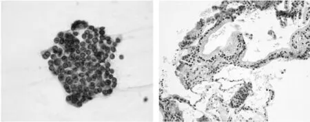
患者男性,65岁,2013年11月患者因爬山劳累受凉后出现咳痰,为白色泡沫状,量多,咳嗽不明显,无发热、鼻塞、流涕,无咳血,无胸闷胸痛等症状,就诊于当地医院,诊断为“上呼吸道感染”,予抗炎治疗,无效,2天后痰量增多,就诊于上级医院,行胸片示:肺气肿改变,右下肺炎,右侧胸腔少量积液。予以抗炎治疗痰略有减少,效果不佳,遂来我院就诊,于呼吸内科住院治疗,经抗感染治疗痰量未见减少。送检毛刷细胞涂片结果示:查见核异质细胞(图1左)。行CT引导下穿刺,病理结果示:腹壁生长为主的黏液腺癌可能性大(图1右)。住院期间行PET-CT检查,结果示:右肺下叶及中叶局部实变影,轻度葡萄糖代谢增高,不除外炎性肺癌可能,建议结合CT穿刺进一步检查。遂转入胸外科,于2014年1月5日行右肺下叶切除+右肺中叶外侧段切除术。标本送病理。
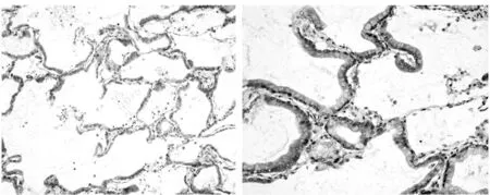
手术标本大体检查:右肺下叶体积21 cm×11 cm×11 cm,胸膜面光滑,灰白色,支气管断端直径2 cm,长0.5 cm,大部分黏膜光滑,部分区域可见散在灰白色斑块样,肺组织切面灰黄色,呈胶冻样,富于黏液,细腻,质软,病变几乎累及整个肺叶,紧邻胸膜,距支气管断端最近距离0.5 cm,周围仅见极少量正常肺组织。镜下特征:肿瘤细胞沿细支气管肺泡壁排列,部分增生呈小乳头状,大部分细胞呈单层柱状或杯状,部分为3-4层,胞浆透亮呈分泌状态,含大量黏液,核位于基底,细胞轻度异型,核分裂像罕见(图2)。免疫组化结果显示:CK5/6(-),CK7(+),KP-1(-),Napsin A(+),TTF-1(+),P63(-)。

图1 毛刷涂片HE染色(左图)(×400);CT穿刺组织学
病理诊断:(右肺下叶)黏液腺癌(弥漫分布于整个肺叶),未侵犯胸膜。

图2 手术标本HE染色(左图×100,右图×200)
2讨论
非小细胞肺癌是全世界导致肿瘤相关性死亡的主要原因[1]。而腺癌是最常见的非小细胞肺癌的病理类型,病发率呈上升趋势。在2011 年肺腺癌国际多学科分类中[2],将原分类中的黏液性细支气管肺泡癌取消,而浸润性黏液腺癌被提出作为独立的肺腺癌亚型。黏液腺癌可以发生在多种器官,单纯的肺黏液腺癌在肺腺癌中比较少见,需排除其它部位转移,才能做出原发性诊断,本例经PET-CT核实无其他部位病灶,符合原发性。本例癌变累及整个肺叶,实属罕见,国内仅见2例报道。黏液性腺癌主要有两种组织形态学表现,一种是具有丰富的细胞内黏液的柱状或杯状细胞,另一种是具有丰富的细胞外黏液,伴有间质浸润的浸润性腺癌成分。发生在肺的浸润性黏液腺癌也具有以上两种组织学表现。与其他肺腺癌亚型相比,浸润性黏液腺癌具有不同的免疫组化和分子生物特征,以及不同的预后。浸润性黏液腺癌在东亚、欧洲及美国占肺腺癌的2%-10%[3-5],比其他常见的肺腺癌亚型(如附壁生长型和腺泡型)更具侵袭性[6],对于浸润性黏液腺癌的预后,部分学者认为黏液腺癌的预后比非黏液性腺癌差[7],部分学者认为黏液腺癌不能作为预后差的指标[8]。最近有学者报道,将浸润性黏液腺癌根据以上所说的两种组织形态学特点分为细胞内黏液性腺癌及细胞外黏液性腺癌,据研究结果显示,细胞内黏液性腺癌与非黏液性腺癌相比较,预后无明显差异;而细胞外黏液性腺癌,与非黏液性腺癌相比预后明显差。而且他们将细胞外黏液性腺癌和细胞内黏液性腺癌相比较,细胞外黏液性腺癌预后更差,其淋巴结更易转移。本例为单纯性细胞内黏液腺癌,细胞为柱状或杯状细胞,分化好,未见黏液湖及间质浸润区,无淋巴结转移。
肺原发性细胞内黏液腺癌,临床症状出现较晚,病程较长,可达数年以上。临床之所以将本例病变误诊为肺炎,因其病变累及整个肺叶,影像学表现为大片均匀实变高密度影或实变病灶,始终未见实质性肿块,无特征性影像学变化,易误诊为大叶性肺炎,导致较长时间内在多家医院误诊。在此,提醒临床医师注意,对于肺黏液腺癌,单凭影像学改变难以作出诊断,要尽早设法取得病理学资料,以便作出正确诊断,争取治疗时间。
参考文献:
[1]Jemal A,Bray F,Center MM,et al.Global cancer statistics[J].CA Cancer J Clin,2011,61(2):69.
[2]Travis WD,Brambilla E,Noguchi M,et al.International association for the study of lung cancer/American thoracic society/european respiratory society international multidisciplinary classification of lung adenocarcinoma[J].J Thorac Oncol,2011,6(2):244.
[3]Warth A,Muley T,Meister M,et al.The novel histologic International Association for the Study of Lung Cancer/American Thoracic Society/European Respiratory Society classification system of lung adeno-carcinoma is a stage-independent predictor of survival[J].J Clin Oncol,2012,30(13):1438.
[4]Tsuta K,Kawago M,Inoue E,et al.The utility of the proposed IASLC/ATS/ERS lung adenocarcinoma subtypes for disease prognosis and correlation of driver gene alterations[J].Lung Cancer,2013,81(3):371.
[5]Yoshizawa A,Motoi N,Riely GJ,et al.Impact of proposed IASLC/ATS/ERS classification of lung adenocarcinoma: prognostic subgroups and implications for further revision of staging based on analysis of 514 stage I cases[J].Mod Pathol,2011,24(5):653.
[6]Cadranel J,Quoix E,Baudrin L,et al.IFCT-0401 Trial Group.IFCT-0401 Trial: a phase II study of gefitinib administered as first-line treat-ment in advanced adenocarcinoma with bronchioloalveolar carcinoma subtype[J].J Thorac Oncol,2009,4(9):1126.
[7]Russell PA,Wainer Z,Wright GM,et al.Does lung adenocarcinoma subtype predict patient survival:A clinicopathologic study based on the new International Association for the Study of Lung Cancer/American Thoracic Society/European Respiratory Society international multidisciplinary lung adenocarcinoma classification[J].J Thorac Oncol,2011,6(9):1496.
[8]Westaway DD,Toon CW,Farzin M,et al.The International Association for the Study of Lung Cancer/American Thoracic Society/European Respiratory Society grading system has limited prognostic significance in advanced resected pulmonary adenocarcinoma[J].Pathology,2013,45(6):553.
[9]Deng Cai,Hang Li.Comparison of clinical features,molecular alterations,and prognosis in morphological subgroups of lung invasive mucinous adenocarcinoma[J].Onco Targets and Therapy,2014,7:2127.
收稿日期:(2014-08-09)
文章编号:1007-4287(2015)04-0677-02
通讯作者*

