趋化因子受体CXCR4在结肠癌中的表达及其临床意义
钱 益,施 文,孙晋洁,孙永强
趋化因子受体CXCR4在结肠癌中的表达及其临床意义
钱 益,施 文,孙晋洁,孙永强
南通大学第二附属医院普外科,南通 226001
目的探讨趋化因子受体4(chemokine receptor4,CXCR4)在结肠癌组织中的表达及其临床意义。方法选择2012年南通大学第二附属医院普外科施行的结肠癌手术组织标本及对应癌旁组织,应用免疫组织化学方法检测不同临床分期结肠癌组织(50例)及癌旁组织(10例)CXCR4表达。结果 结肠癌CXCR4阳性表达率明显高于癌旁组织[72.0%(36/50)vs.10%(1/10),P<0.05];其表达水平在肿瘤侵犯浆膜层、淋巴结转移,TNM高分期明显增高(P<0.05);但与结肠癌患者性别、年龄、肿瘤部位、肿瘤大小、组织学类型无关。结论 CXCR4蛋白在结肠癌高表达,并与肿瘤恶性程度和病人预后相关。
结肠癌;趋化因子受体4;免疫组化
结肠癌是常见的消化系统恶性肿瘤之一,在我国其发病率呈逐年上升趋势[1-3]。外科根治性手术是主要的治疗措施,但临床上仍有相当病例在术后发生复发转移,特别是肝转移,预后欠佳[5]。如何准确判断结肠癌预后并采取相应的治疗方法成为当前的研究热点。趋化因子受体4(CXCR4)属于趋化因子受体家族成员,在正常细胞不表达或微弱表达,但存在于多种人类肿瘤细胞表面,且表达明显上调[6]。研究发现CXCR4在多种肿瘤的发生进展中起重要作用[7-8]。本文运用免疫组织化学方法检测50例结肠癌组织标本CXCR4蛋白表达,探讨CXCR4与结肠癌生物学行为的关系。
1 材料和方法
1.1 标本来源
选取2012年1月至2014年12月南通大学第二附属医院普外科手术切除的结肠癌标本50例,其中男37例,女13例,年龄43~81岁(中位年龄64岁);高、中、低分化腺癌各8例、15例、27例;按AJCC/ UICC推荐TNM分期属Ⅰ、Ⅱ、Ⅲ、Ⅳ期各6例、14例、24例、6例。并取其中10例结肠癌标本的癌旁组织(距肿瘤组织超过5 cm)。所有病例均经南通大学第二附属医院病理科诊断确认。所有患者均进行了电话随访,随访时间12~40个月(中位32个月)。按随访结果分复发组17例、删失组(研究结束仍存活或因其他疾病死亡或随访过程中失访)33例。
1.2 免疫组化分析
所有组织标本4%甲醛固定,制作6μm石蜡切片;经脱蜡水化,柠檬酸盐高温修复抗原,PBS缓冲液漂洗后以3%过氧化氢-甲醇孵育,清除内源性过氧化物酶活性,PBS漂洗后滴加正常山驴羊血清封闭,室温孵育,PBS冲洗,滴加1∶200兔CXCR4多克隆抗体(abcam公司),4℃冰箱孵育过夜,PBS漂洗后再滴加二抗,室温孵育30m in,PBS漂洗,DAB显色,苏木精复染,脱水,二甲苯透明,中性树胶封片,PBS代替一抗作阴性对照。
结果判定,CXCR4阳性判定标准为细胞浆及细胞膜内出现淡黄色至棕褐色染色为CXCR4阳性阳性细胞,随机选择5个高倍镜视野(×400)进行判断。阳性细胞的染色强度按无着色、淡黄色、棕黄色和棕褐色分别记为0、1、2、3分,CXCR4阳性细胞率按<5%、6%~25%、26%~50%、>50%分别记为0、1、2、3分,然后两项记分相乘,得出结果判断:大于等于1分的记为阳性,小于1分为阴性(其中一项计分为0即阴性)。
1.3 统计学分析
采用SPSS17.0软件。定性资料用χ2检验或Fisher参数精确检验,单因素分析用Kaplan-Meier法及Log-Rank检验,以P<0.05为差异有统计学意义。
2 结果
2.1 CXCR4蛋白在结肠癌及癌旁组织中表达
如图1~2所示,CXCR4主要表达于细胞浆和细胞膜,细胞核不表达;结肠癌细胞有CXCR4阳性表达,肿瘤分期低者表达较弱呈浅黄或黄色,分期高呈棕褐色且阳性细胞数较多;CXCR4在结肠癌组织中阳性率为72.0%(36/50),癌旁正常结肠组织仅1 例CXCR4表达阳性(1/10,10%),两者CXCR4表达阳性率差异有统计学意义(P<0.01,表1)。

表1 CXCR4在结肠癌及癌旁组织中表达Tab.1 The expression of CXCR4 in the colon cancer and paracancerous tissue
2.2 结肠癌CXCR4表达与临床病理的关系
如表2,在有、无淋巴结转移,CXCR4表达阳性率存在差异(52.6%vs.83.9%,P<0.05)。在肿瘤未侵犯与侵犯浆膜层(50%vs.82.4%,P<0.05)、结肠癌Ⅰ、Ⅱ期和Ⅲ、Ⅳ期(40%vs.93.3%,P<0.05),CXCR4表达阳性率均有统计学意义差异(P<0.05),前者提示CXCR4阳性肿瘤具有较高侵袭性,可能与患者复发转移不良预后有关,后者表明CXCR4阳性表达随结肠癌分期增加而升高。但CXCR4表达与患者年龄、性别、肿瘤大小、肿瘤部位、分化程度及手术方式无关。
2.3 结肠癌CXCR4表达等与预后的关系
本组患者1年、3年未复发分别为31.3%、92%。复发时间为6~40月,中位复发时间为25个月。经Kaplan-Meier法单因素生存分析及Log-Rank检验,CXCR4表达、淋巴结转移、肿瘤大小、浸润深度和临床分期对结肠癌患者1年及3年复发率有显著影响(P<0.05,表3;图3)。
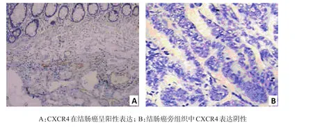
图1 CXCR4在结肠癌及其癌旁组织中的表达A:The positiveexpression of CXCR4 in colon cancers;B:The expression of CXCR4 in paracancerous colon tissueswasnegativeFig.1 The exp ression of CXCR4 in colon cancersand paracancerous colon tissues
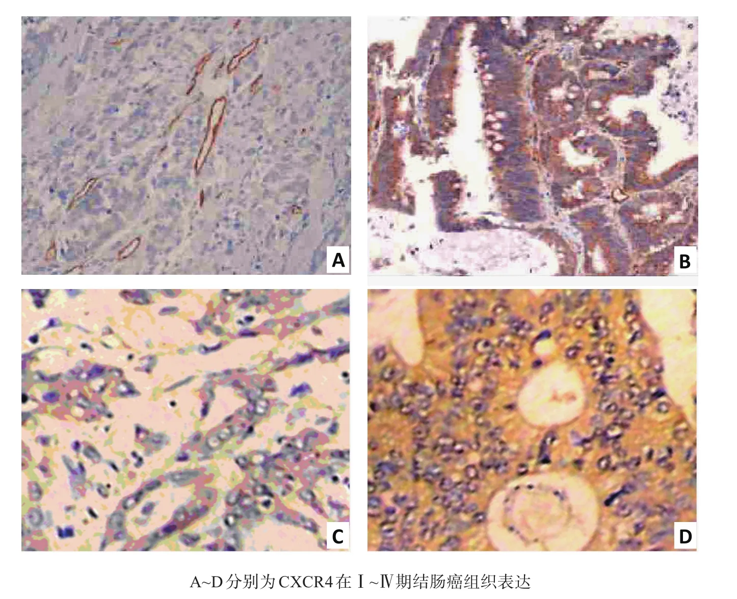
图2 CXCR4在不同分期结肠癌表达A~D is theexpression of CXCR4 inⅠ~Ⅳstage of coton cancer respectivelyFig.2 The expression of CXCR 4 in different colon cancer stage
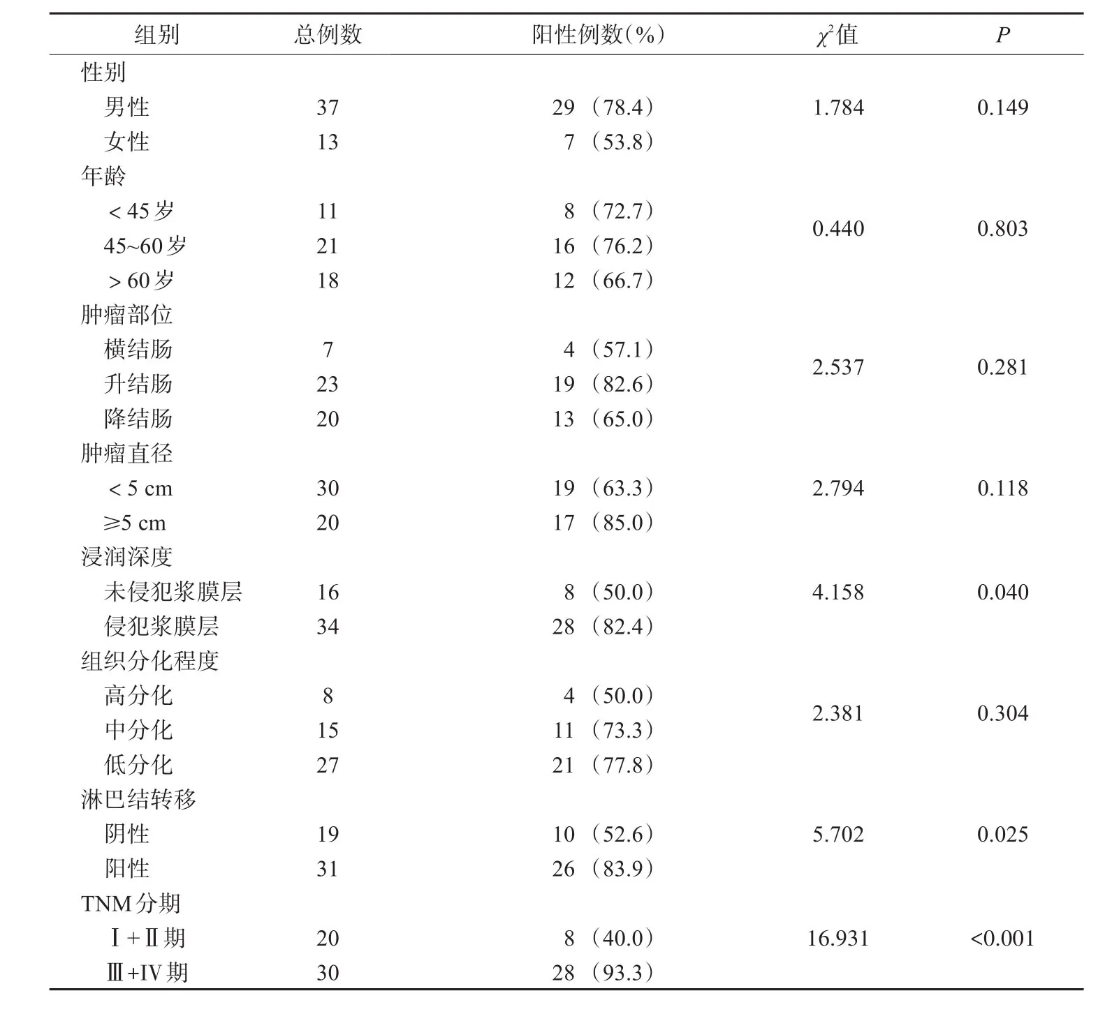
表2 结肠癌CXCR4表达与临床病理参数的关系Tab.2 Relationship between CXCR4 exp ression and clinicopathological factors in colon cancer
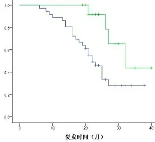
图3 CXCR4表达与患者复发时间关系Fig.3 Relationship between CXCR4 expression and recurrence time
3 讨论
趋化因子及其受体的主要功能是参与炎症、胚胎发育、造血、血管生成及动脉粥样硬化等病理生理过程,因受体与配体间的结合产生“归巢”效应发挥作用故而得名[8]。研究表明,CXCR4在多种人类肿瘤细胞表面表达,在乳腺癌、肺癌、神经胶质瘤、前列腺癌等肿瘤细胞的迁移、增殖、侵袭、转移过程中发挥重要作用[9-11]。
本文应用免疫组化检测CXCR4在结肠癌高表达,癌旁组织阴性或表达低(1/10例弱阳性);随结肠癌分期增高其表达上调(P<0.05),有淋巴结转移、侵犯结肠浆膜层组CXCR4阳性率明显高于无淋巴结转移、未侵犯浆膜组(P<0.05);但患者年龄、性别、肿瘤部位、大小、分化程度无关。提示CXCR4阳性表达肿瘤具有较高的侵袭、转移特性,与结肠癌TNM分期、肿瘤进展相关;CXCR4可能在结肠癌的发生、发展中起重要作用,参与结肠癌病理进展过程,与结肠癌的恶性行为密切相关。
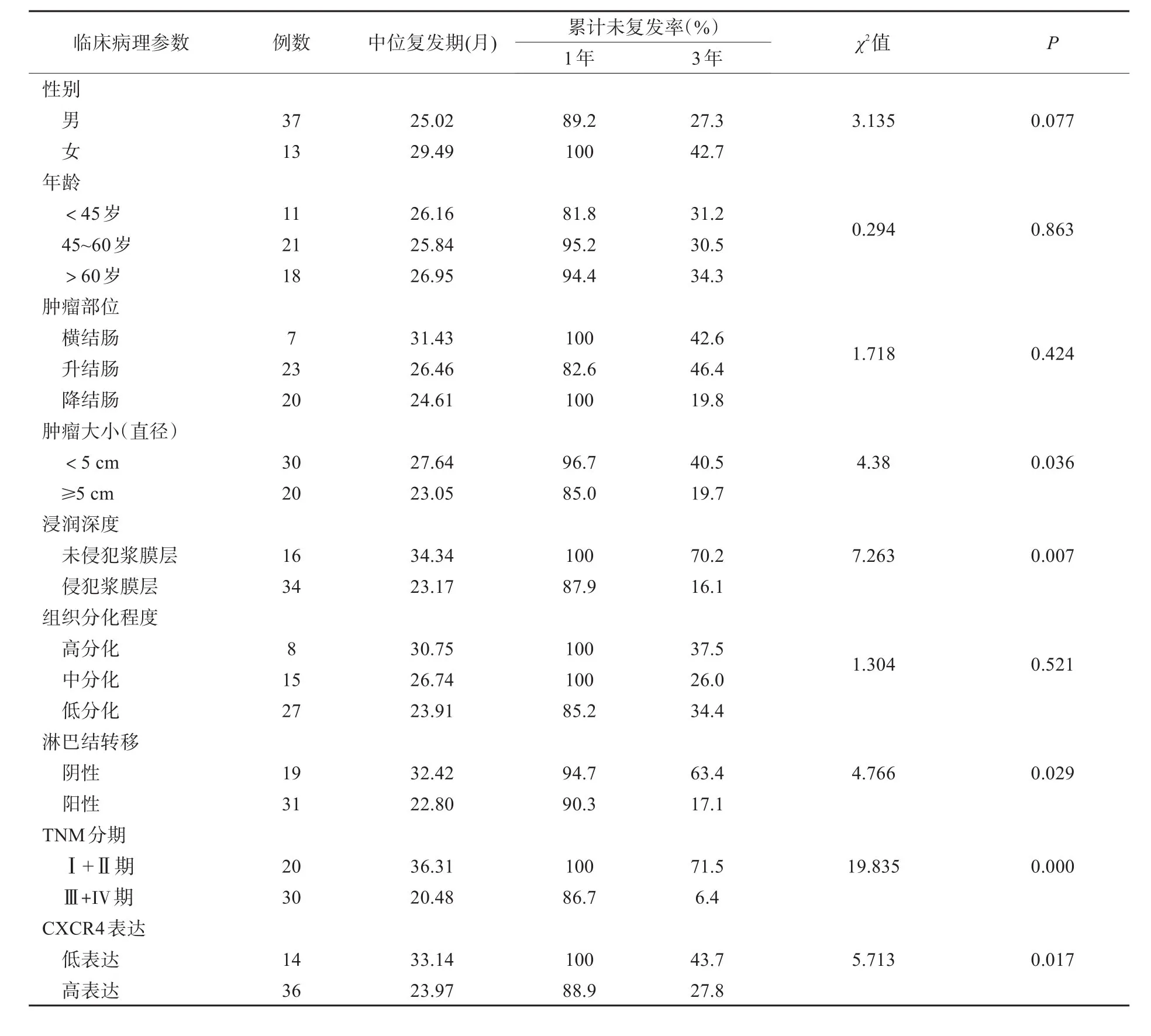
表3 结肠癌CXCR4表达等与预后的关系Tab.3 Relationship between CXCR4 exp ression et aland p rognosis in colon cancer
随着CXCR4及其他趋化因子受体家族成员在结肠癌发生发展过程中的研究深入和CXCR74相关信号传导通路及各配体间协同关系的逐渐清晰[12-15],靶向沉默CXCR4及其相关趋化因子受体基因表达,可能为将来结肠癌防治靶点提供理论基础和实验依据。
[1] Meincke M.Near-infrared molecular imaging of tumors via chemokine receptors CXCR4 and CXCR7[J].Clin Exp Metastasis,2011,28(8):713-720.
[2] M iyoshi K.Close correlation between CXCR4 and VEGF expression and frequent CXCR7 expression in rhabdomyosarcoma[J].Hum Pathol,2014,45(9):1900-1909.
[3] Hattermann K,M entlein R.An infernal trio:the chemokine CXCL12 and its receptors CXCR4 and CXCR7 in tumor biology[J].Ann Anat,2013,195(2):103-110.
[4]邱明远,李健文,郑民华.CXCR4和CXCR7在肿瘤中的研究进展[J].中国癌症杂志,2010,20(3):222-226.
[5] Sun X.CXCL12/CXCR4/CXCR7 chemokine axis and cancer progression[J].Cancer M etastasis Rev,2010,29(4):709-722.
[6] Boudot A.Differential estrogen-regulation of CXCL12 chemokine receptors,CXCR4 and CXCR7,contributes to the grow th effect of estrogens in breast cancer cells[J].PLoSOne, 2011,6(6):e20898.
[7] Walentow icz-Sadlecka M.Stromal derived factor-1(SDF-1)and its receptors CXCR4 and CXCR7 in endometrial cancer patients[J].PLoSOne,2014,9(1):e84629.
[8] Romain B.Hypoxia differentially regulated CXCR4 and CXCR7 signaling in colon cancer[J].Mol Cancer,2014,13 (98):58.
[9] M iyoshi K.Close correlation between CXCR4 and VEGF expression and frequent CXCR7 expression in rhabdomyosarcoma[J].Hum Pathol,2014,45(9):1900-1909.
[10] D'A lterio C.Concom itant CXCR4 and CXCR7 expression predicts poor prognosis in renal cancer[J].Curr Cancer Drug Targets,2010,10(7):772-781.
[11] Choi YH.CXCR4,but not CXCR7,discrim inates metastatic behavior in non-small cell lung cancer cells[J].Mol Cancer Res,2014,12(1):38-47.
[12] Wurth R.CXCL12 modulation of CXCR4 and CXCR7 activity in human glioblastoma stem-like cells and regulation of the tumormicroenvironment[J].Front Cell Neurosci,2014,8(28):144.
[13] Liu C.Expression and functional heterogeneity of chemokine receptors CXCR4 and CXCR7 in primary patient-derived glioblastoma cells[J].PLoSOne,2013,8(3):e59750.
[14] Beider K.CXCR4 antagonist 4F-benzoyl-TN14003 inhibits leukem ia and multiple myeloma tumor grow th[J].Exp Hematol,2011,39(3):282-292.
[15] Jin J,Zhao WC,Yuan F.CXCR7/CXCR4/CXCL12 axis regulates the proliferation,m igration,survival and tube formation of choroid-retinal endothelial cells[J].Ophthalm ic Res,2013,50(1):6-12.
Expression of CXCR4 in colon cancersand its clinicalvalue
QIAN Yi,SHIWen,SUN Jinjie,SUN Yongqiang
DepartmentofSurgery,the Second Affiliated Hospital,Nantong University,Nantong 226001,China
Objective To observe the expression of chemokine receptor 4(CXCR4)in colon cancer and its clinical value.M ethods The expression of CXCR4 protein in 50 specimens of colon cancer and 23 specimens of paracancerous from The 2nd A ffiliated Hospital of Nantong University were analyzed by immunohistochem ical staining and its relationship with clinicopathological features and prognosiswas observed.Results The positive rate of CXCR4 expression in colon cancerswas significantly higher than thatof paracancerous tissues[72.0%(36/50)vs.10%(1/10),P<0.05];and CXCR4 expression was significantly upregulated in colon serosa invasion,lymph nodemetastasis and high TNM stage(P<0.05).But CXCR4 expression was not related to the gender,age,tumor location,tumor size and histological type of colon cancer of patients.Conclusion The expression of CXCR4 protein in colon cancers is upregulated and related to colon cancermalignancy and prognosis.
Colon cancer;chemokine receptor 4;Immunohistochem istry
R656
A
2095-378X(2015)03-0153-05
10.3969/j.issn.2095-378X.2015.03.004
江苏省南通市科技局社会事业科技创新项目(HS2013031)作者简介:钱 益(1980—),男,主治医师,从事胃肠外科
孙永强,电子信箱:sunyq505@sina.com

