慢性冠脉综合征患者血尿酸及钙化积分对CT-FFR的影响
徐彧 杜丰夷 袁小佳 赵天佐 陈正光


[摘要] 目的:探讨血尿酸及钙化积分对深度学习算法下冠脉综合征(CCS)患者血流储备分数(CT-FFR)的影响。方法:回顾性分析150例冠状动脉(冠脉)CTA(CCTA)示靶血管狭窄的慢性CCS患者,采用深度学习法计算其CT-FFR,取3支靶血管中最低值作为该患者CT-FFR,按CT-FFR>0.8及CT-FFR≤0.8分为正常组77例、异常组73例,比较2组血尿酸值及总钙化积分;采用SPSS 26.0统计软件对CT-FFR、血尿酸、钙化积分行Pearson分析;使用logistic回归分析且通过ROC曲线评估其诊断效能。结果:2组在钙化积分上差异有统计学意义(Z=-5.958,P<0.05),而血尿酸差异无统计学意义(t=-0.448,P>0.05);Pearson相关分析得出,钙化积分与CT-FFR呈显著负相关(r=-0.556,P<0.05);ROC曲线的AUC为0.782,且达到病变程度(CT-FFR≤0.8)的钙化积分阈值为313.05分,约登指数为0.464,其诊断准确率为78.2%,敏感度为68.5%,特异度为77.9%。结论:冠脉钙化积分可能影响CT-FFR,可预测功能性缺血的发生,即钙化积分越高,血管功能越差,CT-FFR越低,缺血越严重。CCS患者血尿酸是否與CT-FFR存在相关性有待进一步研究。
[关键词] 血尿酸;钙化积分;血流储备分数;冠状动脉;心肌缺血
Effect of blood uric acid and calcification scores to CT-based noninvasive fractional flow reserve (CT-FFR) in patients with chronic coronary syndrome
[Abstract] Objective:To explore the effect of blood uric acid and calcification score to CT-based noninvasive fractional flow reserve (CT-FFR) in patients with chronic coronary syndrome (CCS). Methods:A total of 150 CCS patients were enrolled,all patients underwent coronary CTA (CCTA) and stenosis in the target vessels was detected. and the serum uric acid concentration was measured during hospitalization. CT-FFR was calculated by deep learning method,and the lowest value among the three target vessels was taken as the CT-FFR value. According to the CT-FFR value,all patients were divided into the normal group (CT-FFR>0.8,77 cases) and the abnormal group (CT-FFR≤0.8,73 cases),and the serum uric acid and the total calcification score were compared between the two groups. Pearson correlation analysis was performed for CT-FFR and serum uric acid,total calcification score,logistic regression analysis was performed,and the diagnostic accuracy was analyzed by ROC curve. Results:There was a statistically significant difference in calcification score between the two groups (Z=-5.958,P<0.05) and no significant difference in serum uric acid (t=-0.448,P>0.05). Pearson correlation analysis revealed a significant negative correlation between calcification score and CT-FFR (r=-0.556,P<0.05). The AUC was 0.782,and the calcification score threshold for pathological level (CT-FFR≤0.8) was 313.05 points,with a Youden index of 0.464,an accuracy of 78.2%,a sensitivity of 68.5% and a specificity of 77.9%. Conclusions:The calcification score of coronary artery can influence the CT-FFR value and can predict the occurrence of functional ischemia. The greater the calcification score,the lower the CT-FFR. Whether uric acid has a corrrelation with CT-FFR in CCS patients needs the further study.
[Key words] Serum uric acid;Calcification score;Fractional flow reserve;Coronary artery;Myocardial ischaemia
冠状动脉(冠脉)造影下的血流储备分数(fractional flow reserve,FFR)作为诊断的冠脉血流动力学的金标准[1],存在费用高、有创等局限。而基于冠脉CTA(coronary computed tomography angiography,CCTA)的血流储备分数(computed tomography derived fractional flow reserve,CT-FFR)因具有无创、便捷快速、诊断效能高等特点[2-3]被应用于临床。
既往大量研究表明,高血尿酸血症(hyperuricemia,HUA)是冠状动脉粥样硬化性心脏病(冠心病)的危险因素[4]。临床诊断冠脉狭窄往往基于其解剖学的结构狭窄,忽视了管腔狭窄后血管自身代偿及侧支循环建立后血管所支配的心肌供血情况,不能客观反映是否存在心肌缺血[5],且高浓度血尿酸对血管内皮影响可导致血管不同程度钙化,从而影响血管弹性[6-8]。因此,本研究以CCS患者的CT-FFR、血尿酸及钙化积分为研究对象,探讨血尿酸及钙化积分是否对CT-FFR产生影响。
1 资料与方法
1.1 一般资料
回顾性分析2018年6月至2021年6月于北京中医药大学东直门医院行CCTA的慢性冠脉综合征(chronic coronary syndrome,CCS)[9]患者,筛选其CCTA结果存在狭窄且有血尿酸检测结果者。排除标准:①既往行冠脉血运重建;②存在心肌桥;③CTA图像质量不合格(呼吸错层伪影、断层、层厚>1 mm等);④有其他可导致血尿酸代谢异常的血液系统性疾病;⑤近30 d曾服可致血尿酸含量增加的药物。
最终纳入150例,年龄34~88岁,其中男87例,平均年龄(65.33±9.89)岁;女63例,平均年龄(70.27±10.18)岁。根据2017年《中国高血尿酸血症相关疾病诊疗多学科专家共识》[10],以男性血尿酸值>420 μmol/L、女性>360 μmol/L诊断为HUA,以CT-FFR=0.8為冠脉缺血的临界值[11],>0.8为正常组(77例),≤0.8为异常组(73例)。所有患者均签署知情同意书。
1.2 仪器与方法
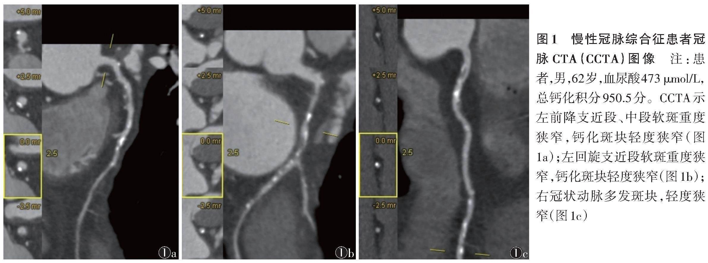
1.2.1 CCTA扫描 使用Siemens新双源CT对冠脉进行扫描,患者心率≤90次/min。扫描参数:100~120 kV,350~450 mA,准直器宽度128×0.625 mm,层厚、层距均为0.5 mm,X线管转速0.27 s/r,矩阵512×512。采用前瞻性心电门控及对比剂跟踪技术,经肘正中静脉注射对比剂碘佛醇注射液,流率4~5 mL/s。行MPR、MIP、CPR及VR,同时按Agatston等[12]积分法计算钙化积分(图1)。
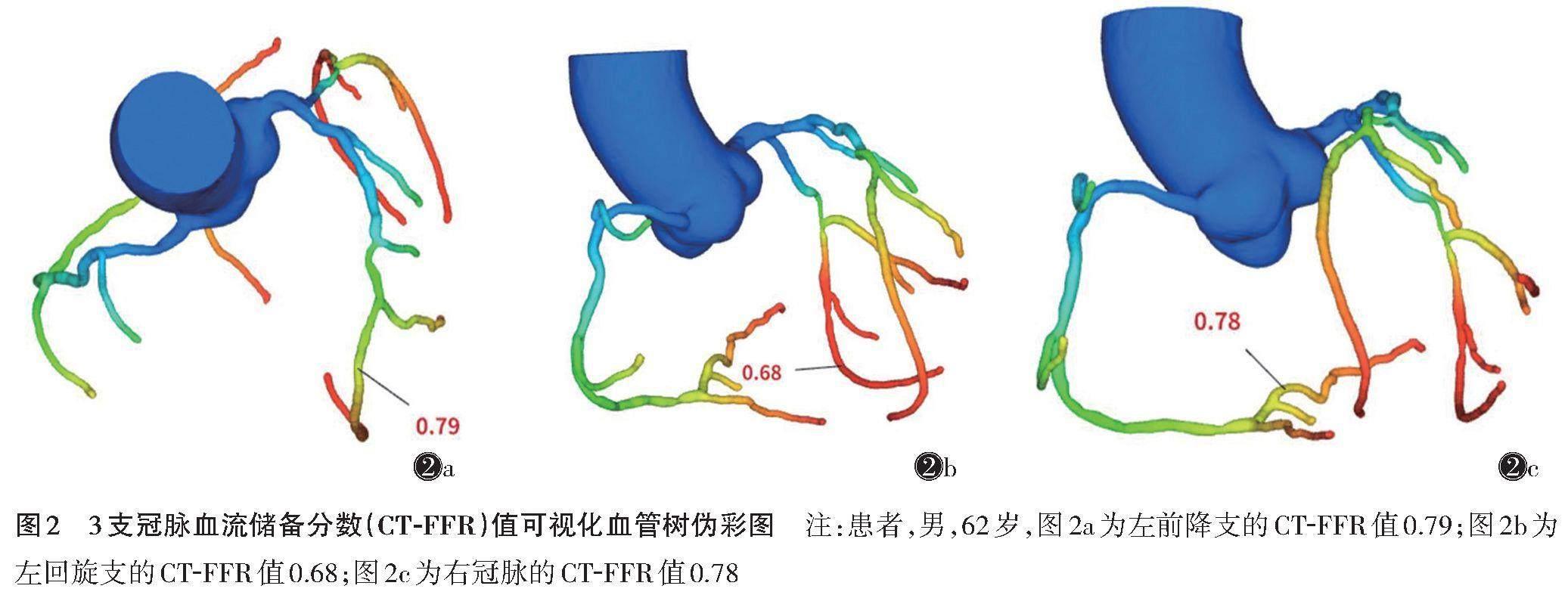
1.2.2 CT-FFR的测量 将扫描后的图像以MPR/断面层DICOM格式导入专业CT-FFR计算软件(深脉分数?-DVFFR,科亚医疗,中国)。软件自动识别图像中冠脉的管腔及中心线,提取其算法所需几何特征,对其进行基于Poiseuille定律和Navier-Stokes方程计算,使流体力学特征融合深度学习模拟算法。分析冠脉左前降支、左回旋支、右冠脉的CT-FFR及数值可视化的冠脉血管树,以最低值作为该患者的冠脉CT-FFR(图2)。
1.3 统计学方法
使用SPSS 26.0软件对数据进行统计分析,对连续性变量行正态性检验,符合正态分布的计量资料用[x]±s表示,非正态分布用M(Q1,Q2)表示,计数资料用频数和率表示。正态性数据组间比较行独立样本t检验,非正态及等级变量行Mann-Whitney U检验非参数秩和检验。对CT-FFR、血尿酸值、钙化积分等行Pearson相关分析。通过logistic回归分析进行预测,通过ROC曲线分析钙化积分、血尿酸和CT-FFR对缺血性狭窄的诊断价值。以P<0.05为差异有统计学意义。
2 结果
2组性别、年龄及血尿酸比较,差异均无统计学意义(均P>0.05);而2组钙化积分差异有统计学意义(Z=-5.958,P<0.05)(表1),即血管无功能学狭窄患者的钙化积分更低。
钙化积分与CT-FFR呈显著负相关(r=-0.556,P<0.001),即钙化积分越高,CT-FFR越低;而血尿酸与CT-FFR、钙化积分均无相关性(r=0.078,0.019;均P>0.05)。
logistic回归分析显示,2组仅钙化积分差异有统计学意义(β=0.003,P<0.001)。绘制ROC曲线(图3),计算AUC为0.782,即钙化积分预测冠脉缺血的准确率为78.2%,敏感度为68.5%,特异度为77.9%,且异常组的钙化积分阈值为313.05分,约登指数为0.464。
3 讨论
本研究以CT-FFR作为冠脉狭窄的评估指标,探讨血尿酸、总钙化积分对CT-FFR的影响。研究发现,血尿酸水平与冠脉狭窄缺血无相关性,但钙化积分与CT-FFR呈显著负相关,即钙化积分越高,冠脉狭窄造成的心肌缺血越严重,且当钙化积分>313.05分时,CT-FFR≤0.8,其预测准确率为78.2%,敏感度为68.5%,特异度为77.9%。既往关于血尿酸与冠脉狭窄的研究[13]多基于CCTA下的冠脉管腔解剖学狭窄情况,而因侧支循环及自身代偿等因素导致管腔解剖学的狭窄程度常与真实的缺血程度存在一定差异。多项研究表明,CT-FFR与FFR在冠脉狭窄血流动力学评估方面一致性较好[14-16]。本研究应用CT-FFR作为评价冠脉狭窄所致心肌缺血的功能性评价指标,将其与血尿酸及钙化积分联合进行分析,为CCS合并HUA及冠脉高钙化积分患者的冠脉狭窄缺血事件发生的预防提供一定依据。
研究表明,HUA与冠脉疾病密切相关[4,17],血尿酸越高,血管狭窄程度越重[18]。而本研究中血尿酸與CT-FFR并无相关性,原因可能是由于血尿酸产生的炎性反应、血小板激化、血管内皮功能紊乱等[19-20]机制需在一定时间下促使管腔狭窄,早期HUA可能并未对管腔狭窄及血管弹性产生影响。DAI等[21]发现,当血尿酸>467 μmol/L时,3支病变的风险显著增加。因此,血尿酸在超过一定阈值时对冠脉血流动力学产生明确的影响,但这个阈值可能并非临床诊断HUA的数值。后续需行大样本量临床研究及基础试验进一步明确此阈值。后期研究中应考虑患者HUA的病程及是否有尿酸盐结晶等因素,同时将血尿酸进行分层研究。
Qi等[22]的研究表明,钙化积分与血管狭窄存在相关性,钙化积分越高,血管狭窄越严重。本研究中钙化积分与CT-FFR呈显著负相关,即钙化积分越高,CT-FFR越低,冠脉功能缺血狭窄越严重,当钙化积分>313.05分时,CT-FFR≤0.8,其预测的准确率为78.2%,敏感度为68.5%,特异度为77.9%。本研究进一步证实钙化积分与冠脉功能学缺血狭窄亦有关,其准确率尚可,敏感度一般。冠脉钙化多因钙盐沉积于血管壁使中膜及内膜受损,导致血管硬化,从而影响冠脉血流,与本研究高钙化积分患者CT-FFR低的结果一致。既往研究多以斑块性质为切入点,探讨不同斑块的钙化积分对心功能的影响[23-25],本研究从血管钙化影响血流动力学的角度论证高钙化积分对心血管产生的影响,在理论上虽然血管钙化对血管弹性的影响可能导致CT-FFR测量值存在偏倚,但目前研究认为血管钙化并未影响CT-FFR的诊断效能[26]。
本研究存在的不足:样本量较小;所用钙化积分为总钙化积分,反映的是血管整体的斑块负荷情况,而总钙化积分并不能完全等同于靶血管缺血部位的钙化程度,因此下一步有待扩大样本量针对各靶血管的钙化积分进行细化分析。
综上所述,CCS患者钙化积分与CT-FFR呈显著负相关,可预测冠脉狭窄所致的心肌缺血情况;而CCS患者血尿酸是否与CT-FFR存在相关性,需进一步研究。
[参考文献]
[1] PIJLS N H,VAN SON J A,KIRKEEIDE R L,et al. Experimental basis of determining maximum coronary,myocardial,and collateral blood flow by pressure measurements for assessing functional stenosis severity before and after percutaneous transluminal coronary angioplasty[J]. Circulation,1993,87(4):1354-1367.
[2] RAMASAMY A,JIN C,TUFARO V,et al. Computerised methodologies for non-invasive angiography-derived fractional flow reserve assessment:a critical review[J]. J Interv Cardiol,2020,2020:6381637.
[3] NORGAARD B L,LEIPSIC J,GAUR S,et al. Diagnostic performance of noninvasive fractional flow reserve derived from coronary computed tomography angiography in suspected coronary artery disease:the NXT trial (Analysis of Coronary Blood Flow Using CT Angiography:Next Steps)[J]. J Am Coll Cardiol,2014,63(12):1145-1155.
[4] NDREPEPA G. Uric acid and cardiovascular disease[J]. Clin Chim Acta,2018,484:150-163.
[5] TONINO P A,FEARON W F,DE BRUYNE B,et al. Angiographic versus functional severity of coronary artery stenoses in the FAME study fractional flow reserve versus angiography in multivessel evaluation[J]. J Am Coll Cardiol,2010,55(25):2816-2821.
[6] KEAN R B,SPITSIN S V,MIKHEEVA T,et al. The peroxynitrite scavenger uric acid prevents inflammatory cell invasion into the central nervous system in experimental allergic encephalomyelitis through maintenance of blood-central nervous system barrier integrity[J]. J Immunol,2000,165(11):6511-6518.
[7] YU W,CHENG J D. Uric acid and cardiovascular disease:an update from molecular mechanism to clinical perspective[J]. Front Pharmacol,2020,11:582680.
[8] LIEW G,CHOW C,VAN PELT N,et al. Cardiac society of Australia and New Zealand position statement:coronary artery calcium scoring[J]. Heart,Lung and Circulation,2017,26(12):1239-1251.
[9] KNUUTI J,WIJNS W,SARASTE A,et al. 2019 ESC Guidelines for the diagnosis and management of chronic coronary syndromes[J]. Eur Heart J,2020,41(3):407-477.
[10] 高尿酸血癥相关疾病诊疗多学科共识专家组. 中国高尿酸血症相关疾病诊疗多学科专家共识[J]. 中华内科杂志,2017,56(3):235-248.
[11] KOO B K,ERGLIS A,DOH J H,et al. Diagnosis of ischemia-causing coronary stenoses by noninvasive fractional flow reserve computed from coronary computed tomographic angiograms. Results from the prospective multicenter DISCOVER-FLOW (Diagnosis of Ischemia-Causing Stenoses Obtained Via Noninvasive Fractional Flow Reserve) study[J]. J Am Coll Cardiol,2011,58(19):1989-1997.
[12] AGATSTON A S,JANOWITZ W R,HILDNER F J. Quantification of coronary artery calcium using ultrafast computed tomography[J]. J Am Coll Cardiol,1990,15(4):827-832.
[13] LIM D H,LEE Y,PARK G M,et al. Serum uric acid level and subclinical coronary atherosclerosis in asymptomatic individuals:an observational cohort study[J].
Atherosclerosis,2019,288:112-117.
[14] TESCHE C,DE CECCO C N,ALBRECHT M H,et al. Coronary CT angiography-derived fractional flow reserve[J]. Radiology,2017,285(1):17-33.
[15] TESCHE C,DE CECCO C N,BAUMANN S,et al. Coronary CT angiography-derived fractional flow reserve:machine learning algorithm versus computational fluid dynamics modeling[J]. Radiology,2018,288(1):64-72.
[16] MIYAJIMA K,MOTOYAMA S,SARAI M,et al. On-site assessment of computed tomography-derived fractional flow reserve in comparison with myocardial perfusion imaging and invasive fractional flow reserve[J]. Heart Vessels,2020,35(10):1331-1340.
[17] SAITO Y,TANAKA A,NODE K,et al. Uric acid and cardiovascular disease:a clinical review[J]. J Cardiol,2021,78(1):51-57.
[18] CASIGLIA E,TIKHONOFF V,VIRDIS A,et al. Serum uric acid and fatal myocardial infarction:detection of prognostic cut-off values:the URRAH (Uric Acid Right for Heart Health) study[J]. J Hypertens,2020,38(3):412-419.
[19] MARUHASHI T,HISATOME I,KIHARA Y,et al. Hyperuricemia and endothelial function:from molecular background to clinical perspectives[J]. Atherosclerosis,2018,278:226-231.
[20] SAITO Y,KITAHARA H,NAKAYAMA T,et al. Relation of elevated serum uric acid level to endothelial dysfunction in patients with acute coronary syndrome[J].
J Atheroscler Thromb,2019,26(4):362-367.
[21] DAI X M,WEI L,MA L L,et al. Serum uric acid and its relationship with cardiovascular risk profile in Chinese patients with early-onset coronary artery disease[J]. Clin Rheumatol,2015,34(9):1605-1611.
[22] QI L,SHI K,LI C,et al. Coronary computed tomography (CT) angiography characteristics of high-risk plaque:correlation with stress myocardial perfusion imaging in patients with moderate coronary stenosis[J]. Med Sci Monit,2020,26:920950.
[23] 袁宏涛,陈宝锦,顾慧,等. 冠状动脉钙化积分与基于CT特征追踪技术的左心室心肌应变的关系[J]. 医学影像学杂志,2023,33(7):1181-1185.
[24] 朱楠,刘亚,蒋双燕,等. 第2代追踪冻结技术下冠状动脉钙化积分、CAD-RADS评分及CT血流储备分数的相关性分析[J]. 中国中西医结合影像学杂志,2023,21(5):503-507.
[25] 孙一,刘博,孙秀彬,等. CCTA-AI联合FFR-CT诊断冠状动脉狭窄病变的价值[J]. 医学影像学杂志,2022,32(12):2081-2085.
[26] 刘春雨,李建华,唐春香,等. 钙化对FFR_(CT)诊断冠状动脉疾病准确性影响的研究[J]. 国际医学放射学杂志,2018,41(3):277-281.

