Evaluation of retinal and choroidal thickness changes in overweight and obese adults without ocular symptoms by swept-source optical coherence tomography
Qing-Jian Li, Sheng-Mei Zhou, Ling-Yu Zhang, An-Ni Lin, Yang Zhang, Jing Jiang,Xin Che, Yi-Wen Qian, Yan Liu, Zhi-Liang Wang
1Department of Ophthalmology, Huashan Hospital, Fudan University, Shanghai 200040, China
2Eye Institute of Xiamen University, Xiamen University School of Medicine, Xiamen 361005, Fujian Province, China
3Department of Gastroenterology, Huashan Hospital, Fudan University, Shanghai 200040, China
Abstract
· KEYWORDS: overweight; obesity; body mass index;choroidal thickness; retinal thickness; swept-source optical coherence tomography
INTRODUCTION
Overweight and obesity are defined as abnormal or excessive fat accumulation that may damage health.In the past 30y, the number has increased by 50%-80%[1].At present, it is estimated that approximately 39% of global adults are overweight or obese[2].The rising prevalence of overweight and obesity is currently being described as a global pandemic.Despite some variability, the prevalence is increasing across sex, age, and socioeconomic conditions.
Both overweight and obesity may affect the structure and function of human organs, increasing the risk of morbidity and mortality[3-4].There has been evidence that excess body weight is a risk factor for diabetes mellitus, hypertension,dyslipidemia, and certain cancers[5-6].As one of the most sensitive organs, the eye is vulnerable to metabolic disorders,inflammatory infiltration, and vascular anomaly.Currently, the associations of overweight or obesity with ocular diseases, such as age-related macular degeneration, epiretinal membranes,and glaucoma, have been shown in varying degrees[7-10].
As a metabolically active tissue, the retina consumes many nutrients and oxygen[11].Among the body’s blood vessels, thechoroid has the highest blood flow per unit of tissue weight,which is responsible for supplying oxygen and nutrients to the retina[12-13].Physiological conditions and systemic disorders can affect the retina or choroid[14-15].Additionally, several ocular diseases, including age-related macular degeneration, epiretinal membranes, and glaucoma, have also been associated with changes in the retinal or choroidal thickness[16-19].

Table 1 Demographic characteristics
Imaging the eye is crucial as it provides essential information for the diagnosis and management of ocular diseases.It allows non-invasive, microstructural investigations to gather information regarding the systemic vascular and central nervous system status.Ocular imaging goes beyond the eye and represents a multidisciplinary toolbox with applications in neurology, rheumatology, metabolic disorders, and many other disciplines.As the most important structures of the eyeball, the retina and choroid can be captured and measured by sweptsource optical coherence tomography (SS-OCT).
As is well known, the evidence on the associations of overweight and obesity with retinal and choroidal thickness is inconsistent.Therefore, this research aims at evaluating whether the thickness of the retina or choroid is affected by the status of overweight and obesity in adults.
SUBJECTS AND METHODS
Ethical ApprovalThis cross-sectional research was conducted at Huashan Hospital affiliated to Fudan University from January 2020 to October 2020, following the principles of the Declaration of Helsinki.It was approved by the Institutional Review Board of Huashan Hospital (No.KY2016-274).The informed consent was obtained from the subjects.
Study Design and ParticipantsThe participants received ophthalmological examinations, including best-corrected visual acuity (BCVA), slit-lamp biomicroscopy, funduscopy,and SS-OCT scan.As an indicator of the subjects’ nutritional status, the body mass index (BMI) was calculated as the ratio of body weight (kg) and height2(m2).According to the World Health Organization (WHO) recommendation, the nutritional condition was categorized into normal weight(18.50≤BMI<25.00 kg/m2), overweight (25.00≤BMI<30.00 kg/m2),and obesity (BMI≥30.00 kg/m2).
Exclusion CriteriaAmong the exclusion criteria were: 1)age<18 or >70y; 2) BCVA worse than 0.1 logMAR; 3) a history of retinal or choroidal diseases; 4) a history of ocular surgeries; 5) poor OCT images; 6) a history of diabetes,hypertension, or thyroid diseases.
Swept-Source Optical Coherence Tomography ImagingOwing to its greater scanning depth, SS-OCT (Version 9.31, Topcon Co., Tokyo, Japan) provides an advantage in visualizing the choroid[20-21].According to previous articles[22],the retinal and choroidal thicknesses have been defined respectively.Thickness maps were created automatically by the standard Early Treatment Diabetic Retinopathy Study(ETDRS) subfield[23].The thickness of the retina and choroid was measured at 8-10a.m.to reduce diurnal variations[24].
Statistical AnalysisSPSS software (Version 24.0, IBM Inc., Chicago, IL, USA) was used for statistical analysis.Continuous data were presented as the mean±standard deviation (SD).Categorical data were expressed as frequency(percentage).The one-way ANOVA was used for continuous variable comparisons among the three groups.The Chi-square test was used to compare categorical variables.Pearson’s correlation analysis was used to evaluate the relationships between the measured variables.Statistical significance was defined as a 2-tailedP<0.05.
RESULTS
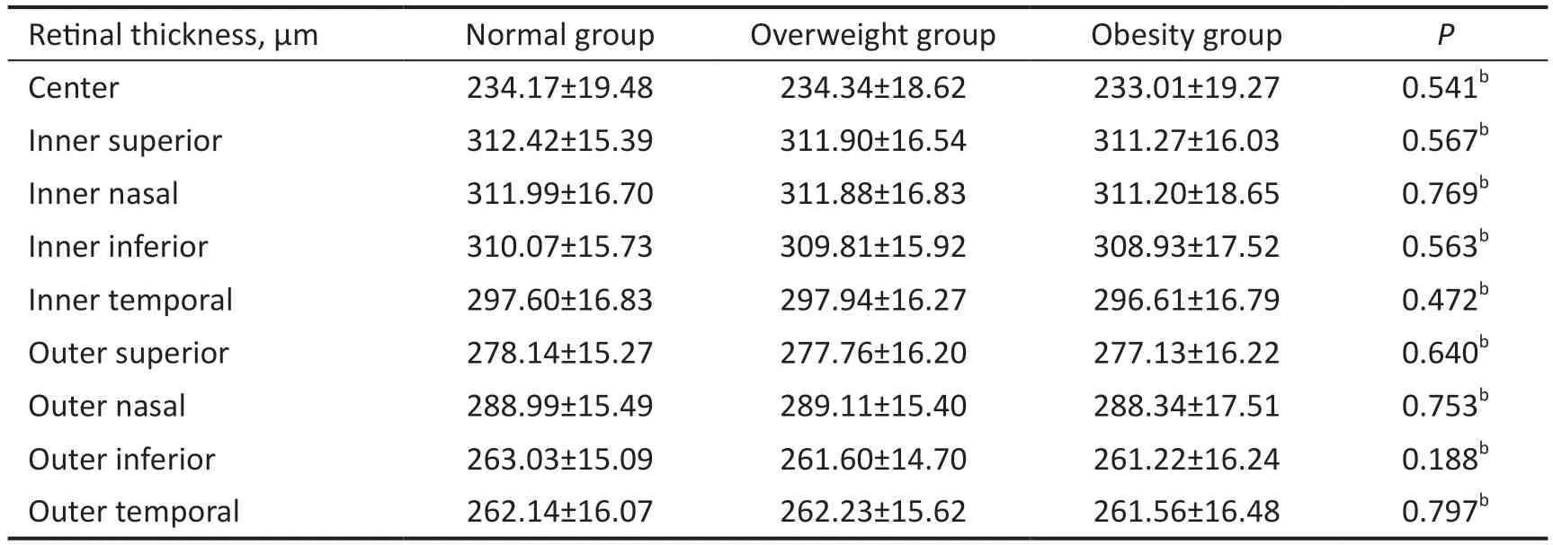
This research covered the left eyes of 3 groups of 434 age- and sex-matched subjects each: normal, overweight, and obesity.The demographic characteristics are presented in Table 1.The male-to-female ratio was 326:108 in the normal, overweight,and obesity groups.The mean age was 40.51±10.27y (range,20-69y) among the three groups.The mean BMI was 22.20±1.67 kg/m2(range, 18.50-24.90 kg/m2) in the normal group, 26.82±1.38 kg/m2(range, 25.00-29.80 kg/m2) in the overweight group, and 32.21±2.35 kg/m2(range, 30.00-42.60 kg/m2)in the obesity group.Table 2 details the retinal thickness for the three groups.The retinal thickness in each sector of the ETDRS grid showed no significant differences among the normal, overweight, and obesity groups.

Table 2 The retinal thickness of nine sectors of the ETDRS grid n=434, mean±SD

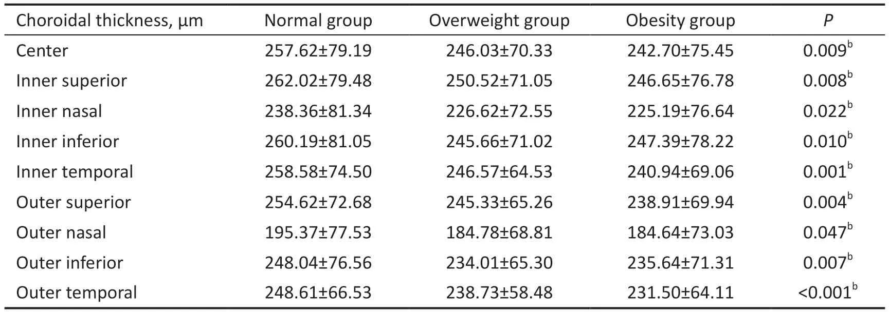
Table 3 The choroidal thickness of nine sectors of the ETDRS grid n=434, mean±SD
Table 3 details the choroidal thickness for the three groups.The choroid in both the overweight group and the obesity group was significantly thinner than that in the normal group,while the choroidal thickness showed no significant differences between the overweight group and the obesity group.
Pearson’s correlation analysis concluded that BMI was significantly negatively correlated with choroidal thickness in all areas of the ETDRS grid, but no significant correlation was observed between BMI and retinal thickness (Tables 4 and 5).
DISCUSSION
In the study, we compared macular retinal and choroidal thickness among the normal, overweight, and obesity groups by SS-OCT.The research indicated that the choroid in both the overweight group and the obesity group was significantly thinner than that in the normal group, but there was no same result in the retina.Additionally, choroidal thickness was significantly negatively correlated with BMI.These suggest that choroidal thickness is affected by overweight and obesity.To date, the relationship between overweight or obesity and retinal thickness remains inconsistent.Wonget al[25]assessed the association of retinal thickness with BMI using OCT and found that retinal thickness was significantly positively correlated with BMI.Moreover, in a study by Panonet al[26],a significant difference between overweight and normal weight groups was found in terms of retinal thickness.On the contrary, Teberiket al[27]suggested that retinal thickness was significantly reduced in obese subjects compared to control subjects by spectral-domain OCT (SD-OCT).In our study,there was no significant relationship between retinal thickness and BMI.
Similarly, the association of overweight or obesity with choroidal thickness remains conflicting.Some studies showed that choroidal thickness was significantly negatively correlated with BMI[27-30].Similar to these researches, our study revealed a thinner choroidal thickness in overweight or obese subjects compared to those with a normal weight.On the contrary,Yumusaket al[7]and Buluset al[13]compared the choroidal thickness of obese subjects and normal weight subjects using enhanced depth imaging (EDI) OCT and discovered that choroidal thickness was significantly positively correlated with BMI.A study evaluated the effect of obesity on choroidal thickness using SD-OCT in children, and the results showed no significant difference between the obesity and control groups[31].The different OCT types may lead to conflicting observations.Typically, choroidal thickness is measured only at one or several points with OCT.The measurements are influencedby focal thickening or thinning of the choroid because some eyes have an irregular chorioscleral interface (CSI)[32-33].In addition, the measurements may also change due to different subjects measuring manually the choroid.There are potential advantages to SS-OCT that could overcome these deficiencies[34].By SS-OCT, the CSI is precisely detected in 100% of eyes compared with 73.6% of cases using EDIOCT[35].Besides, Copeteet al[20]reported that SS-OCT was able to reproducibly measure the choroidal thickness in 100%of eyes versus 74.4% with SD-OCT.In our research, the retinal and choroidal thickness were measured by SS-OCT and averaged following the ETDRS subfield automatically with confirmed reliability.

Table 4 Correlation analysis between retinal thickness and weight, height, or BMI

Table 5 Correlation analysis between choroidal thickness and weight, height, or BMI
The results of our study showed that the choroid was significantly thinner in both the overweight group and the obesity group compared to the normal group.The choroid is a highly vascularized structure and receives more than 70% of ocular blood flow[14,36].The effects of overweight or obesity on the vascular system, both morphologically and functionally, have been previously reported[6,37].Endothelial dysfunction and vascular damage due to obesity may have a particularly deleterious impact on the ocular vasculature.Besides, overweight or obesity plays a part in the increased rate of atherosclerosis which is associated with decreased vessel density and blood flow area of the choroid[38-39].A high BMI may be responsible for affecting the choroidal thickness.The evaluation of the choroid provides important outcomes in the assessment of the ocular vasculature.
The choroid in the overweight and obesity group was significantly thinner than that in the normal group, but no similar results were found in the retina.The retinal tissue receives about 4% of ocular blood flow, significantly lower than the choroidal tissue[14,36].Moreover, the retina is protected by the blood-retinal barrier that is formed by tight junctions of retinal vascular endothelial cells and retinal pigment epithelial cells.The differences in blood flow and barrier function may explain why the retina isn’t influenced by BMI.
Being overweight or obese affects choroidal thickness.Meanwhile, the changes in choroidal thickness are related to specific ocular diseases.Therefore, the choroid may serve as a link between overweight or obesity and certain ocular pathologies.Our study showed that the choroid was affected before the emergence of ocular symptoms in both overweight and obese subjects.A decreased choroidal thickness may be an important clue to prevent choroidal diseases that may develop later in life if overweight or obesity is not addressed.
The study has several weaknesses.First, the present research is a cross-sectional study.Changes in the retina and choroid during fat accumulation are not available.We are currently conducting a longitudinal study with an extended cohort.Second, participants with subclinical diseases that may affect the retina or choroid were included, despite all subjects being assessed in detail.
In summary, choroidal thickness may have been affected by overweight and obesity prior to ocular symptom onset.The reduction in choroidal thickness in overweight and obese adults is likely to serve as an early warning sign of specific ocular disorders.
ACKNOWLEDGEMENTS
Foundations:Supported by the Science and Technology Commission of Shanghai Municipality (No.20Y11910800).
Conflicts of Interest: Li QJ, None;Zhou SM,None;Zhang LY,None;Lin AN,None;Zhang Y,None;Jiang J,None;Che X,None;Qian YW,None;Liu Y,None;Wang ZL,None.
 International Journal of Ophthalmology2024年4期
- International Journal of Ophthalmology的其它文章
- Algorithm of automatic identification of diabetic retinopathy foci based on ultra-widefield scanning laser ophthalmoscopy
- CD3ε of a pan T cell marker involved in mouse Aspergillus fumigatus keratitis
- Neuroprotective effects of acteoside in a glaucoma mouse model by targeting Serta domain-containing protein 4
- Neuroprotective and anti-inflammatory effects of eicosane on glutamate and NMDA-induced retinal ganglion cell injury
- Bone morphogenetic protein-6 suppresses TGF-β2-induced epithelial-mesenchymal transition in retinal pigment epithelium
- Dry eye rate and its relationship with disease stage in patients with primary hypertension: a cross-sectional study in Vietnam
