The generation and properties of human cortical organoids as a disease model for malformations of cortical development
Xiu-Ping Zhang, Xi-Yuan Wang, Shu-Na Wang, Chao-Yu Miao
AbstractAs three-dimensional“organ-like”aggregates, human cortical organoids have emerged as powerful models for studying human brain evolution and brain disorders with unique advantages of humanspecificity, fidelity and manipulation.Human cortical organoids derived from human pluripotent stem cells can elaborately replicate many of the key properties of human cortical development at the molecular, cellular, structural, and functional levels, including the anatomy, functional neural network,and interaction among different brain regions, thus facilitating the discovery of brain development and evolution.In addition to studying the neuro-electrophysiological features of brain cortex development, human cortical organoids have been widely used to mimic the pathophysiological features of cortical-related disease, especially in mimicking malformations of cortical development,thus revealing pathological mechanism and identifying effective drugs.In this review, we provide an overview of the generation of human cortical organoids and the properties of recapitulated cortical development and further outline their applications in modeling malformations of cortical development including pathological phenotype, underlying mechanisms and rescue strategies.
Key Words:cortical development; disease models; human cortical organoids; human cortical spheroids; human pluripotent stem cells; malformations of cortical development; telencephalon organoids; whole brain organoids
Introduction
During the evolution of the human brain, the cerebral cortex is a key brain region that plays a key role in the enhanced behavioral, cognitive and higher intellectual capacity of human as compared to non-human species (Bystron et al., 2008; Geschwind and Rakic, 2013).The development of the cerebral cortex involves a series of elaborately coordinated developmental events;the disruption of these events can directly cause malformations of cortical development or other brain disorders (Barkovich et al., 2012).Different types ofin vivoandin vitronon-human models have been used to study the properties of human cortical development and disease pathology, thus helping us to construct our existing knowledge framework of the cerebral cortex.However, these models are associated with certain limitations,including species differences and insufficient fidelity.Over recent years,human cortical organoids (hCOs) have emerged as a novel research model to study human cortical development under normal or abnormal conditions(Taverna et al., 2014; Di Lullo and Kriegstein, 2017).
Human brain organoids are three-dimensional“organ-like”tissues that self-organize from human pluripotent stem cells (hPSCs), such as human embryonic stem cells and human induced pluripotent stem cells (Qian et al.,2019).Human brain organoids can form elaborately organized regions of the cerebral cortex within tissue and are known as hCOs; these can effectively recapitulate the key properties of cortical development, such as diverse cell types, organized architecture, neural activities, functionality and interaction between different regions (Agboola et al., 2021; Fernandes et al., 2021).Based on these key features, hCOs predominantly include whole brain organoids (WBOs), telencephalon organoids, and human cortical spheroids(hCSs) (Kadoshima et al., 2013; Lancaster et al., 2013; Pasca et al., 2015).Of these, WBOs can spontaneously form forebrain, midbrain and hindbrain regions and more refined sub-regions including the cortex, hippocampus,ventral forebrain, choroid plexus and immature retina.Telencephalic organoids contain regions with a ventral identity similar to the lateral ganglionic eminences as well as a dorsal identity that is specific to the cerebral cortex.hCSs almost exclusively generate the cortical region, in addition to expressing a few choroid plexus cells.Many researchers have used hCOs to unravel the roles of genes, transcription factors, secreted proteins, receptors,and hormones in brain developmentin vivoto shed light on the mechanisms underlying brain development (Kyrousi et al., 2021; Kelava et al., 2022).With the introduction of reprogramming and genomic engineering technologies,hCOs have been increasingly used to mimic multifarious brain disease-specific models which can facilitate the study of human-specific disease pathogenesis and the pharmacological assessment of drugs that can restore or alleviate different disease phenotypes (Lancaster and Huch, 2019; Sidhaye and Knoblich, 2021).Moreover, hCOs are powerful tools for studying embryology,evolution, toxicology, and regenerative medicine (Tang et al., 2022).Our laboratory has established a research platform using WBOs and performed a series of studies in the field of brain injury, including the feasibility of WBOs for rescuing traumatic brain injury and ischemic stroke as a form of transplantation regenerative therapy and to generate an ischemic stroke model for discovering potential anti-stroke drugs (Wang et al., 2017, 2020b,c, 2022).In this narrative review, we provide a comprehensive overview of different types of hCOs in terms of their generation, cortical developmental properties, and their application in malformations of cortical development.
Retrieval Strategy
We searched the existing literature using the PubMed database with the following keywords to select articles for evaluation focusing on brain organoids, cerebral organoids, forebrain organoids, telencephalon organoids,and cortical organoids.Most of the selected studies (85% of all references)were published between 2013 and 2022.
The Generation of Human Cortical Organoids
Three classical methodologies for the generation of hCOs

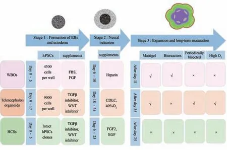
Figure 1| Schematic diagram depicting the generation of hCOs.
The generation of hCOs is a multi-step process that can be divided into three stages: (1) embryoid bodies and ectoderm formation, (2) neural induction, and (3) expansion and long-term maturation, as shown in Figure 1(Kadoshima et al., 2013; Lancaster et al., 2013; Pasca et al., 2015).The main roles of chemical supplements and physical culture conditions at different developmental stages are summarized in Table 1.First, dissociated hPSCs or intact hPSCs clones self-organize into three-dimensional aggregates referred to as embryoid bodies in ultra-low-binding plates; this is followed by ectoderm differentiation.This step represents the initiation of neural specification and factors that may contribute to non-neural fates (such as mesoderm and endoderm differentiation) are often eliminated or inhibited, such as serum,transforming growth factor-β (TGFβ) and WNT signaling factors (Ying et al.,2003; Di-Gregorio et al., 2007).Furthermore, a selective Rho-associated kinase inhibitor, Y27632, needs to be provided as a supplement for the first several days in order to reduce the apoptosis of dissociated hPSCs (Watanabe et al., 2007).Then, the ectodermal layer undergoes neural induction; this is a critical period for determining the fates of neural cells in different brain regions.Finally, various supplements and physical culture conditions that mimic thein vivohuman environment are employed to promote the expansion and maturation of hCOs.The culture methodologies used for WBOs, telencephalon organoids, and hCSs, can be divided into three stages, as mentioned previously.Although there are many similarities in terms of culture systems, different types of hCOs are associated with differences in the culture timeline, medium composition,and physical culture conditions; these factors are important for the formation of individual cellular diversity and brain region composition (Kadoshima et al., 2013; Lancaster et al., 2013; Pasca et al., 2015).The culture system used for WBOs does not introduce exogenous TGFβ inhibitors and WNT inhibitors in the initial regulation of germ layer differentiation.In contrast, these inhibitors are supplemented in telencephalon organoids and hCSs culture methods to enhance neuronal conversion and telencephalon fate (Eiraku et al., 2008; Kadoshima et al., 2013; Lancaster and Knoblich, 2014; Pasca et al.,2015).TGFβ inhibitors, also known as SMAD inhibitors, commonly include bone morphogenetic proteins inhibitors (such as Noggin, Dorsomorphin,LDN193189, and BMPRIA-Fc), and Activin/Nodal inhibitors (such as Lefty and SB431542) (Zhou et al., 2010; Surmacz et al., 2012).Common WNT inhibitors include DKK1, IWR1, and IWP2, while WNT activators include WNT3a and CHIR99021 (Eiraku et al., 2008; Bagley et al., 2017).In contrast,the initial medium used for the culture of WBOs also contains fetal bovine serum and basic fibroblast growth factor, in which fibroblast growth factor may stabilize the ectoderm.Moreover, fetal bovine serum may be a key factor for the generation of mesoderm-derived cells in WBOs, as a previous study demonstrated that hPSCs were converted into cells derived from the mesoderm, endoderm and ectoderm in the presence of serum (Watanabe et al., 2005; Dang and Tropepe, 2006; Lupo et al., 2014).Following neural induction, the methods used to culture WBOs and telencephalon organoids are supplemented with Matrigel by bedding organoids or directly adding to the medium to provide a scaffold for neuroepithelial expansion, thus mimicking the basement membranein vivo(Sato et al., 2009).Finally, the WBOs culture method adopts spinning bioreactors or alternative orbital shakers to promote the exchange of nutrients and oxygen, thus enabling long-term culture for up to 10 months.Telencephalon organoids need to be periodically cut into half size under hyperoxia culture conditions to facilitate the uptake of oxygen and nutrients to enable long-term culture over 13 weeks (Kadoshima et al., 2013).In contrast, the hCSs culture method, established by Pasca’s group, simplified the generation process without the need for extracellular matrix bedding,hyperoxia and rotational culture, thus achieving long-term culture for more than 24 months (Sloan et al., 2018; Yoon et al., 2019).
Modifications and optimizations of the method used to generate hCOs
Currently, there is no standardized method for the generation of hCOs.However, the classical methods have been modified and optimized to overcome certain limitations and adapt to different research purposes.
Studies have shown that only the superficial areas within a depth of 200 μm are fully permeable to oxygen and nutrients and that a lack of oxygen and nutrients inside the organoids can limit the late differentiation and maturation of hCOs (Pham et al., 2018; Qian et al., 2020).Therefore,developmental methods employed certain strategies during expansion and long-term maturation to improve the culture environment and promote differentiation or maturation.The most common optimization strategy is to supplement neurotrophic factors brain-derived neurotrophic factor and glial cell-derived neurotrophic factor, TGFβ and cyclic adenosine monophosphate,as well as dissolved Matrigel into the medium to promote the differentiation and maturation of hCOs (Mariani et al., 2012; Li et al., 2017a; Miura et al.,2020; Xiang et al., 2021).Another small molecule, CHIR99021, plays a slightly different role in different culture stages and can promote neuroepithelial expansion in the long-term culture stage; this factor also effectively reduces cell death when adding during the Matrigel embedding stage (Lancaster et al.,2017; Qian et al., 2018).Moreover, the addition of leukemia inhibitory factor activates STAT3 signaling, increases the generation of outer radial glia cells and astrocytes, and improves the formation of basement membrane (Watanabe et al., 2017).Some studies replaced basic DMEM/F12 and neurobasal medium with BrainPhys medium which supports neuronal functions such as action potential firing and synaptic activity (Kim et al., 2019; Fair et al., 2020; Dalgin et al., 2021).Orbital agitation or spinning bioreactors can also be replaced by hyperoxia culture and physical shearing in the telencephalon organoids culture method; this strategy extends long-term culture to at least 6 months(Velasco et al., 2019).Furthermore, periodic slicing the cultured hCOs into disk shapes in orbital shakers can ensure the effective diffusion of oxygen and nutrients (Qian et al., 2020).Another modification strategy is intended to directionally induce specific cell fates and regional specification of brain.The WBOs culture method can also incorporate TGFβ inhibitors and optional WNT inhibitors to enhance neural fate (Albanese et al., 2020).A previous study demonstrated that the addition of dual SMAD inhibitors and WNT inhibitors during the early induction stage can effectively inhibit non-neural fate and enhance cortical identity (Rosebrock et al., 2022).Notably, WNT signaling factors direct cell fates towards dorsal telencephalon specification during the neural induction stage in a manner that is distinct from their role in the ectodermal formation stage (Wang et al., 2021).Typically, the WNT activator or sonic hedgehog (SHH) antagonist cyclopamine A is supplemented during the neural induction stage to enhance dorsal telencephalon identity, while the WNT inhibitor and SHH activator are added to the medium during the same stage to induce ventral forebrain identity (Qian et al., 2016; Bagley et al., 2017; Birey et al., 2017).In addition,reducing the concentration of heparin in the neural induction medium used for the culture of WBOs can enhance the expression of mesoderm-derived microglia (Ormel et al., 2018).In addition, several studies have complemented oligodendrocyte lineage cells by adding platelet-derived growth factor AA and insulin-like growth factor 1 to expand the formation of oligodendrocyte progenitor cells and promote oligodendrocyte differentiation and myelination(Madhavan et al., 2018; Marton et al., 2019).

Table 1 |The role of chemical supplements and physical culture conditions in the generation of human cortical organoids
Recapitulation of Cortical Developmental Properties in Human Cortical Organoids
Generally, one specific culture method is used to generate hCOs in an individual study, rather than generating different types of hCOs by the application of various culture methods.However, the cortical properties of different types of hCOs can differ.Thus, understanding the cortical developmental properties of different types of hCOs is vital when choosing an appropriate culture method to address the specific scientific question at hand.Overall, hCOs can mimic early human brain development spanning 8–24 weeks post-conception (Kelava and Lancaster, 2016).The three types of hCOs can exhibit similar cortical cell types and organized cortical architecture but with different neural activities and interactions within different brain regions.
Cell composition and organized structure
All types of hCOs preserve basic cortical cell types and organized structures similar to those of the human embryonic cortex, as shown in Figure 2(Arlotta and Pasca, 2019).The cortex in hCOs is composed of diverse layerspecific radial glia cells, intermediate progenitor cells, outer radial glia cells,excitatory neurons and Cajal-Retzius cells, which are stereotypically organized into cortical laminar structures of the ventricular zone, subventricular zone,cortical plate, and marginal zone from the apical to the basal direction(Kadoshima et al., 2013; Quadrato et al., 2017; Yoon et al., 2019).In addition, telencephalon organoids recapitulate a cell-sparse intermediate zone and a Calretinin-marked subplate structure, whereas WBOs and hCSs do not generate these two zones.The major neural progenitor population of radial glia cells exhibits interkinetic nuclear migration behavior and undergoes symmetrical divisions to generate daughter neural progenitors and asymmetrical divisions to generate daughter neural progenitors and neurons(Kadoshima et al., 2013; Lancaster et al., 2013; Pasca et al., 2015), which are consistent with that observed inin vivostudies (Hartfuss et al., 2001).Astrocytes are an essential non-neuronal cellular component of the cortex which support the formation of neurons and synapses, and participate in neural circuits and the blood-brain barrierin vivo(Olsen et al., 2015; Appelt-Menzel et al., 2017).All hCOs can express markers of astrocytes.In particular,the astrocytes in hCSs exhibit a higher level of maturity and key functional properties of synaptic phagocytosis after long-term culture beyond 20 months(Sloan et al., 2017).Another important cortical cell composition, which is essentially absent in hCOs, is oligodendrocytes.Telencephalon organoids lack the cell cluster of oligodendrocyte precursors (Velasco et al., 2019).Although WBOs rarely express oligodendrocyte progenitor cells, there is no expression of oligodendrocytes (Quadrato et al., 2017; Ziffra et al., 2021).hCSs induced by specific growth factors and hormones are capable of expressing oligodendrocyte progenitor cells and oligodendrocytes, and can form compact myelin in axons (Madhavan et al., 2018; Marton et al., 2019).Overall, nondirected WBOs exhibit more diverse cellular types, such as mesodermal cells and mesenchymal cells (Lancaster et al., 2013; Camp et al., 2015; Quadrato et al., 2017).It is worth mentioning that the cellular diversity of WBOs can sacrifice inter-organoid reproducibility, at least to some extent (Quadrato et al., 2017).When compared with WBOs, individual telencephalon organoids and hCSs are relatively homogeneous and can reproducibly generate cortical cells (Velasco et al., 2019; Yoon et al., 2019).
Thein vivogeneration of diverse cell types follows a temporal sequence,and newborn excitatory glutamatergic neurons can migrate radially to their destined positions in an“inside-out”pattern (Dehay and Kennedy, 2007;Silbereis et al., 2016).WBOs can reproduce the temporal sequence of generation for a diverse range of cortical cells.For example, Cajal-Retzius cells in WBOs are first generated and then migrate into the most superficial layer;subsequently, progenitors sequentially generate deep-layer neurons that express CTIP2 and a superficial layer of neurons that express SATB2; finally,the expression of astrocytes is enhanced after approximately 100 days of culture (Molyneaux et al., 2007; Renner et al., 2017).Neurogenesis in hCOs can also reproduce the stereotypic“inside-out”pattern in which late-born cortical neurons migrate to the more superficial side with early-born neurons located inside (Kadoshima et al., 2013; Lancaster et al., 2013).

Figure 2| The cortical lamination structure of human embryonic cortex and hCOs.
Neural activity and function
Neural activity and functional neural networks underlie brain functionality and are indispensable for modeling brain disorders.There are action potentials in hCOs that can be blocked by the voltage-gated sodiumchannel antagonist tetrodotoxin.Variations in neural network activity can be assessed by electrophysiological means via calcium imaging, patch clamping and multi-electrode arrays; this activity is directly related to the dynamic development and maturation of multiple neurons and glial cells(Lancaster et al., 2013; Pasca et al., 2015; Watanabe et al., 2017; Fair et al.,2020).When cultured for 76 to 104 days, telencephalon organoids exhibit spontaneous excitatory postsynaptic currents and spontaneous calcium dynamics, but with little synchronized activity in asynchronous calcium transients, thus implying relatively immature neural network functionality (Li et al., 2017a; Sakaguchi et al., 2019).WBOs not only exhibit calcium surges,they also possess a spontaneous active neuronal network in eight-monthold organoids (Quadrato et al., 2017).The electrophysiological properties of WBOs is dynamically mature, with weak spiking activities appeared after 1 month of culture, and increased synchronized burst firings from 120 to 161 days (Fair et al., 2020).WBOs also exhibit theta frequency oscillations that are synchronized with neuronal population bursts (Sharf et al., 2022).Synchronized burst firing and oscillatory activity with distinct frequencies are typical features of functional neural networks.hCSs also exhibit functional neural network activities that accompany developmental programming(Pasca et al., 2015; Trujillo et al., 2019).Research has also shown that 130-day-old hCSs exhibit spontaneous calcium spikes and that 180-day-old hCSs exhibit abundant synaptogenesis with spontaneous synaptic activity that can be completely blocked by pharmacological exposure to glutamate receptor antagonists (Pasca et al., 2015).hCSs exhibit spontaneous network events with developmental oscillations that transform from periodic and regular nested oscillations to more complex and variable oscillations, and the synchronous network events are similar to some features that have been observed in the electroencephalography of human preterm infants (Trujillo et al., 2019).Furthermore, fused organoids composed of hCOs and ganglionic eminence organoids exhibit neural oscillations with multiple frequencies that do not occur in unfused hCOs, thus indicating the important role of inhibitory interneurons in neural network activity (Samarasinghe et al., 2021).
Interaction between the cerebral cortex and other brain regions
Neural interactions between distinct brain regions, such as interneuron migration and axon tract extension, are very important for brain development and functionality (Bagley et al., 2017).Cortical interneurons primarily originate from the ventral telencephalon and integrate into the cortical neural network after long distance migration (Wonders and Anderson, 2006).The dorsal region of WBOs express positive immunostaining for interneurons,thus indicating the potential for interneuron migration (Lancaster et al.,2013).However, considering the variability and random generation of the ventral forebrain, WBOs are not an ideal model for detailed analysis of the interactions between different brain regions (Lancaster et al., 2013).Furthermore, the classic method for hCS culture only produces excitatory glutamatergic neurons, which also restricts the study of interneuron migration(Pasca et al., 2015).A novel modular strategy applied a fusion paradigm that fuses hCSs with ventral human forebrain spheroids or more refined human medial ganglionic eminence organoids, thus recapitulating the saltatory directional migration of GABAergic interneurons from the ventral forebrain to the dorsal forebrain and the formation of an integrated neural network (Bagley et al., 2017; Birey et al., 2017; Xiang et al., 2017).Another important structural basis for interaction between the cerebral cortex and other brain regions is the axon tract which is responsible for connecting different brain regions and transmitting information (Swanson et al., 2017).Glutamatergic neurons in the cerebral cortex project to other brain regions and form functional circuits, such as the cortico-striatal circuits that are important for the regulation of motivated behaviors and movements, and the corticothalamic pathways that are important in transmitting sensorimotor information (Lopez-Bendito and Molnar, 2003; Stiles and Jernigan, 2010).Cortico-striatal assembloids can recapitulate YFP+projections oriented from cortical neurons in the hCSs to neurons in human striatal spheroids connected by functional synapses (Miura et al., 2020).Research has shown that mCherry-labeled thalamus-like organoids and green fluorescent proteinlabeled hCO assembloids can recapitulate reciprocal axon projections in the presence of specific cell feeders (Xiang et al., 2019).In addition,cortical glutamatergic neurons connect with the hindbrain and spinal cord through long-distance axons, thus forming functional cortico-motor circuits that activate muscles and produce movement (Shim et al., 2012; Kiehn,2016).Recently, more complex three-component assembloids, containing hCSs, human spinal spheroids and human skeletal muscle spheroids, have been constructed by Pasca’s group, which can recapitulate the activation of muscle contractions by cortical neurons under glutamate uncaging photo-stimulation or optogenetic stimulation (Andersen et al., 2020).In addition to these interactions among brain organoids, one recent study has reported functional interactions between post-transplanted hCOs and the somatosensory cortex of rats (Revah et al., 2022).These post-transplanted hCOs can receive thalamocortical and corticocortical projections that could be activated by surrounding stimuli, and also extend axons to the brain that could drive reward-seeking behavior.Overall, hCOs not only provide an ideal model for investigating neural crosstalk and neural connections during brain development; they also promote the exploration and validation of specific actions or other manifestations controlled by specific regions of the brain cortex.
Application of Human Cortical Organoids in Modeling Malformations of Cortical Development
Abnormal cortical development caused by genetic, infectious, vascular, and metabolic factors, will lead to malformations of cortical development, a class of heterogeneous brain disorders that manifest as developmental delay,cerebral palsy or seizures (Severino et al., 2020).Specifically, malformations of cortical development mainly exhibit three abnormal phenotypes, including abnormal neuronal and glial proliferation or apoptosis, abnormal neuronal migration, and abnormal post-migration development (Barkovich et al.,2012).Since three-dimensional hCOs can efficiently reproduce the key developmental properties of the cortex, including progenitor proliferation,neuronal migration and organized structure, patient-derived or gene-edited hCOs have been used to investigate pathological phenotypes, underlying pathogenic mechanisms, and rescue strategies, for different types of malformations of cortical development, as shown in Table 2.
Microcephaly
Primary microcephaly is a genetically heterogeneous disorder with several identified associated mutations of genetic loci, includingMCPH1,WDR62,CDK5RAP2, andASPM(Passemard et al., 2013).HCOs carrying specific genetic mutations of primary microcephaly exhibit a reduced size, a key phenotype of primary microcephaly.WBOs derived from patients withCDK5RAP2mutations exhibit smaller neural tissues and reduced neuroepithelial tissues, with a reduced number of radial glia cells and an increased number of neurons;these manifestations are indicative of the premature neural differentiation caused by the loss of CDK5RAP2 protein (Lancaster et al., 2013).Another microcephalic model of WBOs derived from patients with ASPM mutation orASPM-knockdown lines also exhibit a severe reduction in size, the absence of a cortical laminar structure, and defective progenitor proliferation with the loss of neural progenitors (Li et al., 2017a).In addition, functional dysregulation was observed and manifested by reduced calcium activity and asynchronized neuronal activity (Li et al., 2017a).Telencephalon organoids derived from patients with microcephaly andNARS1mutations also had a reduced organoid size with reduced proliferation of radial glia cells (Wang et al., 2020a).
In addition to genetic mutations, viral infections can also cause microcephaly,including ZIKV virus, cytomegalovirus, herpes simplex virus, and rubella virus(Devakumar et al., 2018).HCOs exposed to ZIKV virus and cytomegalovirus also exhibit a microcephalic phenotype.ZIKV virus has been shown to invade hCSs and predominantly target neuron progenitors, thus resulting in reduced thickness of the ventricular zone and neuronal layer and an enlarged lumen,possibly associated with a reduction in the number of proliferating cells and increased cell death (Qian et al., 2016).Watanabe et al.(2017) also reported dramatically increased cell death and the reduced overall size of ZIKV-infected telencephalon organoids.They also proposed that innate immune responses may contribute to ZIKV-triggered programmed cell death and tested whether multiple drugs could effectively ameliorate the impaired phenotype.Cytomegalovirus-infected WBOs exhibited severe neurodevelopmental disorders and neural function defects, including a reduced number of proliferating cells, an increased number of apoptotic cells, impairment in structural organization, and reduced calcium signaling and neural network activity; these phenotypes were effectively prevented by Nabs, an antibody that specifically targets the cytomegalovirus pentamer complex (Sison et al.,2019; Sun et al., 2020).Transcriptional analysis also demonstrated that human cytomegalovirus can cause the downregulation of cortical development and functional genes in WBOs (O’Brien et al., 2022).
Macrocephaly
Macrocephaly refers to a general increase in head size with an occipitalfrontal circumference that exceeds the mean for age and gender by at least two standard deviations (Severino et al., 2020).A previous study reported thatRAB39b-mutated WBOs successfully reproduced the key phenotype of human macrocephaly of increasing head size and proliferation (Zhang et al.,2020).The underlying mechanism may be thatRAB39bdeletion promotes PI3K-AKT-mTOR signaling in neuronal progenitors; furthermore, the inhibition of AKT signaling was shown to rescue these disease phenotypes.
Pretzel syndrome
Pretzel syndrome, also known as polyhydramnios, megalocephaly, and symptomatic epilepsy syndrome, is caused by homozygous germline mutations in theSTRADAgene (Puffenberger et al., 2007).HCSs derived from patients withSTRADAmutation show an increased size and other phenotypes such as delayed neurogenesis, increased proliferation, disrupted primary cilia architecture, along with a reduced number of outer radial glia cells (Dang et al., 2021).
Tuberous sclerosis complex
Tuberous sclerosis complex is a multisystem disorder with the hallmark pathological feature of cortical tubers and is caused by germline heterozygous mutations in theTSC1orTSC2genes that regulate hyperactivation of the mTOR pathway (Blair and Bateup, 2020).hCSs possessing the homozygous deletion ofTSC1orTSC2exhibit the reduced expression of neuronal markers or delayed neurogenesis, an increased expression of glial cell markers, and morphological changes (Blair et al., 2018).Rapamycin can reverse these phenotypes by blocking mTORC1 signaling at key developmental stages.
Periventricular heterotopia
Periventricular heterotopia is characterized by the dysregulated migration of newborn neurons located in the lateral ventricles (Romero et al., 2018).Several gene mutations are implicated in this pathology, includingFLNA,ARFGEF2,DCHS1, andFAT4(Stouffer et al., 2016).WBOs derived from patients withDCHS1andFAT4mutations or from isogenic knockout lines can successfully recapitulate periventricular heterotopia-related phenotypes of disrupted morphology in neural progenitors and the abnormal migration dynamics in a subset of neurons (Klaus et al., 2019).Relatively high expression levels of theGNG5gene may be an underlying factor that contributes to the abnormal phenotypes of periventricular heterotopia (Klaus et al., 2019; Ayo-Martin et al., 2020).
Miller Dieker syndrome
Miller Dieker syndrome is a severe form of classical lissencephaly that is caused by microdeletion within chromosome 17p13.3 and exhibits symptoms of intellectual disability, seizures, and craniofacial dysmorphisms (Blazejewski et al., 2018).Three-dimensional telencephalon organoids derived from patients with Miller Dieker syndrome have helped to investigate diseaserelated phenotype defects, including increased apoptosis and reduced vertical cell divisions of neural progenitors, abnormal neuronal migration,as well as the prolonged mitotic cycle of outer radial glia cells (Bershteyn et al., 2017).Iefremova et al.(2017) reproduced consistent Miller Dieker syndrome-related phenotypes in a similar organoid system, including reduced cortical expansion, altered organization in the ventricular niche and adhesion molecules, as well as non-cell-autonomous disruption in the N-cadherin/β-catenin/WNT signaling axis.Furthermore, administration of the WNT activator CHIR99021 was shown to rescue the reduced expansion phenotype(Iefremova et al., 2017).
Rett syndrome
Rett syndrome, a rare neurodevelopmental disorder, is primarily caused by X-linked gene mutations in theMECP2gene with the characteristic symptoms of loss of acquired speech and motor skills, repetitive hand movements and seizures (Kyle et al., 2018).Three-dimensional WBOs carryingMECP2mutations exhibited an increased ventricular area, decreased radial thickness,altered neurogenesis and neuronal differentiation, and migration defects;these effects can be affected by the upregulation of miR-199 and miR-214 to regulate ERK and AKT signaling (Mellios et al., 2018).hCSs and ventral forebrain organoids and their assembloids derived from female patients with Rett syndrome withMECP2mutations exhibited a reduced number of neural progenitors, the premature differentiation of immature neurons,and defects in interneuronal migration (Gomes et al., 2020).MECP2-mutant assembloids of hCOs and human medial ganglionic eminence organoids exhibited synapse formation defects, hyperexcitability and hypersynchrony, as well as epileptiform-like spikes and high frequency oscillations; these effects could be rescued by Pifithrin-α, a putative TP53 target inhibitor (Samarasinghe et al., 2021).MECP2-mutant interneurons have been shown to be the major cause of dysfunction in the neural network, asMECP2-mutant interneurons exhibited abnormalities in molecular and functional maturation in subsequent studies (Xiang et al., 2020; Samarasinghe et al., 2021).
Angelman syndrome
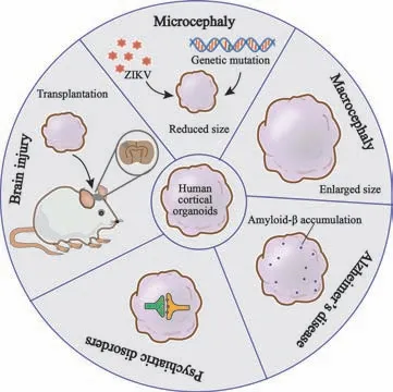
Angelman syndrome is a complex neurodevelopmental disorder that is characterized by delayed development, seizures, and intellectual disability;this condition is predominantly caused by the loss of ubiquitin ligase E3 UBE3A(Lopez et al., 2018).WBOs derived from maternalUBE3A-deleted Angelman syndrome patients replicated the early silencing of the UBE3A protein with higher calcium transient frequency; these effects could be partially rescued by topoisomerase inhibitors (Sen et al., 2020).Another form of UBE3A knockout hCSs exhibited augmented neuronal excitability and elevated afterhyperpolarization due to the augmentation of large potassium channels; the administration of the large potassium channel antagonist paxilline was shown to restore normal neuronal excitability and network activity (Sun et al., 2019).In addition to malformations of cortical development modeled by hCOs as outlined in this review, hCOs have allowed the generation of a broad range of model systems for diseases such as psychiatric disorders, neurodegenerative diseases, and other brain disorders caused by trauma, infection, or cancer,thus providing a powerful platform for studying complex disease pathology as well as for drug development, as shown in Figure 3.For instance, hCOs exposed to severe acute respiratory syndrome coronavirus 2 have been applied to interrogate the infection mechanisms and neurotoxicity associated with the current global outbreak of corona virus disease 2019 (Ramani et al., 2020).Furthermore, hCOs are candidate sources for transplantation therapy in regenerative medicine with a sufficient and diverse range of cells and functional neural networks, thus contributing to the regeneration and reconstruction of functional neural networksin vivo.

Table 2 |Application of hCOs in modeling malformations of cortical development
Limitations and Prospects
Although hCOs have been widely used to mimic human brain development and the modeling of different types of brain disorders, there are still unsolved limitations in with regards to the application of hCOs.For example, we do not yet have a full understanding of hCOs with regards to the fidelity of cell types,subtypes, and tissue structures.

Figure 3|Overview of the application of hCOs in different disease models.
The cell lineages of hCOs are largely derived from the ectoderm and rarely from the mesoderm; this means that hCOs are not expressed in endothelial cells and rarely expressed in microglia (Tanaka et al., 2020).Co-culture strategies involving the culture of hCOs with microglia or endothelial cells has been proposed to complement these missing cell types (Bejoy et al., 2019;Song et al., 2019).In addition, thein vivotransplantation of organoids into animals can provide a blood supply and nutritional support for organoids; this may represent a powerful means of constructing primary blood vessels within organoids as anin vivoorganoid research model (Mansour et al., 2018; Pham et al., 2018; Shi et al., 2020).A previous study constructed vascularized hCOs with certain blood-brain barrier properties by employing vascular endothelial growth factor to induce vasculogenesis without impairing neurogenesis (Ham et al., 2020).The enhanced expression of endothelial signature proteoglycan clusters may generate precursor cells of endothelial lineage which may represent a key target for inducing angiogenesis (Tanaka et al., 2020).
Furthermore, the cortical structure of hCOs cannot convincingly replicate the subplate layer and subsequent canonical six layered structure of the cortical plate; neither can these structures preserve gyrencephalic folding and the complete spatial topography patterned by morphogen gradients.The subplate is a transient layer in the process of cortical development and plays crucial roles in cortical connections, microcircuitry, and synaptogenesis (Molnar et al., 2019).Organotypic culture of fetal human brain has contributed to investigations of subplate-related synaptic networks (McLeod et al., 2022).Transcription factors and growth factors have been demonstrated to play essential roles in regulating cortical expansion and folding (Rash et al., 2013;Lui et al., 2014).A genetically modified hCO system that deletes thePTENgene and sequentially enhances the PTEN-AKT signaling pathway has been used to reproduce cortical expansion and continuous folding (Li et al., 2017b).A previous study also engineered a hCO system possessing a triggered SHH protein gradient to refine the patterning and topographical organization of the forebrain region (Cederquist et al., 2019).
In addition, heterogeneity among individual organoids, even in one culture batch, remains an obstacle that still needs to be overcome.Studies have shown that multiple factors such as hPSCs linage, signaling factors, extracellular matrix, and spinning bioreactors are responsible for the variability observed in hCOs, and that variations mostly occur in the neural induction stage (Yoon et al., 2019; Strano et al., 2020).In the future,developing new signaling factors and a matrix with definite components is of great significance for the development of hCOs.The transcriptomic trajectories of the human brain developingin vivoprovide a path to explore key signaling factors to pattern hCOs more specifically.In addition, hyaluronic acid, a major extracellular matrix in the human brain, may be considered as an alternative extracellular matrix that may activate WNT signaling and exhibit a caudalizing effect when combined with heparin (Bejoy et al., 2018; Yi et al., 2021).The combination of organoids with biomaterials, nanotechnology,and bioengineering strategies such as three-dimensional (3D) printers and microfluidic devices, may ameliorate these limitations and drive the development and refinement of the organoids field.
Conclusions
As a novel versatile model with unique advantages, hCOs possess tremendous potential for uncovering unknown mechanisms related to brain development and even the trajectory of embryonic development, thus making it possible to investigate early brain developmental events and disease mechanisms in the human brain.In addition to the applications mentioned earlier, broader applications of organoids, including hCOs, are already being investigated.For example, culturing individual specific hCOs in combination with genomeediting technology or cell trans-differentiation may help to identify defects in fetal brain development by introducing whole genome sequencing and proteomics; these strategies might be important for reproductive optimization.There is no doubt that extensive study in the future will enable hCOs to better mimic human brain structure and function, thereby strengthening basic and clinical studies involving hCOs.
Author contributions:XPZ and XYW retrieved and analyzed concerned literatures.XPZ and SNW designed and wrote the manuscript.CYM designed and revised the manuscript.All the authors approved the final version of the manuscript.
Conflicts of interest:The authors declare no conflict of interest.
Data availability statement:No additional data are available.
Open access statement:This is an open access journal, and articles are distributed under the terms of the Creative Commons AttributionNonCommercial-ShareAlike 4.0 License, which allows others to remix, tweak, and build upon the work non-commercially, as long as appropriate credit is given and the new creations are licensed under the identical terms.
- 中国神经再生研究(英文版)的其它文章
- From static to dynamic: live observation of the support system after ischemic stroke by two photon-excited fluorescence laser-scanning microscopy
- MicroRNAs in mouse and rat models of experimental epilepsy and potential therapeutic targets
- Nanotechnology-based gene therapy as a credible tool in the treatment of Alzheimer’s disease
- Detection of Alzheimer’s disease onset using MRI and PET neuroimaging: longitudinal data analysis and machine learning
- A pancreatic player in dementia: pathological role for islet amyloid polypeptide accumulation in the brain
- The role of fibronectin in multiple sclerosis and the effect of drug delivery across the blood-brain barrier

