Danggui Buxue decoction promotes proliferation and differentiation of Caco-2 cells and antagonizes obesity by stimulating apolipoprotein-IV expression
Zi-Kai Chen, Ya-Qun Liu, Zhen-Xia Zhang, Ying-Qing Du, Pei-Kui Yang, Guang-Cai Zha, Yao-Lin Huang, Yu-Zhong Zheng*
1Department of Cell Biology,Hanshan Normal University,Chaozhou,Guangdong 521041,China; 2Department of Urology,Chaozhou Central Hospital Affiliated to Southern Medical University,Chaozhou 521000,China.
Abstract Background: Chinese medicine has been proposed as a novel approach to the prevention of metabolic disorders such as obesity.Danggui Buxue decoction, a decoction prepared from Huangqi (Astragali Radix) and Danggui(Angelicae Sinensis Radix), has been used to nourish vitality and enhance blood circulation in traditional Chinese medicine.However, the effect of Danggui Buxue decoction on obesity is still primarily unknown.Methods: Cell proliferation, differentiation, and apolipoprotein-IV transcription were investigated to explore the function of Danggui Buxue decoction by methyl thiazolyl tetrazolium assay, alkaline phosphatase assay, and luciferase assay,respectively.Results: Danggui Buxue decoction promoted cell growth by up to 15% (P= 0.034) and induced cell differentiation by up to 38% (P = 0.006) at 0.3 mg/mL.Moreover, Danggui Buxue decoction enhanced the transcription of apolipoprotein-IV by 2.4 times (P = 0.027), activating its promoter by 28.9% (P = 0.031) at 0.3 mg/mL.In addition,Danggui Buxue decoction functioned in a dose-dependent manner.Conclusion:These results suggest that Danggui Buxue decoction promotes cell differentiation by enhancing apolipoprotein-IV transcription and alkaline phosphatase activity, and it may also have a potential anti-obesity effect because apolipoprotein-IV transcription is closely related to the reduction of food intake.
Keywords:Danggui Buxue decoction,Angelicae Sinensis Radix,Astragali Radix,Apolipoprotein-IV,Anti-obesity
Background
Obesity is a medical conditionwherein excess body fat accumulation causes serious health issues including metabolic syndrome, type 2 diabetes, and cardiovascular diseases.The incidence of obesity has increased dramatically in China [1, 2].According to the 2020 Global Nutrition Report released by the Global Alliance for the Improvement of Nutrition,more than 2 billion people worldwide are overweight and obese, and 38.3 million of them are children [3].Thus,treatment of obesity is urgently needed.
Most anti-obesity Western medicine has side-effects,however, and some are even life threatening [4].For example, MediatorTM, composed primarily of benfluorex, an appetite inhibitor, was produced by the French pharmaceutical enterprise Servier in 1976.It was banned by the French government in 2009 because of damaging effects on the heart.According to official French statistics, approximately 1,300 people have died as a result of this medication [5].Chinese medicine provides an option to decrease body lipid by transcriptional activation of apolipoprotein-IV(ApoA-IV) and reduction of triglycerides using Danggui (Angelicae Sinensis Radix, ASR) and Huangqi(Alismatis Rhizoma,AR)[4,6].
ApoA-IV is a recycled glycoprotein that is synthesized during fat absorption.It is predominantly expressed in the small intestine and has been shown to inhibit food intake in rats [7].ApoA-IV accelerated cholesterol efflux through the high-density lipoprotein receptor metabolic pathway and promoted the secretion of apolipoprotein B-mediated triglycerides in enterocytes [8, 9].ApoA-IV expression, sorting, and secretion by the intestinal cells could be induced by lipid exposure [10].Its mRNA level was highly induced in the remnant ileum of small intestine-resected rats, indicating a potential role in intestinal repair [11].Thus, the transcription of ApoA-IV has been linked to cell differentiation [12].Alkaline phosphatase (ALP) is an enzyme that catalyzes the hydrolysis of p-nitrophenyl phosphate in the alkaline environment.Its activity has also been used as an indicator of Caco-2 differentiation[13].
Danggui Buxue decoction(DBT)originates from Li Dongyuan’sDifferentiation on Endogenousin 1247 C.E.In this book, he wrote: “this prescription can be used to replenishQiand generate blood (Qiand blood are two kinds of basic substances in human body that play an important role in human life activities).The dosage of AR is five times as that of ASR in DBT”.The compatibility of DBT is simple and effective.It is one of the representative compounds in traditional Chinese medicine research.In addition to its traditional effects, DBT also regulates immune function, protects cardiovascular and cerebrovascular vessels, promotes anti-atherosclerosis and anti-tumor development, reduces diabetic nephropathy, stimulates anti-fibrosis activity, and promotes lipid metabolism[14–18].The concentrations of active ingredients in DBT,ligustilide and ferulic acid,were higher than that ofAstragali Radix(AR) alone [19].It was reported that DBT, when combined with ginger, could activate brown fat-specific genes, such asPPARγ,UCP1,PCG1α,CPT1A, andHSL, which indicated that DBT may enhance the anti-obesity effect of ginger,although the anti-obesity function of DBT itself is not yet fully understood[20].
In this study,we show that DBT benefits the growth and differentiation of human intestinal epithelial Caco-2 cells, in an in vitro model for intestinal lipoprotein synthesis and secretion.Moreover, we show that DBT stimulates ApoA-IV transcription by activating the ApoA-IV promoter, and thus, may be used to antagonize lipid absorption and treat obesity.
Methods
DBT preparation
Three-year-old AR was collected from Shanxi province, and 2-year-old ASR was collected from Gansu province (Figure 1).The plant material had been authenticated by Dr.Tina Dong at Hong Kong University of Science and Technology.A total of 25 g of sliced AR and 5 g of sliced ASR were mixed and boiled in 240 mL of water for 2 hours(w:v =1:8),and the residues reboiled in 180 mL of water for 1 hour(w:v = 1:6).The decoction was combined, condensed at 100°C,filtered,lyophilized at −20°C,and stored at−20 °C before use.Respectively,0.03 mg, 0.1 mg, 0.3 mg, 0.5 mg, and 1 mg of DBT powder was dissolved in pure water to obtain the indicated concentrations at the final volume of 1 mL[19].
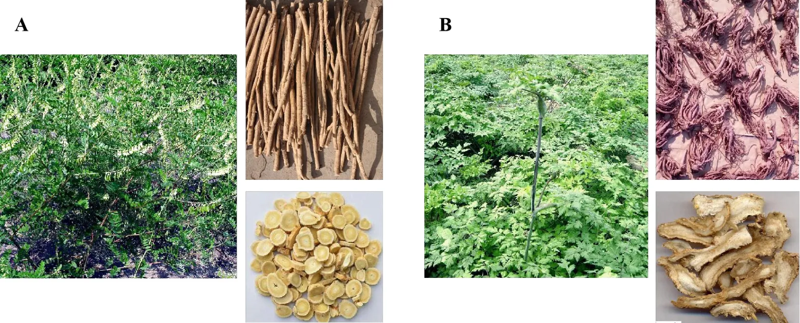
Figure 1 Herbs plant, root, and root pieces.A: Astragali Radix plant, root, and root pieces; B: Angelicae Sinensis Radix plant,root,and root pieces.
Quantification of active ingredients in DBT
The quantitative analysis was performed as described[21].Calycosin (> 99%), formononetin (> 99%), and ferulic acid (> 99%) were obtained from the National Institute for the Control of Pharmaceutical and Biological Products (China).Ligustilide (> 99%) was provided by Professor Pengfei Tu, Medical College of Peking University (China).The purities of these compounds were tested by high-performance liquid chromatography (HPLC).HPLC-grade reagents were obtained from Merck corporation (Germany).An HPLC system(Waters corporation,USA)consisting of a 600 pump, a 717 auto-sampler, and a UV/VIS photodiode array 2996 detector, coupled with an Alltech ELSD 2000 evaporative light scanning detector, was used for all analyses.Chromatographic separations were carried out on an Agilent Eclipse XDB-C18 column (4.6 mm × 250 mm, 5 µm) with 0.1% formic acid (as solvent A) and acetonitrile (as solvent B) at a flow rate of 1.0 mL/min at the room temperature.A linear gradient elution was applied from 15%to 20%B at 0 to 10 minutes,20%B at 10 to 20 minutes,20%to 34%B at 20 to 45 minutes,34%to 48%B at 45 to 55 minutes, 48%to 65%B at 55 to 70 minutes, and 65% to 80% B at 70 to 80 minutes.The re-equilibration time of the gradient elution was 10 minutes.A 10-µL (after a 0.45 µm Millipore filter)sample was injected, and the signals were detected at 254 nm with UV detection.
Calibration curves for calycosin, formononetin,ferulic acid, and ligustilide were constructed by plotting the peak area (x) vs the concentration of each analyte (y) of the standard.Each calibration curve was derived from 6 data points (n = 3).The concentration of each analyte in DBT was then calculated with the measurement of the peak area of the compound in the sample(Table 1).

Table 1 Calibration curves for calycosin,formononetin,ferulic acid,and ligustilide
Cell culture
Human intestinal epithelial cells Caco-2, obtained from the American type culture collection, was cultured in DMEM, supplemented with 20% fetal bovine serum, 1 mM nonessential amino acids, 100 units/mL penicillin, and streptomycin.The cells were maintained in a humidified CO2(5%) incubator at 37 °C.The culture medium was replaced every other day until the cell morphology resembled that of the enterocyte.
Methyl thiazolyl tetrazolium(MTT)assay
Caco-2 cell proliferation was measured by MTT assay.Briefly, 5 × 10⁴cells were cultured in a 96-well plate for 24 hours and treated with sodium butyrate (0–1 mM) or DBT (0–1 mg/mL) for 48 hours.MTT (0.5 mg/mL)was added to the cultures,followed by DMSO extraction.The absorbance was measured at 570 nm.DBT powder is highly soluble in water, and the solution was almost colorless.The solution was removed before DMSO extraction.In general, the color of DBT did not affect results in the MTT assay.
ALP assay
Cells (2 × 10⁵) were cultured in 24-well plate for 24 hours and treated with sodium butyrate (0–1 mM) or DBT (0–1 mg/mL) for 48 hours.The cells were then collected with phosphate buffered saline containing 0.2% Triton X-100 (pH = 7.4).ALP activity was measured by adding ALP buffer (pH = 10.4)containing 5 mM p-nitrophenyl phosphate, 0.1 M glycine, 1 mM MgCl2, and 1 mM ZnCl2.After incubation at 37 °C for 1 hour, 50 μL of 2 M NaOH was added to stop the reaction,and the absorbance was measured at 405 nm.
Luciferase assay
The ApoA-IV promoter was inserted to pBI-GL vectors upstream of the luciferase reporter gene.Caco-2 cells were transfected 1 day after the cells were plated in 24-well-plate,and 60 μL of 2.5 M CaCl2was combined with 540 μL of DNA solution containing 6 μg of the plasmid.The solution was mixed with 600 μL of 2×HEPES buffered saline(50 mM HEPES;pH,7.05; 140 mM NaCl; 1.5 mM Na2HPO4) and added into the cells.The transfected cells were treated with sodium butyrate (0–1 mM) or DBT (0–1 mg/mL) for 48 hours.The cells were then collected and resuspended in lysis buffer containing 0.2% Triton X-100,1 mM DTT,and 100 mM potassium phosphate(pH = 7.8).The samples were incubated at 37 °C for 30 minutes and tested for luciferase activity with a commercial kit(Tropix Inc,USA).Substrates(100 μL)were added to 20 μL of lysate in a 96-well plate, and the luminescence was immediately measured at 550 nm by the Tropix TR717 microplate luminometer, and the total protein amount was measured with a protein assay kit (Bio-Rad Laboratories, USA).The luciferase activity was normalized to the total protein amount for each sample.
Real-time quantitative polymerase chain reaction
Total RNA from Caco-2 cells was isolated with TRIzol(Invitrogen, USA) and reverse-transcribed to cDNA with reverse transcriptase (Invitrogen,USA)according to the manufacturer’s instructions.The primers used for qPCR were ApoA-IV-F, 5'-ATG TTC CTG AAG GCC GTG G-3';ApoA-IV-R, 5'-TGC AGG TCA CCT GCG TAA G-3';18S rRNA-F,5'-TGT GAT GCC CTT AGA TGT CC-3'; and 18S rRNA-R, 5'-GAT AGT CAA GTT CGA CCG TC-3'.Quantitative real-time PCR was performed on a PCR amplifier(Mx3000PTM, Stratagene, USA).The relative expression level of ApoA-IV was normalized to that of 18S rRNA.The PCR products were analyzed by gel electrophoresis and melting curve analysis to confirm the specific amplification.
Statistical analysis
One-way analysis of variance was used to determine whether the difference between two groups of data was significant using SPSS software version 19.0 (SPSS,USA).All data shown were determined with four independent replicates and presented as the mean ±standard deviation.
Results
DBT promotes cell proliferation of Caco-2
To guarantee the quality of the homemade DBT, we quantified the active ingredients with the dry powder.Calycosin, formononetin, ferulic acid, and ligustilide were 0.186 mg/g, 0.155 mg/g, 0.351 mg/g, and 0.204 mg/g, respectively (Figure 2).To evaluate the effect of DBT on cell proliferation, Caco-2 cells were treated with DBT at different concentrations for 48 hours,and the cell viability was determined by MTT assay.As a control, sodium butyrate suppressed Caco-2 cell proliferation in a dose-dependent manner as expected(Figure 3A).On the contrary, DBT promoted cell proliferation, even at a low concentration (< 0.25 mg/mL).It promoted cell growth by up to 15% (P=0.034) at 0.3 mg/mL(Figure 3B).This result confirms that DBT is beneficial to cell growth.

Figure 2 HPLC to detect calycosin, formononetin, ferulic acid, and ligustilide.The numbers above the peaks indicates the peak area for each analyte.DBT, Danggui Buxue decoction; HPLC, high-performance liquid chromatography.
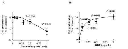
Figure 3 DBT promotes Caco-2 cell proliferation.A, growth of Caco-2 cells is dose-dependently inhibited by sodium butyrate (0–1 mM) after drug treatment for 48 h.B, growth of Caco-2 cells is dose-dependently increased by DBT(0–1 mg/mL)after drug treatment for 48 h.Data are shown as mean±standard deviation(n=4).The drug treated sample at each dose was compared with the untreated sample.DBT,Danggui Buxue decoction.
DBT induces Caco-2 cell differentiation
Caco-2 spontaneously differentiates into polarized enterocytes with gradually increased activity of ALP,which can be used as a reliable marker to evaluate the extent of cell differentiation [13].The cells were treated with different concentrations of sodium butyrate or DBT and applied to the ALP activity measurement.The activity of ALP enhanced after sodium butyrate treatment only at high concentration(Figure 4A) but increased after DBT treatment even at a low concentration (Figure 4B).Moreover, the effect of DBT was dose dependent.DBT induced the differentiation of Caco-2 cells by up to 38% (P=0.006) at 0.3 mg/mL, indicating that it may help to repair and protect the intestine.
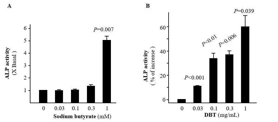
Figure 4 DBT stimulates ALP activity in Caco-2 cells.The ALP activity is dose-dependently stimulated by sodium butyrate (0–1 mM) (A)or DBT(0–1 mg/mL)(B)after drug treatment for 48 h.Data are shown as mean ±standard deviation (n = 4).The drug-treated sample at each dose was compared with the untreated sample.DBT,Danggui Buxue decoction;ALP,alkaline phosphatase.
DBT induces ApoA-IV mRNA transcription in Caco-2
To investigate the anti-obesity function of DBT, we monitored the transcription of human ApoA-IV, which plays an important role in modulating food intake.The expression level of ApoA-IV mRNA was significantly increased by both sodium butyrate and DBT in a dose-dependent manner(Figure 5).It was enhanced by DBT by 2.4 times at 0.3 mg/mL (P= 0.027).These results indicate that DBT may antagonize obesity by stimulating ApoA-IV transcription.
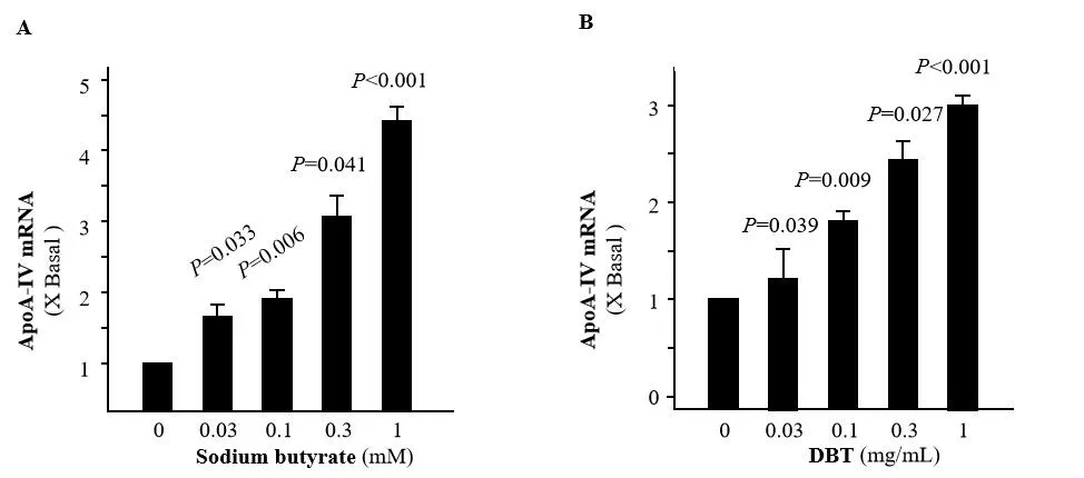
Figure 5 DBT induces ApoA-IV mRNA expression in Caco-2 cells.The transcriptional level of ApoA-IV is dose-dependently induced by sodium butyrate (0–1 mM)(A)or DBT(0–1 mg/mL)(B)after drug treatment for 48 h.Data are shown as mean ± standard deviation (n=4).The drug-treated sample at each dose was compared with the untreated sample.DBT,Danggui Buxue decoction;ApoA-IV,apolipoprotein-IV.
DBT induces expression of ApoA-IV by activating its promoter
To explore the mechanism of the Apop-IV transcriptional activation by DBT, we fused its promoter (230 bp) with a luciferase gene and constructed a plasmid containing this reporter system.The cells were transfected with the plasmid, treated with sodium butyrate or DBT for 48 hours, and harvested for the luciferase assay.As expected,sodium butyrate significantly enhanced the transcriptional activity of the ApoA-IV promoter in a concentration-dependent manner (Figure 6A).Similarly, the promoter was dose-dependently activated by DBT after the 48-hour treatment, with an increase of 28.9% (P= 0.031) at 0.3 mg/mL (Figure 6B).This result suggests that DBT activates the human ApoA-IV promoter,thus promoting gene transcription.
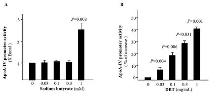
Figure 6 DBT activates the ApoA-IV promoter in Caco-2 cells.A, luciferase reporter assay to determine ApoA-IV promoter activity after sodium butyrate (0–1 mM) treatment for 48 h.B, luciferase reporter assay to determine ApoA-IV promoter activity after DBT (0–1 mg/mL) treatment for 48 h.Data are shown as mean ±standard deviation (n = 4).The drug-treated sample at each dose was compared with the untreated sample.DBT,Danggui Buxue decoction;ApoA-IV,apolipoprotein-IV.
Discussion
DBT produces many pharmacological activities, such as enhancement of cell adherence and migration,modulation of mitochondrial bioenergetics, and acting on a variety of pathways and molecular targets[22–24].However, the functions of DBT on cell proliferation were controversial, depending on different cell types.For instance, DBT could reduce the size of melanoma and enhance anti-tumor immune response, but serum treatment showed no direct effect on the growth of tumor cells [25].Conversely, it suppressed high glucose-induced proliferation of mesangial cells [26].Caco-2 is an excellent in vitro model for the investigation of intestinal lipoprotein metabolism [27].Sodium butyrate has been shown to regulate the ApoA-IV mRNA expression in Caco-2 cells and served as a positive control in this study [28].DBT could protect Caco-2 cytomembrane, but its effects on Caco-2 cell growth have not yet been reported [4, 29].In this study, we showed that DBT promoted proliferation of Caco-2, indicating that DBT vitalizes intestinal cells and may be beneficial for gut maintenance.
DBT induced the formation of capillaries and blood vessels in a rat model of liver fibrosis [30].It also promoted differentiation of endothelial progenitor cells to the mature red blood cells in vitro [25].When combined with gelatin and β-tricalcium phosphate,DBT stimulated bone regeneration after injury [31].Moreover, the intake of glucosides in DBT protected lungs in rate from bleomycin-induced fibrosis by inhibiting NOX4 [32].In a recent study, DBT was found to restore the gut by reversing a metabolic disorder caused by antibiotics [33].We showed here that DBT induced ApopA-IV transcription and increased ALP activity, both of which are closely related to cell differentiation [11–13].Therefore, DBT could promote the differentiation of Caco-2 at concentrations as low as 0.3 mg/mL, suggesting that it may be beneficial for gut renewal.In addition, DBT contains calycosin, formononetin, ferulic acid, and ligustilide, all of which have been reported to be related to cell differentiation.Calycosin and formononetin stimulated the osteogenic differentiation via activation of the IGF1R/PI3K/Akt signaling pathway and the JAK-STAT/Smad-1/5/8 signaling pathways, respectively [34, 35].Ferulic acid and ligustilide contributed to the differentiation of mesenchymal stem cells to osteoblast, via the Wnt signaling pathway and the GPR30/EGFR pathway,respectively [36, 37].Ferulic acid and ligustilide may also contribute to Caco-2 differentiation in this study.
Previous studies showed that in a diabetic Goto-Kakizaki rat model, DBT remarkably reduced the serum concentrations of triglyceride, cholesterol,and high-density lipoprotein.In addition, DBT modulated lipid metabolism by downregulating the lipogenic genes,such as monocyte chemotactic protein 1 and intercellular adhesion molecule 1 [38].The fat absorption-related glycoprotein ApoA-IV was modulated by AR, one of the components of DBT [4].Pharmacologically, DBT should contain more active ingredients that function to lower lipids within the cells [19].In this study, we confirmed that DBT activates the promoter of ApoA-IV, induces its expression, and subsequently regulates lipid metabolism.
In DBT,AR and ASR are mixed in the ratio of 5:1,which has the optimal biological activity to supplement blood.However, the optimal ratio to antagonize obesity is still unknown.Moreover, Dong et al.showed that alcohol-processed ASR provided better DBT, whereas Guo et al.showed that water as the solvent of DBT was much more effective than alcohol in activating ApoA-IV promoter [4, 19].Therefore,optimization of drug ratio and the solvent is needed to evaluate the anti-obesity efficacy of DBT in future studies.
Conclusion
DBT promotes growth and differentiation of intestinal cells in a dose-dependent manner.It may antagonize obesity by stimulating ApoA-IV transcription, which results in the reduction of food intake.
 Traditional Medicine Research2021年5期
- Traditional Medicine Research的其它文章
- The protective effect of a standardized hydroalcoholic extract of Prosopis farcta(Banks&Sol.)J.F.Macbr.fruit in a rat model for experimental ulcerative colitis
- Neuroprotective effect and mechanism of daidzein in oxygen-glucose deprivation/reperfusion injury based on experimental approaches and network pharmacology
- Understanding the prevention and cure of plagues in Daoist medicine
- Efficacy of Xianglian pill for antibiotic-associated diarrhea:a protocol for systematic review and meta-analysis
- Effects of Dendrobium candidum polysaccharides on microRNA-125b and mitogen-activated protein kinase signaling pathways in diabetic cataract rats
- Research advances concerning the mechanism of glucocorticoid resistance in relation to traditional Chinese medicine for patients with chronic obstructive pulmonary disease
