Helicobacter pylori promotes invasion and metastasis of gastric cancer by enhancing heparanase expression
Li-Ping Liu, Xi-Ping Sheng, Tian-Kui Shuai, Yong-Xun Zhao, Bin Li, Yu-Min Li
Abstract Correction to "Liu LP, Sheng XP, Shuai TK, Zhao YX, Li B, Li YM. Helicobacter pylori promotes invasion and metastasis of gastric cancer by enhancing heparanase expression. World J Gastroenterol 2018 ; 24 : 4565 -4577 [PMID: 30386106 DOI: 10 .3748 /wjg.v24 .i40 .4565 ]." In this article, we have identified some of the images in Figure 2 A, C, E, G, and I are identical to the images in Figures 1 B, 2 A,3 B, 3 E, and 3 G of another paper entitled "Liu L, Zhao Y, Fan G, Shuai T, Li B, Li Y.Helicobacter pylori infection enhances heparanase leading to cell proliferation via mitogenactivated protein kinase signalling in human gastric cancer cells.", which was published by us in the Molecular Medicine Reports in December, 2018 [PMID:30320396 DOI: 10 .3892 /mmr.2018 .9558 ]. The reason why we asked to replace the pictures was that when we were simultaneously preparing to submit our two different articles to the World Journal of Gastroenterology (WJG) and Molecular Medicine Reports, we uploaded the wrong pictures to the WJG, which were same as those submitted to the Molecular Medicine Reports. We apologize for this negligence and any inconvenience that this may cause. We would be grateful if you could replace the wrong pictures with the correct ones attached.
Key Words: Correction; Replace the wrong pictures; Gastric cancer; Helicobacter pylori
TO THE EDITOR
Correction
Correction to: Figure 2 A, C, E, G and I. Liu LP, Sheng XP, Shuai TK, Zhao YX, Li B, Li YM.Helicobacter pyloripromotes invasion and metastasis of gastric cancer by enhancing heparanase expression.World J Gastroenterol2018 ; 24 : 4565 -4577 [PMID:30386106 DOI: 10 .3748 /wjg.v24 .i40 .4565 ].
In this article[1 ], we have identified some of the images in Figure 2 A, C, E, G, and I are identical to the images in Figures 1 B, 2 A, 3 B, 3 E, and 3 G of another paper entitled"Liu L, Zhao Y, Fan G, Shuai T, Li B, Li Y.Helicobacter pyloriinfection enhances heparanase leading to cell proliferationviamitogenactivated protein kinase signalling in human gastric cancer cells"[2 ], which was published by us in theMolecular Medicine Reportsin December, 2018 .
The reason why we asked to replace the pictures was that when we were simultaneously preparing to submit our two different articles to theWorld Journal of Gastroenterology(WJG) andMolecular Medicine Reports, we uploaded the wrong pictures to theWJG, which were same as those submitted to theMolecular Medicine Reports.
We apologize for this negligence and any inconvenience that this may cause.
We would be grateful if you could replace the wrong pictures with the correct ones attached (Figure 1 ).


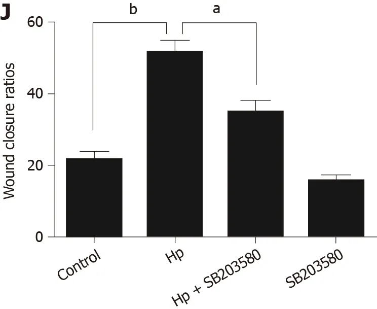
Figure 1 Heparanase protein expression following Helicobacter pylori infection in MKN-45 gastric cancer cells via the mitogen-activated protein kinase signaling pathway. A: Heparanase (HPA) expression was determined by Western blot at 0 , 6 , 12 , 24 , and 48 h after Helicobacter pylori (H.pylori) infection; B: Quantitative Western blot results of HPA; C: p-p38 mitogen-activated protein kinase (MAPK) expression was determined by Western blot at 0 , 30 ,60 , 120 , and 480 min after H. pylori infection; D: Quantitative Western blot results of p-p38 MAPK; E: HPA expression when the MAPK inhibitor SB203580 was given to MKN-45 cells before H. pylori infection; F: Quantitative Western blot results of HPA when the MAPK inhibitor SB203580 was given. bP < 0 .01 compared with the value at 0 h; G and H: Cell invasion rates in the three groups detected using a Transwell invasion assay; I and J: Migration rates in the three groups detected using a scratch migration assay. aP < 0 .05 , bP < 0 .01 . HPA: Heparanase; MAPK: Mitogen-activated protein kinase; H. pylori: Helicobacter pylori. Figure 2 in the original manuscript: Liu LP, Sheng XP, Shuai TK, Zhao YX, Li B, Li YM. Helicobacter pylori promotes invasion and metastasis of gastric cancer by enhancing heparanase expression. World J Gastroenterol 2018 ; 24 : 4565 -4577 [PMID: 30386106 DOI: 10 .3748 /wjg.v24 .i40 .4565 ].
ACKNOWLEDGEMENTS
在过去很长一段时间中,传统高校图书馆资源形式大多为纸质书籍、报刊、文献等。但在大数据时代到来以后,这种传统高校图书馆资源形式被完全打破,高校图书馆的资源形式也变得越来越多。使用大数据技术后的高校图书馆,不仅具备馆藏纸质文献等传统资源,还在此基础上增设了云上存储资源、网络资源等,使高校图书馆资源形式不再局限于纸质化。与此同时,高校图书馆在使用大数据技术之后,用户在浏览高校图书馆网站之后产生的信息也将成为高校图书馆大数据资源的组成部分。
We thank Dr. Zhong-Tian Bai for his critical reading of this manuscript. The authors appreciate Wen-Ting He for technical assistance.
 World Journal of Gastroenterology2021年22期
- World Journal of Gastroenterology的其它文章
- Fecal microbiota transplantation for irritable bowel syndrome: An intervention for the 21 st century
- Hepatocellular carcinoma in viral and autoimmune liver diseases: Role of CD4 + CD25 + Foxp3 + regulatory T cells in the immune microenvironment
- Application of artificial intelligence-driven endoscopic screening and diagnosis of gastric cancer
- Mucosal lesions of the upper gastrointestinal tract in patients with ulcerative colitis: A review
- Preservation of superior rectal artery in laparoscopically assisted subtotal colectomy with ileorectal anastomosis for slow transit constipation
- Early serum albumin changes in patients with ulcerative colitis treated with tacrolimus will predict clinical outcome
