
Comparison of Diagnostic Effects of T2-Weighted lmaging,DWl,SWl,and DTl in Acute Cerebral lnfarction
2021-05-12 06:14
School of Biomedical Engineering,Xinhua College of Sun Yat-Sen University,Guangzhou,510520,China
Abstract Objective:To achieve precision medicine,the use of imaging methods to help the clinical detection of cerebral infarction is conducive to the clinical development of a treatment plan and increase of the cure rate and improvement of the prognosis of patients.Methods: In this work,T2-weighted imaging (T2WI),dif fusion-weighted imaging (DWI),susceptibility-weighted imaging (SWI),and diffusion tensor imaging (DTI) examinations were performed on 34 patients with clinically diagnosed cerebral infarction to measure the difference in signal intensity between the lesion and its mirror area and make a comparative analysis by means of the Student-Newman-Keuls method.Results:The detection rate of T2WI was 79% (27/34),the detection rate of DWI was 97% (33/34),the detection rate of SWI was 88% (30/34),and the detection rate of DTI was 94% (32/34).Conclusion:The imaging performance was in the order DWI >D TI>S WI>T2WI for the diagnosis of cerebral infarction,and combined imaging is better than single imaging.
Keywords:T2-weighted imaging;susceptibility-weighted imaging;diffusion tensor imaging;diffusion-weighted imaging;cerebral infarction
lntroduction
Ischemic cerebral infarction is harmful to human health and quality of life and causes great pain to humans [1].To realize ef fective treatment of cer -ebral infarction,ensure the safety of patients with cerebral infarction,and determine the prognosis,accurate diagnosis of cerebral infarction has irreplaceable value in clinical practice.
It is important to choose the best examination method for accurate diagnosis of cerebral infarction to realize ef fective treatment.In this work,34 patients with cerebral infarction were examined by T2-weighted imaging (T2WI),dif fusion-weighted imaging (DWI),susceptibility-weighted imaging (SWI),and dif fusion tensor imaging (DTI) to measure the dif ference in signal intensity between the lesion and its mirror area and compare the four kinds of technologies for cerebral infarction diagnosis to help clinicians choose the best method to achieve a precise medical examination.
The advantages of these imaging sequences are as follows.T2WI is often obtained with a fast spin echo to enhance the ef fect of the T2 value on image contrast and to highlight signals from tissues with higher T2 values such as f uids.SWI can collect intensity data and phase data,and can superimpose the processed phase information on intensity information.It is sensitive to paramagnetic components and has advantages in imaging cerebral vascular diseases.Weighted contrasts resulting from dif ferences in voxel dispersion coeff cients caused by the dispersion effect of the magnetic resonance signal (the dispersion movement of the spin nucleus correspondingly gener -ates additional phase shifts) are used in DWI,and DWI has signif cant advantages in the diagnosis of cerebral infarction.DTI is an imaging method that uses the anisotropy of water dispersion to detect the microstructure of tissue.Using a variety of parameters and data processing,DTI can ref ect the change of dif fusion in imaging voxels quantitatively and directionally.
Methods
Under the condition of keeping other variables consistent and appropriate,T2WI,DWI,SWI,and DTI examinations were performed on 34 patients with clinically diagnosed cerebral infarction with use of a 3.0 T Signa Pioneer 2.0 MRI machine,and the differences in the signal intensity of the focal area and the specif c signal value of the mirror area for each imaging technique were compared.The Student-Newman-Keuls method was used to compare the signal intensity statistics in pairs.An MRI technician with more than 10 years of clinical experience performed the entire procedure independently.All image data measurements for patients were completed by one person.
Results
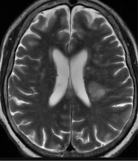
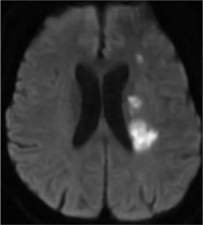
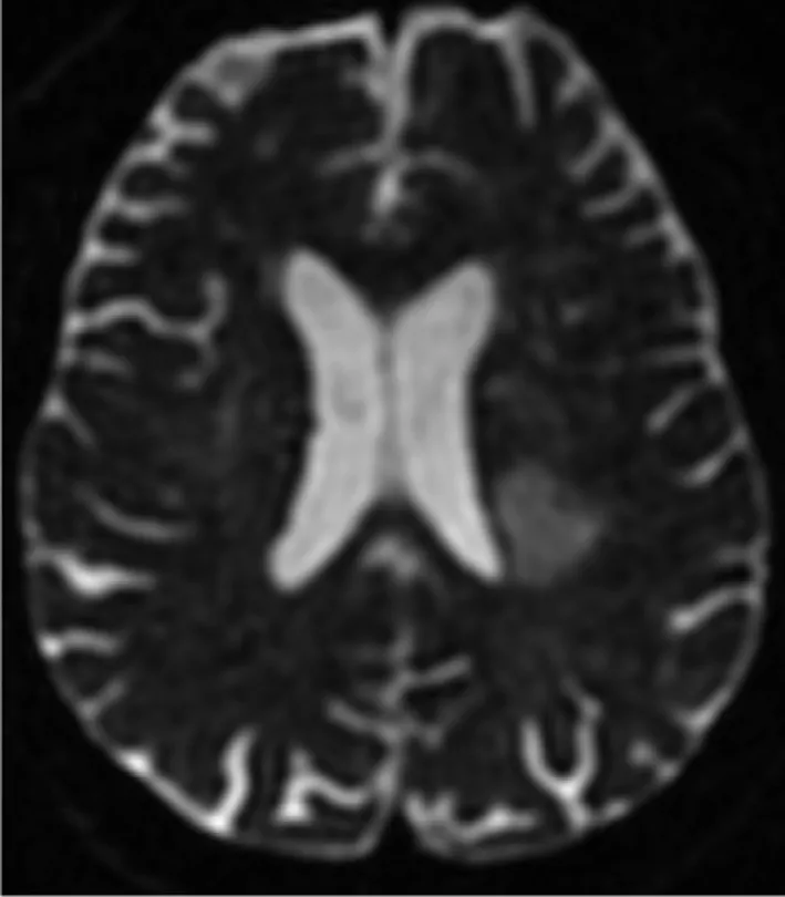
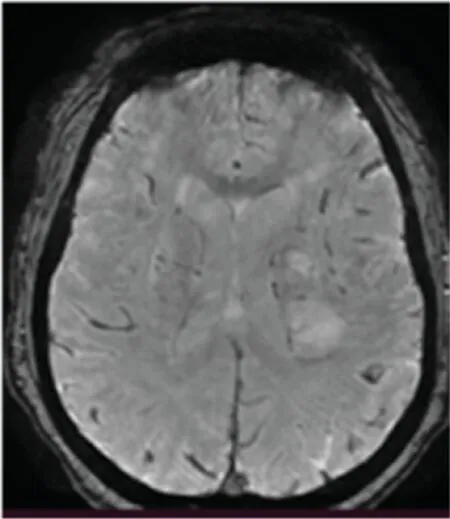
By the Student-Newman-Keuls method,when DWI was compared with T2WI,theqvalue was 6.2614711,P=0.0001,and the dif ference was statistically signif cant.When SWI was compared with T2WI,qwas 3.1814925,0.01 Table1 Comparison of Imaging Methods. Emboli caused by atherosclerosis often appear in human blood.These emboli can enter and block the cerebral vessels with the blood circulation of human body,and f nally lead to cerebral infarction and other diseases.This is the pathogenesis of cerebral infarction.Cerebral infarction seriously affects human health,so it is of great signif cance to carry out precise treatment. DWI can display the information associated with various types of cerebral infarction,and has become the gold standard for the identif cation of the core of cerebral infarction lesions.The application of DWI in clinical practice will be increased [2].Through DWI technology,the area of decreasing dispersion and increasing dispersion can be clearly displayed on the image.Through the analysis of DWI in 3,236 patients with acute ischemic cerebral infarction,the prevalence rate for patients with acute ischemic cerebral infarction under the premise of negative DWI was 6.8% [3]. DWI can ef fectively predict acute middle cer -ebral artery occlusion associated with intracranial atherosclerotic disease [4].Thin-slice DWI has advantages in the diagnosis of small lesions of unilateral localized infarction [5].DWI has the most advantageous diagnostic ef fect in the detection and differentiation of early and very early cerebral infarction and cerebral ischemia,as well as the def nition of the core area of infarction [6].DWI with apparent dif fusion coeff cient (ADC) images has a signif cant advantage in the differential diagnosis of cytotoxic edema (decreased ADC) from vasogenic edema (increased ADC) [7].DWI can be used for recanalization of thrombectomy at the site of cerebral infarction with good results [8].DWI combined with thrombolytic reperfusion therapy is helpful to analyze whether further craniectomy is necessary in patients with cerebral infarction [9].DWI and ADC images have obvious advantages in the dif ferential diagnosis of cerebral infarction and have guiding signif cance for the prognosis of patients with cerebral infarction [10].The differential detection of the lesion area by DWI can add valuable information to the assessment of the progress of the underlying microvascular disease [11].The diagnosis of cerebral infarction by 3D DWI is obviously better than that by conventional DWI,and its combination with angiography has a great advantage in the differentiation of cerebral infarction [12].Combined DWI is helpful to increase the accuracy of prediction of cerebral infarction after a TIA [13].Combined DWI and SWI has obvious advantages in the diagnosis of cerebral infarction,and SWI is helpful to identify intravascular thrombosis in the lesion area,which is helpful for subsequent intravascular thrombolytic therapy [14].However,DWI also has limitations in the diagnosis of cerebral infarction.Even if the best ef forts cannot inhibit the growth of early cerebral infarction lesions,the growth of lesions should be considered and measured in the treatment of patients with cerebral infarction [15]. Figure1 T2-Weighted Imaging of a Patient. Figure2 Diffusion-Weighted Imaging of the Same Patient as in Figure1 at the Same Level. Figure3 Diffusion Tensor Imaging of the Same Patient as in Figure1 at the Same Level. Figure4 Susceptibility-Weighted Imaging of the Same Patient as in Figure1 at the Same Level. SWI and DTI also play a special role in the diagnosis of cerebral infarction.The thrombosis sites and ranges of cerebral infarction lesions detected by SWI within 72 hours have similar thrombosis compositions,which can be considered to predict the severity of clinical lesions and short-term clinical prognosis [16].Changes caused by cerebral infarction can be clearly shown on DTI images[17].DTI can distinguish and judge the severity of cerebral infarction by comparing the cerebral blood f ow (CBF) and average ADC of the cerebral infarction lesion and its mirror area with the CBF in the mismatch area of S(CBF) and S(DWI) [18].It can clearly show the manifestations of tendon injury in each stage of acute ischemic cerebral infarction [19],and distinguish the therapeutic effect of a patient with cerebral infarction [2 0].Moreover,DTI is of great signif cance in the detection and identif cation of early cerebral infarction lesions and the evaluation of neurological recovery after cerebral infarction[21]. From comparison of the signal intensity of lesions,the imaging performance was in the order DWI >D TI>S WI>T2WI for the diagnosis of cer -ebral infarction (see Figures1–4 C o)m.bined imaging is obviously better than single imaging.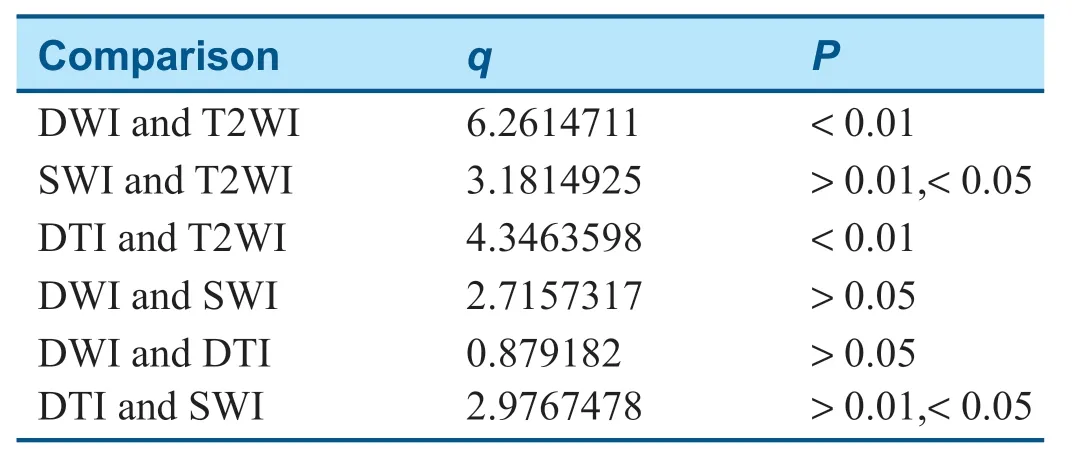
Discussion
Conclusion
 Cardiovascular Innovations and Applications2021年2期
- Cardiovascular Innovations and Applications的其它文章
- Progress in the Study of the Left Atrial Function lndex in Cardiovascular Disease:A Literature Review
- Better Than You Think—Appropriate Use of Implantable Cardioverter-Def ibrillators at a Single Academic Center:A Retrospective Review
- A Nomogram to Predict Patients with Obstructive Coronary Artery Disease:Development and Validation
- Calcium-Sensing Receptor of lmmune Cells and Diseases
- The Relationship between Abnormal Circadian Blood Pressure Rhythm and Risk of Readmission in Patients with Heart Failure with Preserved Ejection Fraction
- A Case of Pediatric Heart Failure Caused by Anomalous Origin of the Left Coronary Artery from the Pulmonary Artery:Case Report and Literature Review