IGF-1对6-OHDA诱导人神经母细胞瘤细胞损伤作用
王晓雯 晏振 陈夙 袁良杰 陈文芳

[摘要] 目的 探讨胰岛素样生长因子-1(IGF-1)对6-羟基多巴胺(6-OHDA)诱导的人神经母细胞瘤细胞(SK-N-SH细胞)神经毒性反应的抑制作用。方法 将SK-N-SH细胞种于96孔板,首先用不同浓度的IGF-1(6.25、12.50、25.00、50.00 μg/L)预处理细胞24 h,然后与6-OHDA(100 μmol/L)共同孵育细胞24 h,采用MTT法检测细胞活力。再将SK-N-SH细胞分为对照组(细胞不处理)、6-OHDA组(用100 μmol/L 6-OHDA处理细胞24 h)、6-OHDA+IGF-1组(细胞先用6.25 μg/L IGF-1预处理24 h,再与100 μmol/L 6-OHDA共同作用24 h),应用Western blot方法检测Bcl-2及Bax蛋白表达。结果 6.25~50.00 μg/L IGF-1均能够明显对抗6-OHDA诱导的神经毒性作用,提高细胞的存活率(F=11.53,q=4.795~5.556,P<0.05)。与对照组相比较,6-OHDA组Bcl-2蛋白的表达水平明显降低,而Bax蛋白的表达水平明显升高,差异有显著性(F=22.92、35.80,q=8.223、8.727,P<0.01);6.25 μg/L IGF-1能够明显对抗6-OHDA诱导的Bcl-2蛋白表达的下调和Bax蛋白表达的上调(q=8.361、11.450,P<0.01)。结论 IGF-1可对抗神经毒素6-OHDA诱导的SK-N-SH细胞凋亡相关蛋白Bcl-2及Bax的蛋白表达变化,发挥神经保护作用。
[关键词] 胰岛素样生长因子1;细胞凋亡;帕金森病;神经母细胞瘤
[中图分类号] R742.5;R338 [文献标志码] A [文章编号] 2096-5532(2020)02-0161-04
doi:10.11712/jms.2096-5532.2020.56.090 [開放科学(资源服务)标识码(OSID)]
[网络出版] http://kns.cnki.net/kcms/detail/37.1517.R.20200519.1433.007.html;2020-05-19 17:24
[ABSTRACT] Objective To investigate the inhibitory effect of insulin-like growth factor-1 (IGF-1) on the neurotoxic response of SK-N-SH human neuroblastoma cells induced by 6-hydroxydopamine (6-OHDA). Methods SK-N-SH cells were ino-culated into 96-well plates and pretreated with different concentrations of IGF-1 (6.25,12.50,25.00, and 50.00 μg/L) for 24 h. After that, the cells were co-incubated with 6-OHDA (100 μmol/L) for another 24 h. MTT assay was performed to determine the cell viability. In order to explore the protective mechanisms of IGF-1 on dopaminergic neurons, SK-N-SH cells were divided into control group (cells were left untreated), 6-OHDA group (cells were treated with 100 μmol/L 6-OHDA for 24 h), and 6-OHDA+IGF-1 group (cells were pre-treated with 6.25 μg/L IGF-1 for 24 h and then co-incubated with 100 μmol/L 6-OHDA for 24 h). Western blot was used to measure the expression of anti-apoptotic protein Bcl-2 and pro-apoptotic protein Bax in the SK-N-SH cells. Results Compared with the 6-OHDA group, 6.25-50.00 μg/L IGF-1 could significantly protect against the neurotoxic effects of 6-OHDA and improve the survival rate of SK-N-SH cells (F=11.53,q=4.795-5.556,P<0.05). Compared with the control group, the 6-OHDA group had significantly down-regulated protein expression of Bcl-2 and significantly up-regulated protein expression of Bax (F=22.92,35.80;q=8.223,8.727;P<0.01); 6.25 μg/L IGF-1 could significantly protect against 6-OHDA-induced down-regulation of Bcl-2 protein expression and up-regulation of Bax protein expression (q=8.361,11.450;P<0.01). Conclusion IGF-1 can protect against the neurotoxin 6-OHDA-induced apoptosis-related Bcl-2 and Bax protein expression changes in SK-N-SH cells, and exert a neuroprotective effect.
[KEY WORDS] insulin-like growth factorⅠ; apoptosis; Parkinson disease; neuroblastoma
帕金森病(PD)又名震颤麻痹,是一种中枢神经系统退行性疾病[1]。PD的主要临床表现为运动症状,如静止性震颤、肌强直、运动迟缓和姿势不稳等;其次为非运动症状,比如嗅觉功能异常、睡眠异常、肠道功能障碍等[2]。PD的主要病理表现是中脑黑质多巴胺(DA)能神经元的渐进性缺失,纹状体投射神经纤维的丢失以及嗜酸性包涵体路易小体(LBs)沉积[3-4]。导致PD病理改变的原因极其复杂,主要有环境因素、免疫炎症反应、遗传因素、线粒体功能异常、自噬功能的紊乱等[5-7]。一些神经毒素,如1-甲基-4-苯基-1,2,3,6-四氢吡啶(MPTP)、6-羟基多巴胺(6-OHDA)、百草枯和鱼藤酮等被用于PD模型的制备,这些药物大多与增强细胞凋亡有关[8]。6-OHDA属于DA与去甲肾上腺素类似物,其结构与儿茶酚胺相似,有研究显示其可以损毁神经末梢,导致递质减少,被广泛用于制备PD的细胞和动物模型[9]。胰岛素样生长因子-1(IGF-1)是一种含有70个氨基酸的多肽激素,其通过细胞膜表面的IGF-1受体(IGF-1R)发挥生物学作用[10]。IGF-1通过促进不同类型细胞的存活和增殖在中枢神经系统的发育和成熟中起着关键作用[11-12]。已有研究显示,血清IGF-1水平随着年龄增加而降低,PD病人早期血清IGF-1水平升高,随着病情的进展,IGF-1水平明显下降,提示其与PD的发病有关[13]。因此,探讨IGF-1的神经保护作用能够为防治PD奠定实验基础。本研究在前期研究的基础上,采用分子生物学技术,探讨IGF-1对6-OHDA诱导的人神经母细胞瘤细胞(SK-N-SH细胞)多巴胺能细胞损伤的作用。现将结果报告如下。
1 材料与方法
1.1 试剂及其来源
SK-N-SH细胞由中国科学院上海细胞库提供;IGF-1购自BioVision公司,用生理盐水配制成1 g/L的溶液;DMEM购自Gibco公司;青霉素/链霉素储存液购自新华制药厂,分装后,-20 ℃保存备用;胎牛血清购自Hyclone公司,分装后,-40 ℃保存备用;二甲基亚砜(DMSO)购自Sigma公司;兔抗-Bcl-2抗体购自CST公司;兔抗-Bax抗体购自CST公司;6-OHDA由Sigma公司提供;BCA试剂盒由碧云天公司提供。
1.2 细胞培养
将SK-N-SH细胞接种于25 cm2的细胞培养瓶中,加入含有体积分数0.10胎牛血清、100 mg/L链霉素和100 kU/L青霉素的DMEM培養液,置于37 ℃含体积分数0.05 CO2的细胞培养箱中。当细胞达到80%~90%融合时进行实验。
1.3 MTT法检测细胞活力
将SK-N-SH细胞接种于96孔板上,每孔加细胞悬浮液100 μL(含6×10⁴个细胞),放置于细胞培养箱中培养至细胞达80%~90%融合时,开始加药处理。先加入不同浓度的IGF-1(6.25、12.50、25.00、50.00 μg/L)预保护SK-N-SH细胞24 h,然后再与100 μmol/L 6-OHDA共同作用24 h。弃去培养液,每孔加入20 μL MTT溶液(5 g/L),继续避光培养4 h。小心吸除多余MTT,每孔加100 μL 的DMSO,摇床80~90 r/min避光振荡10 min,使结晶物充分溶解。应用酶标仪(490 nm)检测各孔的吸光度(A)值。细胞活力以实验组A值/对照组A值表示。
1.4 Bcl-2和Bax蛋白表达检测
应用Western blot方法。将SK-N-SH细胞接种于96孔板中,分为对照组、损伤组、保护药组。对照组细胞不处理;损伤组细胞应用100 μmol/L的6-OHDA处理24 h;保护药组细胞先用6.25 μg/L的IGF-1预保护24 h,再与100 μmol/L 6-OHDA共同作用24 h。弃去细胞培养液,每孔加入裂解液(lysis∶PMSF=99∶1)100 μL,冰上裂解30 min,然后用细胞刮轻轻刮下细胞,收集至1.5 mL的EP管中[14]。置于离心机中离心20 min (4 ℃,12 000 r/min),吸取80 μL上清,用BCA法检测蛋白浓度。电泳30~40 min (80 V稳压),上样量为15 μg;待蛋白Maker的不同分子量条带分离开时,将电压改为120 V,继续电泳,根据所检测蛋白分子量的大小来确定具体的电泳结束时间。然后,将胶和PVDF膜置于转膜夹中进行转膜,条件为300 mA、90 min。转膜结束后将PVDF膜使用50~100 g/L的脱脂奶粉封闭1 h,清洗掉残留的奶粉加一抗,4 ℃摇床过夜后洗膜3次,每次10 min;二抗孵育1~2 h后洗膜3次,每次10 min,以发光液显影。检测Bcl-2、Bax与β-actin蛋白A值,以Bcl-2、Bax与β-actin的A值比值表示蛋白表达[15]。
1.5 统计学处理
应用Graph Pad Prism 5.0统计软件进行数据分析,计量资料结果以±s表示,数据间比较用单因素方差分析(One-Way ANOVA),并继以Tukey法进行两两比较。P<0.05表示差异有显著性。
2 结 果
2.1 不同浓度IGF-1对6-OHDA诱导SK-N-SH细胞活力影响
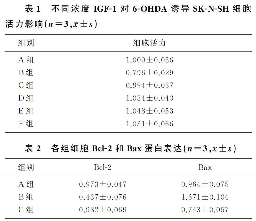
与对照组(A组)相比,6-OHDA组(B组)的细胞活力明显降低 (F=11.53,q=5.967,P<0.05);6.25 μg/L(C组)、12.50 μg/L(D组)、25.00 μg/L(E组)和50.00 μg/L(F组) IGF-1均可对抗6-OHDA的神经毒性作用,提高SK-N-SH细胞的活力(q=4.857~5.556,P<0.05)。见表1。
2.2 IGF-1對6-OHDA诱导SK-N-SH细胞Bcl-2和Bax蛋白表达的影响
与对照组(A组)相比较,6-OHDA组(B组)SK-N-SH细胞Bcl-2蛋白表达水平明显降低(F=22.92,q=8.223,P<0.01),Bax蛋白表达明显升高(F=35.80,q=8.727,P<0.01);与6-OHDA组(B组)相比,IGF-1+6-OHDA组(C组)Bcl-2蛋白的表达明显上升(q=8.361,P<0.01),Bax蛋白表达明显下降(q=11.45,P<0.001)。见表2。
3 讨 论
PD是一种迟发性、进行性、神经退行性的运动障碍疾病,占65岁及以上人口的1%[16-17],随着中国人口老龄化状况的日益加剧,PD的发病率也逐年增高,造成的家庭和社会负担也日益严重[18]。目前,PD病人尚无有效的治疗方法,开发研制有效防治PD的药物迫在眉睫。6-OHDA是目前常用的建立PD模型的神经毒素,能够引起DA能神经元的大量变性死亡,继而导致锥体外系运动功能障碍,如震颤、强直、运动过缓等表现[19]。
IGF-1是一种天然存在于中枢神经系统的强效神经营养和抗凋亡因子,可以促进不同类型细胞的发育分化[20-22]。IGF-1主要是通过IGF-1R信号通路,促进细胞的生长、分化和存活,发挥神经保护作用。有研究显示,IGF-1R在脑组织内广泛表达,其中在黑质几乎所有的DA能神经元以及多数的胶质细胞内均存在[23]。许多研究表明,人体内IGF-1水平与年龄相关,幼儿期含量相对较低,成年后达到高峰,之后随着年龄的增加逐渐降低[24]。临床研究发现,PD早期病人血清中IGF-1的水平升高,随着病情的进展IGF-1的水平逐渐下降。在认知障碍病人中,IGF-1水平亦明显降低[25-26]。
AYADI等[27]的研究显示,IGF-1能够通过激活下游的Ras/ERK1/2和PI3K/Akt信号通路,对抗6-OHDA对大鼠黑质纹状体系统DA能神经元的损伤。在氧葡萄糖剥夺/再灌注SH-SY5Y神经细胞模型中,miR-186-5p可通过降低IGF-1的表达,诱导细胞凋亡[28]。离体细胞实验证明IGF-1通过抗凋亡和抗氧化应激发挥神经元保护功能[29-30]。本研究应用6-OHDA损伤SK-N-SH细胞,制备PD细胞模型,探究IGF-1的神经保护作用及其可能机制。MTT结果显示,6-OHDA明显损伤了SK-N-SH细胞,而IGF-1能够明显对抗6-OHDA的这种神经毒性作用。为了进一步探讨IGF-1的保护作用及其机制,本实验应用Western blot技术,检测了凋亡相关蛋白Bcl-2及Bax蛋白表达情况。实验结果显示,与对照组相比,6-OHDA组抗凋亡蛋白Bcl-2蛋白的表达明显降低,促凋亡蛋白Bax蛋白的表达显著升高;而给予IGF-1预处理细胞后,Bcl-2蛋白表达明显上调,而Bax蛋白的表达明显下调。表明IGF-1神经元保护功能与抑制6-OHDA诱导的细胞凋亡有关。
综上所述,IGF-1可对抗神经毒素6-OHDA诱导的SK-N-SH细胞凋亡相关蛋白Bcl-2及Bax蛋白表达变化,发挥神经保护作用。
[参考文献]
[1] DAWSON T M, DAWSON V L. Molecular pathways of neurodegeneration in Parkinsons disease[J]. Science, 2003,302(5646):819-822.
[2] NEIKRUG A B, MAGLIONE J E, LIU L Q, et al. Effects of sleep disorders on the non-motor symptoms of Parkinson di-sease[J]. Journal of Clinical Sleep Medicine: JCSM, 2013,9(11):1119-1129.
[3] LASHUEL H A, OVERK C R, OUESLATI A, et al. The many faces of α-synuclein: from structure and toxicity to therapeutic target[J]. Nature Reviews Neuroscience, 2013,14(1):38-48.
[4] KALIA L V, LANG A E. Parkinsons disease[J]. The Lancet, 2015,386(9996):896-912.
[5] NING Baile, ZHANG Qinxin, WANG Ninzhen, et al. β-Asarone regulates ER stress and autophagy via inhibition of thePERK/CHOP/Bcl-2/beclin-1 pathway in 6-OHDA-induced Parkinsonian rats[J]. Neurochemical Research, 2019,44(5):1159-1166.
[6] ZHONG Jiahong, XIE Jinfeng, XIAO Jiao, et al. Inhibition of PDE4 by FCPR16 induces AMPK-dependent autophagy and confers neuroprotection in SH-SY5Y cells and neurons exposed to MPP(+)-induced oxidative insult[J]. Free Radical Biology and Medicine, 2019,135:87-101.
[7] LUDTMANN M H R, ABRAMOV A Y. Mitochondrial cal-cium imbalance in Parkinsons disease[J]. Neuroscience Letters, 2018,663:86-90.
[8] SU Lingyan, LI Hao, LV Li, et al. Melatonin attenuates MPTP-induced neurotoxicity via preventing CDK5-mediated autophagy and SNCA/alpha-synuclein aggregation[J]. Autophagy, 2015,11:1745-1759.
[9] GLINKA Y, TIPTON K F, YOUDIM M B. Mechanism of inhibition of mitochondrial respiratory complex I by 6-hydroxydopamine and its prevention by desferrioxamine[J]. European Journal of Pharmacology, 1998,351(1):121-129.
[10] ZHENG W H, QUIRION R. Insulin-like growth factor-1 (IGF-1) induces the activation/phosphorylation of Akt kinase and cAMP response element-binding protein (CREB) by activating different signaling pathways in PC12 cells[J]. BMC Neuroscience, 2006,7:51-61.
[11] BECK K D, POWELL-BRAXTON L, WIDMER H R, et al. IGF1 gene disruption results in reduced brain size, CNS hypomyelination, and loss of hippocampal granule and striatal parvalbumin-containing neurons[J]. Neuron, 1995,14(4):717-730.
[12] RUSSO V C, GLUCKMAN P D, FELDMAN E L, et al. The insulin-like growth factor system and its pleiotropic functions in brain[J]. Endocrine Reviews, 2005,26(7):916-943.
[13] MARCO P D, BARTELLA V, VIVACQUA A, et al. Insulin-like growth factor-Ⅰ regulates GPER expression and function in cancer cells[J]. Oncogene, 2013,32(6):678-688.
[14] CHEN Wenfang, ZHOU Liping, CHEN Lei, et al. Involvement of IGF-Ⅰ receptor and estrogen receptor pathways in the protective effects of ginsenoside Rg1 against A beta(25-35)-induced toxicity in PC12 cells[J]. Neurochemistry International, 2013,62(8):1065-1071.
[15] 任曉璠,孙宪昌,王宇鑫,等. Rg1和GR阻断剂对脂多糖诱导的BV2小胶质细胞iNOS及COX2蛋白表达影响[J]. 青岛大学医学院学报, 2016,52(2):148-152.
[16] BRAAK H, DEL TREDICI K, RB U, et al. Staging of brain pathology related to sporadic Parkinsons disease[J]. Neurobiology of Aging, 2002,24(2):197-211.
[17] JAKOWEC M W, PETZINGER G M. 1-methyl-4-phenyl-1,2,3,6-tetrahydropyridine-lesioned model of Parkinsons di-sease, with emphasis on mice and nonhuman primates[J]. Comparative Medicine, 2004,54(5):497-513.
[18] MICHEL P P, HIRSCH E C, HUNOT S. Understanding dopaminergic cell death pathways in Parkinson disease[J]. Neuron, 2016,90(4):675-691.
[19] TEISMANN P, TIEU K, COHEN O, et al. Pathogenic role of glial cells in Parkinsons disease[J]. Movement Disorders: Official Journal of the Movement Disorder Society, 2003,18(2):121-129.
[20] BENARROCH E E. Insulin-like growth factors in the brain and their potential clinical implications[J]. Neurology, 2012,79(21):2148-2153.
[21] GASPERI M, CASTELLANO A E. Growth hormone/insulin-like growth factor 1 axis in neurodegenerative diseases[J]. Journal of Endocrinological Investigation, 2010,33(8):587-591.
[22] DYER A H, VAHDATPOUR C, SANFELIU A, et al. The role of insulin-like growth factor 1 (IGF-1) in brain development, maturation and neuroplasticity[J]. Neuroscience, 2016,325(14):89-99.
[23] QUESADA A, ROMEO H E, MICEVYCH P. Distribution and localization patterns of estrogen receptor-beta and insulin-like growth factor-1 receptors in neurons and glial cells of the female rat substantia nigra: localization of ERbeta and IGF-1R in substantia nigra[J]. The Journal of Comparative Neurology, 2007,503(1):198-208.
[24] SMITH C P, DUNGER D B, WILLIAMS A J, et al. Relationship between insulin,insulin-like growth factor 1, and dehydroepiandrosterone sulfate concentrations during childhood, puberty, and adult life[J]. The Journal of Clinical Endocrino-logy and Metabolism,1989,68(5):932-937.
[25] GODAU J, HERFURTH M, KATTNER B, et al. Increased serum insulin-like growth factor 1 in early idiopathic Parkinsons disease[J]. Journal of Neurology Neurosurgery and Psychiatry, 2010,81(5):536-538.
[26] GODAU J, KNAUEL K, WEBER K, et al. Serum insulin like growth factor 1 as possible marker for risk and early diagnosis of parkinson disease[J]. Archives of Neurology, 2011,68(7):925-931.
[27] AYADI A E, ZIGMOND M J, SMITH A D. IGF-1 protects dopamine neurons against oxidative stress: association with changes in phosphokinases[J]. Exp. Brain Res, 2016,234(7):1863-1873.
[28] WANG Rui, BAO Hongbo, ZHANG Shihua, et al. miR-186-5p promotesapoptosisby targeting IGF-1 in SH-SY5Y OGD/R model[J]. International Journal of Biological Scirnces, 2018,14(13):1791-1799.
[29] PARK Y G, JEONG J K, MOON M H, at el. Insulin-like growth factor-1 protects against prion peptide-induced cell death in neuronal cellsvia inhibition of Bax translocation[J]. International Journal of Molecular Medicine, 2012,30(5):1069-1074.
[30] WEN Di, CUI Can, DUAN Weisong, et al. The role of insulin-like growth factor 1 in ALS cell and mouse models: a mitochondrial protector[J]. Brain Research Bulletin, 2019,144:1-13.
(本文編辑 黄建乡)

