寰枢关节间撑开技术治疗颅底凹陷症的临床疗效分析
许俊杰,蓝思彬,吴增晖,夏 虹,马向阳,艾福志,章 凯,段明阳,杨善智,陈恩良,梁达轩
临床研究
寰枢关节间撑开技术治疗颅底凹陷症的临床疗效分析
许俊杰,蓝思彬,吴增晖,夏 虹,马向阳,艾福志,章 凯,段明阳,杨善智,陈恩良,梁达轩
目的观察寰枢关节间撑开技术——经口寰枢椎复位钢板内固定(TARP)治疗颅底凹陷症伴难复性寰枢椎脱位的临床疗效,探讨TARP术后神经功能恢复的相关影响因素。方法回顾性分析2010年8月至2012年7月广州军区广州总医院采用TARP手术治疗的22例颅底凹陷症伴难复性寰枢椎脱位患者的临床资料。分别测量术前及末次随访时寰齿间隙(ADI)、Chamberlian线(CL,CT矢状位上硬腭到枕骨大孔的连线)和O-C2角(McGregor线与枢椎椎体下缘交角),计算齿突下移距离(dCL)和O-C2角变化量(dO-C2角);采用日本骨科学会(JOA)神经功能改善率评估患者神经功能恢复程度,根据神经功能改善率将患者分成A组(改善率<50%)和B组(改善率≥50%),其中A组5例,B组17例。结果TARP术后22例患者临床症状均获得不同程度改善。两组ADI、CL、dCL、O-C2角、dO-C2角等影像学参数比较,差异无统计学意义(P>0.05);A组、B组平均病程分别为(106±68)和(45±47)个月,A组明显长于B组(P<0.05)。1例患者术后脑干仍存在持续压迫,予二期经口齿突切除伴后路内固定翻修术,症状无显著缓解。随访期间未见内固定失败病例。结论寰枢关节间撑开技术治疗颅底凹陷症是有效的,不伴齿突切除的TARP技术能有效下移齿突并充分减压椎管;术后神经功能的恢复可能与病程相关。
颅底凹陷症;寰枢关节;脱位;经口手术;齿突尖;减压术,外科;骨板;内固定器
颅底凹陷症是常见的复杂枕颈交界区畸形,其主要病因是齿突向后向上移位直接压迫脑干[1]。目前治疗颅底凹陷症的主要手术方法是一期经口咽寰枢椎减压伴后路融合内固定;亦有术者采用单纯前路或后路术式,可在有效下移齿突的同时减少手术相关并发症[2-4]。Goel[5]率先介绍经后路寰枢关节垫片植入联合后路内固定技术,但在其报道病例中,齿突下移并不充分,寰枢椎脱位亦未完全复位。我们的前期研究表明,经口寰枢椎复位钢板内固定(transoral atlantoaxial reduction and plate fixation,TARP)技术能够有效下移齿突,从而达到椎管减压的目的[6]。本研究总结TARP手术治疗颅底凹陷症伴难复性寰枢椎脱位的临床疗效和影像学评估结果,分析术后神经功能恢复的影响因素,为该病的术式选择及预后评估提供参考依据。
1 资料与方法
1.1 一般资料
将2010年8月至2012年7月我院采用TARP手术治疗的22例颅底凹陷症伴难复性寰枢椎脱位患者纳入本研究,排除枕颈交界区创伤、感染或已行后路内固定者,手术均由同一高年资医师完成。男性9例,女性13例;年龄8~57岁,平均年龄32岁;随访时间55~81个月,平均随访时间67个月;病程6~180个月,平均病程59个月。
1.2 分组
按照日本骨科学会(Japanese Orthopaedic Association,JOA)颈椎评分(17分法)计算,神经功能改善率=(术后JOA-术前JOA)/(17-术前JOA)×100%[7]。根据末次随访时JOA神经功能改善率将22例患者分为A组(改善率<50%)和B组(改善率≥50%)。A组5例患者,男2例,女3例;主要症状为麻木(5例)、肌无力(4例)、枕颈部疼痛(3例)、行走不稳(3例)、斜颈(3例)和小便费力(1例)。B组17例患者,男7例,女10例;主要症状包括有肌无力(13例)、行走失稳(9例)、麻木(8例)、枕颈部疼痛(6例)、斜颈(2例)和呼吸困难(1例)。其中A组1例、B组2例有后颅窝减压手术史。
1.3 测量方法和影像学指标
术前及术后1周、3个月、6个月常规拍摄X线片、CT及MRI,测量以下各影像学指标,同时评估椎管受压、螺钉位置及植骨融合情况。
1.3.1 dO-C2角 通过手术前后颈椎侧位X线片测量McGregor线与枢椎椎体下缘的交角,即O-C2角,然后计算手术前后O-C2角变化量(dO-C2角=术前 O-C2角-术后O-C2角)[8-9]。
1.3.2 dCL首先在CT上测量术前及术后寰齿间隙(atlas-dens interval,ADI)和 Chamberlian 线(Chamberlian line,CL),其中CL为CT矢状位上硬腭到枕骨大孔的连线;然后通过公式dCL=术前CL-术后CL进行计算。当术后ADI≤3 mm、CL≤3 mm时视为达到解剖学复位[10]。
3例患者已行后颅窝减压术,不能直接测量O-C2角及CL,采用Jian等[11]提出的参考线进行相关的数据测量。
1.4 统计学方法
采用SPSS 22.0软件对数据进行处理。计量资料以均数±标准差(±s)表示,两组比较采用两独立样本t检验,计数资料以例表示,比较采用卡方检验或Fisher确切概率法。P<0.05为差异有统计学意义。
2 结果

图1经口寰枢椎复位钢板内固定治疗颅底凹陷症伴难复性寰枢椎脱位手术前后影像学图片(女,34岁)1A术前X线侧位片测量O-C2角为-4.4°1B 术前CT矢状位示寰枕融合后枕骨后缘向下延伸,导致Chamberlian线(CL)向下位移(CL为18.5 mm)1C术前CT矢状位可见侧块关节面向下倾斜 1D术前矢状位MRI提示齿突向后压迫脑干 1E术后1周X线侧位片测量O-C2角为24.6°1F术后1周矢状位MRI提示椎管减压充分 1G 术后6个月CT矢状位可见侧块关节植骨融合,CL为14.8 mm 1H 术后6个月冠状面CT提示双侧侧块关节及齿突周围植骨融合
典型病例见图1。术后ADI均获解剖学复位;8例患者CL达到解剖学复位,14例CL未达解剖学复位。临床症状均获得不同程度的改善。1例患者因齿突尖上方软组织持续压迫脑干,二期行经口齿突切除伴后路植骨内固定术,症状未见显著改善。余21例患者MRI检查提示椎管减压充分。无一例出现内固定失败、椎动脉损伤、脑脊液漏、神经损伤、切口感染并发症。
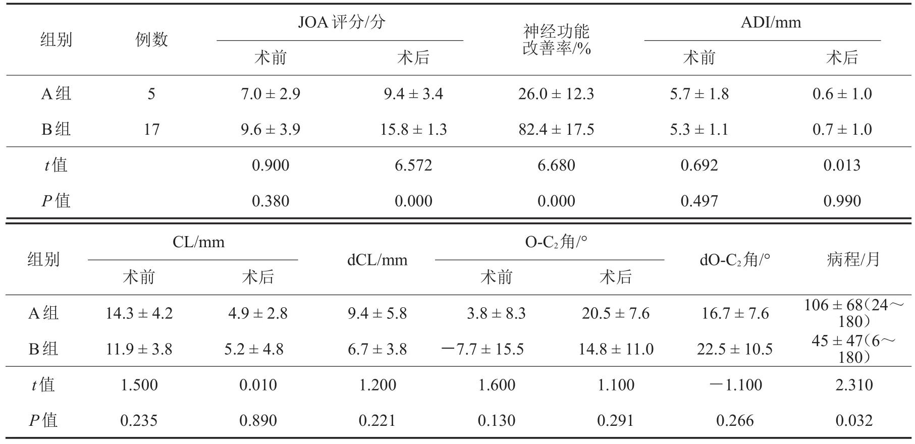
如表1所示,两组性别分布、年龄、随访时间等术前一般资料比较,差异无统计学意义(P>0.05)。如表2所示,术后JOA评分A组明显低于B组;术后两组ADI、CL、dCL、O-C2角、dO-C2角等影像学参数比较,差异均无统计学意义(P>0.05);A组病程明显长于B组,两组比较,差异有统计学意义(P<0.05)。
3 讨论
颅底凹陷症伴难复性寰枢椎脱位属于上颈椎外科难治伤病,该区域解剖结构复杂,毗邻重要神经血管,外科干预极具挑战性。传统的手术方式采用经口齿突切除或松解伴一期后方内固定手术[12],但无论是前方还是后方入路,潜在的手术风险较大,并发症发生率也居高不下。
3.1 手术方式的选择与寰枢椎解剖复位
3.1.1 单纯后路手术 有报道称,单纯后方入路能有效下移齿突,阻止齿突进一步压迫脑干[2],复位效果满意。但对于伴难复性寰枢椎脱位的颅底凹陷症患者而言,单纯后路手术的临床效果如何,业界仍有较大争议[13-15]。有学者指出,齿突及寰枢关节周围增生软组织、挛缩疤痕及寰枢关节间关节囊是阻止寰枢椎脱位解剖复位的原因,术中必须予以清理[15-16];Jian 等[11]认为 C1~C2侧块关节成角可在一定程度上维持复位,然而作为主要的承重关节,侧块关节成角可能导致关节逐渐塌陷及枕颈交界区后凸畸形。因此,经后路手术实现齿突充分下移及寰枢椎脱位的解剖学复位是极其困难的。

表1两组患者一般资料比较
表2神经功能改善程度不同的两组患者临床指标比较(± s)

表2神经功能改善程度不同的两组患者临床指标比较(± s)
注:A组:神经功能改善率<50%;B组:神经功能改善率≥50%;JOA:日本骨科学会;ADI:寰齿间隙;CL:Chamberlian线;dCL:术后齿突下移距离;O-C2角:McGregor线与枢椎椎体下缘的交角;dO-C2角:手术前后O-C2角变化
ADI/mm组别 例数 神经功能改善率/%A组B组t值P值5 17 JOA评分/分术前7.0±2.9 9.6±3.9 0.900 0.380术后9.4±3.4 15.8±1.3 6.572 0.000 26.0±12.3 82.4±17.5 6.680 0.000术前5.7±1.8 5.3±1.1 0.692 0.497术后0.6±1.0 0.7±1.0 0.013 0.990组别dCL/mm dO-C2角/°A组B组t值P值CL/mm术前14.3±4.2 11.9±3.8 1.500 0.235术后4.9±2.8 5.2±4.8 0.010 0.890 9.4±5.8 6.7±3.8 1.200 0.221 O-C2角/°术前3.8±8.3-7.7±15.5 1.600 0.130术后20.5±7.6 14.8±11.0 1.100 0.291 16.7±7.6 22.5±10.5-1.100 0.266病程/月106 ± 68(24~180)45 ± 47(6~180)2.310 0.032
3.1.2 融合器植入手术 一些学者采取将侧块关节间融合器植入侧块关节的方法,迫使齿突下移[5,17],但融合器限制了齿突前倾及寰椎向后复位,复位欠理想;术中为了充分显露以及为融合器植入提供合适位置,必须要切除C2神经根,从而导致C2神经根支配区域出现永久性麻木[5,17];寰枕融合、椎旁静脉丛及寰椎向前滑移也增加了切口的显露难度;术中如果使用过长融合器或融合器植入方向错误,则极易侵犯椎管,导致脊髓或椎动脉损伤。基于以上的手术风险,我们认为植入融合器的过程是不安全的。
3.1.3 TARP手术 本研究中22例患者实施TARP手术,术后影像学结果证实,枕颈交界区序列恢复正常,椎管内减压充分,所有病例ADI均获解剖学复位,其主要原因是TARP手术能使患者前路寰枢椎得到充分松解。本研究中14例患者术后CL未获得解剖学复位,我们推测其原因在于寰枕融合。曾有学者指出,寰枕融合令枕骨后缘向下延伸,导致CL向下产生位移[18]。在这种情况下,齿突过度下移可能会导致术后O-C2角过度增大,引起吞咽困难、呼吸困难、下颈椎滑脱等并发症[9,19]。
本研究中3例患者因Chiari畸形行后颅窝减压术,导致部分枕骨缺如,无法行后路内固定手术;还有7例患者因C2椎弓根狭小而无法植入C2椎弓根螺钉;其他畸形如椎动脉高跨或枕骨发育不良,也不适合采用后路内固定手术[20]。为规避手术风险、降低术后并发症发生率,TARP手术是此类患者的最佳选择。另外,TARP手术还适用于对后路手术失败的患者进行翻修手术,在解决问题的同时极大地减少了手术创伤[3]。
3.2 TARP术后神经功能改善的影响因素
颅底凹陷症外科干预的主要目的在于早期去除来自前方的压迫,提供可靠内固定,但术后脊髓功能的恢复却受年龄、性别、损伤程度、椎管减压程度、病程、MRI信号改变等多种因素影响。本研究根据JOA神经功能改善程度的不同将患者分为A、B两组,患者术后ADI、CL、O-C2角等影像学参数相似,术后随访时间(即神经功能康复时间)亦无显著差异(P>0.05),但在病程方面,神经功能改善差的A组明显长于改善良好的B组(P<0.05)。推测两组患者神经功能改善率存在差异的原因可能与病程相关,病程较短者其术后神经功能恢复较好,病程长者神经功能恢复缓慢。Mazel等[21]报道28例严重颈椎管狭窄症手术患者,结果表明,术后神经功能恢复与病程相关,病程长的患者预后较差,病程短的预后较好;Zhang等[22]对110例脊髓型颈椎病手术患者进行疗效分析,也得出了相同的结论。
目前越来越多的学者开始研究脊髓信号改变与神经功能康复的相关性,但其临床意义存在较大争议。Matsuda等[23]认为脊髓信号改变与神经功能恢复相关,脊髓高信号往往预示脊髓神经功能恢复差;Shin等[24]也认为,脊髓信号等级的高低与患者预后的好坏有明显相关关系。亦有学者持不同看法。Yone等[25]认为脊髓型颈椎病术前症状严重程度及术后神经恢复与脊髓高信号无关;Avadhani等[26]亦认为脊髓信号改变程度与术后Nurick分级无显著性差异。本研究中大部分患者术前MRI高信号改变较明显,无法进行有效的分组比较,也未能得出令人信服的结论。
3.3 手术并发症
尽管TARP手术治疗颅底凹陷症伴难复性寰枢椎脱位效果良好,但仍然存在一定的风险。Yin等[14]在2~4年的随访研究中发现,有2例(6.5%)骨质疏松患者出现螺钉松动;Yang等[3]报道1例患者术后螺钉松动并实施前路翻修手术;Li等[20]的研究结果表明,TARP手术寰椎侧块螺钉及枢椎逆向椎弓根螺钉位置欠佳发生率分别为2.4%(5/212)和13%(27/207),1例患者因椎动脉损害死亡,3例发生手术切口感染,移除前路内固定物后改行后路内固定手术。在前期研究中我们发现,TARP手术C2螺钉松动病例中椎体螺钉松动发生率较高,之后Wu等[27]率先提出经口前路枢椎逆向椎弓根螺钉内固定方法并将其应用于临床,降低了枢椎螺钉松动并发症的发生率,但仍存在损伤椎动脉的风险,因此术前需通过三维CT了解椎动脉变异等异常情况。此外,经口入路术后发生切口感染通常与脑脊液漏相关,术中应仔细操作,避免损伤硬脊膜。
[1]Smith JS,Shaffrey CI,Abel MF,et al.Basilar invagination[J].Neurosurgery,2010,66(3 Suppl):39-47.
[2]Abumi K,Takada T,Shono Y,et al.Posterior occipitocervical reconstruction using cervical pedicle screws and plate-rod systems[J].Spine,1999,24(14):1425-1434.
[3] Yang J,Ma X,Xia H,et al.Transoral anterior revision surgeries for basilar invagination with irreducible atlantoaxial dislocation afterposteriordecompression:a retrospective study of 30 cases[J].Eur Spine J,2014,23(5):1099-1108.
[4]Yin Q,Ai F,Zhang K,et al.Irreducible anterior atlantoaxial dislocation:one-stage treatment with a transoral atlantoaxial reduction plate fixation and fusion:report of 5 cases and review of the literature[J].Spine,2005,30(13):E375-E381.
[5]Goel A.Treatment of basilar invagination by atlantoaxial joint distraction and direct lateral mass fixation[J].J Neurosurg Spine,2004,1(3):281-286.
[6]Xia H,Yin Q,Ai F,et al.Treatment of basilar invagination with atlantoaxial dislocation:atlantoaxial joint distraction and fixation with transoral atlantoaxial reduction plate(TARP)withoutodontoidectomy [J].EurSpine J,2014,23(8):1648-1655.
[7]Fukui M,Chiba K,Kawakami M,et al.Japanese Orthopaedic Association Back Pain Evaluation Questionnaire:part 2:verification of its reliability:the subcommittee on low back pain and cervicalmyelopathy evaluation oftheclinical outcome committee of the Japanese Orthopaedic Association[J].J Orthop Sci,2007,12(6):526-532.
[8]Matsunaga S,Onishi T,Sakou T.Significance of occipitoaxial angle in subaxial lesion after occipitocervical fusion[J].Spine,2001,26(2):161-165.
[9]Miyata M,Neo M,Fujibayashi S,et al.O-C2 angle as a predictor of dyspnea and/or dysphagia after occipitocervical fusion[J].Spine,2009,34(2):184-188.
[10]Shuhui G,Jiagang L,Haifeng C,et al.Surgical management ofadultreducible atlantoaxialdislocation,basilarinvagination and Chiari malformation with syringomyelia[J].Turk neuros,2016,26(4):615-621.
[11]Jian FZ,Chen Z,Wrede KH,et al.Direct posterior reduction and fixation for the treatment of basilar invagination with atlantoaxial dislocation[J].Neurosurgery,2010,66(4):678-687.
[12]Wang C,Yan M,Zhou HT,et al.Open reduction of irreducible atlantoaxialdislocation by transoral anterior atlantoaxial release and posterior internal fixation[J].Spine,2006,31(11):E306-E313.
[13]Wang C,Wang S.Direct posterior reduction and fixation[J].Neurosurgery,2011,68(2):E601-E604.
[14]Yin QS,Ai FZ,Zhang K,et al.Transoral atlantoaxial reduction plate internal fixation for the treatment of irreducible atlantoaxial dislocation:a 2-to 4-year follow-up[J].Orthop Surg,2010,2(2):149-155.
[15]Srivastava SK,Aggarwal RA,Nemade PS,et al.Single-stage anterior release and posterior instrumented fusion for irreducible atlantoaxial dislocation with basilar invagination[J].Spine J,2016,16(1):1-9.
[16]Laheri V,Chaudhary K,Rathod A,et al.Anterior transoral atlantoaxial release and posterior instrumented fusion for irreducible congenital basilar invagination[J].Eur Spine J,2015,24(12):2977-2985.
[17]Chandra PS,Kumar A,Chauhan A,et al.Distraction,compression,and extension reduction of basilar invagination and atlantoaxial dislocation:a novel pilot technique[J].Neurosurgery,2013,72(6):1040-1053.
[18]Ferreira ED,Botelho RV.Atlas assimilation patterns in different types of adult craniocervical junction malformations[J].Spine,2015,40(22):1763-1768.
[19]Takami T,Ichinose T,Ishibashi K,et al.Importance of fixation angle in posterior instrumented occipitocervical fusion[J].Neurol Med Chir,2008,48(6):279-282.
[20]Li X,Ai F,Xia H,et al.Radiographic and clinical assessment on the accuracy and complications of C1 anterior lateral mass and C2 anterior pedicle screw placement in the TARP-III procedure:a study of 106 patients[J].Eur Spine J,2014,23(8):1712-1719.
[21]Mazel C,Trabelsi R,Antonietti P.Systematic circumferential(360 degree)decompression treatment of major arthrotic cervical stenosis[J].Rev Chir Orthop Reparatrice Appar Mot,2002,88(5):449-459.
[22]Zhang JT,Wang LF,Wang S,et al.Risk factors for pooroutcome of surgery for cervical spondylotic myelopathy[J].Spinal Cord,2016,54(12):1127-1131.
[23]Matsuda Y,Miyazaki K,Tada K,et al.Increased MR signal intensity due to cervical myelopathy:analysis of 29 surgical cases[J].J Neurosurg,1991,74(6):887-892.
[24]Shin JJ,Jin BH,Kim KS,et al.Intramedullary high signal intensity and neurological status as prognostic factors in cervical spondylotic myelopathy[J].Acta Neurochir,2010,152(10):1687-1694.
[25]Yone K,Sakou T,Yanase M,et al.Preoperative and postoperative magnetic resonance image evaluations of the spinal cord in cervical myelopathy[J].Spine,1992,17(10 Suppl):S388-S392.
[26]Avadhani A,Rajasekaran S,Shetty AP.Comparison of prognostic value ofdifferentMRIclassifications ofsignal intensity change in cervical spondylotic myelopathy[J].Spine J,2010,10(6):475-485.
[27]Wu ZH,Zheng Y,Yin QS,et al.Anterior pedicle screw fixation of C2:an anatomic analysis of axis morphology and simulated surgical fixation[J].Eur Spine J,2014,23(2):356-361.
Analysis of clinical effect of atlantoaxial distraction technique for the treatment of basilar invagination
XU Junjie,LAN Sibin,WU Zenghui,XIA Hong,MA Xiangyang,AI Fuzhi,ZHANG Kai,DUAN Mingyang,YANG Shanzhi,CHEN Enliang,LIANG Daxuan.Department of Spinal Surgery,Orthopaedics Hospital,Guangzhou General Hospital of Guangzhou Military Command,Institute of Traumatic Orthopaedics of PLA,Key Laboratory of Trauma&Tissue Repair of Tropical Area of PLA,Guangzhou,Guangdong 510010,China
ObjectiveTo investigate the clinical outcomes of atlantoaxial distraction technique,namely transoral atlantoaxial reduction and plate fixation(TARP)without odontoidectomy for the treatment of basilar invagination(BI)with irreducible atlantoaxial dislocation(IAAD),and to explore the influence factors forneurological improvement after TARP surgery.MethodsFrom August 2010 to July 2012,22 consecutive patients with BI and IAAD were treated by TARP surgery in Guangzhou General Hospital of Guangzhou Military Command,and their clinical data were retrospectively analyzed.The pre-and post-operative atlas-dens interval(ADI),distance between the tip of the dens and Chamberlain's line(CL,the line extends from hard palate through foramina magnum in sagittal CT scan),the angle between McGregor's line and the inferior surface line of the axis(O-C2 angle)were measured,the odontoid process descent distance(dCL)and the difference in O-C2 angle(dO-C2 angle)were also calculated.Neurological function was evaluated as neurological improvement of scores of Japanese Orthopaedic Association(JOA),and the patients were assigned to group A(recovered<50%)or group B(recovered>=50%)based on their level of neurological improvement,in which there was 5 patients in group A and 17 patients in group B.ResultsAll patients improved clinically to various degrees.There were no significant differences in the radiographic parameters such as ADI,CL,dCL,O-C2 angle,dO-C2 angle in two groups(P>0.05).The mean preoperative symptom treatment interval(STI)for group A was(106±68)months;for group B,it was(45±47)months(P<0.05).Persistent brainstem compression was observed in one patient.After a revision surgery of transoral odontoidectomy and posterior fixation,the patient's symptoms were not adequately relieved.No fixation failure was observed during the follow-up in all patients.ConclusionsAtlantoaxial distration technique is effective for treating BI,a TARP procedure without odontoidectomy may be able to pull the dens caudally and achieve sufficient decompression of the spinal cord ventrally;Postoperative neurological improvement may be associated with STI.
Basilar invagination;Atlanto-axial joint;Dislocations;Transoral surgery;Odontoid process;Decompression,surgical;Bone plates;Internal fixators
R651.1,R687.32
A
1674-666X(2017)04-197-07
10.3969/j.issn.1674-666X.2017.04.001
广东省自然科学基金项目(2014A030313600);国家自然科学基金(81672178)
510010广州军区广州总医院骨科医院脊柱外科全军创伤骨科研究所全军热区创伤救治与组织修复重点实验室E-mail:gzzyyspinexu@126.com
2017-06-02;
2017-07-10)
(本文编辑:白朝晖)

