自动测量法评估冠状动脉管腔内密度衰减梯度准确性的研究
李娇娇,张 斌,刘 迪,李传贵,李晓东,郎颖涛
(河北北方学院附属第一医院影像科,张家口 075000;*通讯作者,E-mail:dr_lijiaojiao@163.com)
自动测量法评估冠状动脉管腔内密度衰减梯度准确性的研究
李娇娇*,张 斌,刘 迪,李传贵,李晓东,郎颖涛
(河北北方学院附属第一医院影像科,张家口 075000;*通讯作者,E-mail:dr_lijiaojiao@163.com)
目的 评估自动测量法测量管腔内密度衰减梯度(transluminal attenuation gradient,TAG)值的准确性,并评估年龄、性别及不同冠状动脉分支对TAG值的影响。 方法 收集我院采用320排CT对进行冠状动脉CTA检查的连续受检者50例。分别采用自动测量法和手动测量法测量冠状动脉三大主干(LAD、LCx、RCA)的TAG值。同时测量不同年龄、性别及不同冠状动脉分支TAG值的差异性。采用Bland-Altman分析和线性回归分析自动测量法的准确性、观察者间一致性。 结果 女性受检者的TAG值低于男性受检者;老年组受检者的TAG值低于中年组;RCA的TAG值稍高于LAD、LCx。自动测量法测量一支冠状动脉的时间比手动测量法减少64%。Bland-Altman分析显示自动测量法和手动测量法差值的均数较小(-1 HU/cm)。自动测量法和手动测量法均有较高的观察者间一致性。 结论 本研究证实自动测量法对TAG值的测量时间较短,且准确性较高,重复性较好。
320排CT; CT冠状动脉造影; 管腔内密度衰减梯度; CT值
冠状动脉性心脏病(coronary artery heart disease,CHD)简称冠心病,由于其发病率、死亡率高,严重危害着人类的身体健康,早期诊断和治疗对患者至关重要。冠脉CTA作为一种非侵入性检查能够较为准确地评估冠状动脉的解剖学狭窄,且与侵入性冠状动脉造影检查具有很好的相关性,但对于冠状动脉血流动力学意义的评估具有局限性[1]。近年来,由CT冠状动脉造影是获得的管腔内密度衰减梯度(transluminal attenuation gradient,TAG),可以增强冠脉CTA探测功能学信息的能力,从而对冠心病的评估和危险度分层提供了“一站式”的检查[2-4]。TAG定义为冠状动脉管腔内密度衰减和距冠状动脉开口长度之间的线性回归系数[5]。TAG的测量变异较大,受CT扫描参数、对比剂浓度、个体差异等多种因素影响[6]。本研究探究不同年龄、性别、冠状动脉分支对TAG值的影响,为临床预测冠状动脉血流动力学情况提供参考价值,并评估采用自动测量法测量TAG值的准确性。
1 材料和方法
1.1 研究对象
纳入2016-08~2016-12我院进行冠脉CTA检查的连续受检者50例。扫描时心率为(52±4.0)次/min,其中男性26例,女性24例,年龄36-72岁。排除标准:心律失常、呼吸配合不佳。
1.2 CT冠状动脉造影
50例受检者冠脉CTA均由320排CT(Aquilion ONE,Toshiba Medical Systems Corporation,Tochigi,Japan)完成。受检者取仰卧位,屏气扫描。扫描范围:气管分叉下方10-15 mm至膈肌水平。对于心率高于65次/min的患者,在检查前1h舌下给予倍他乐克25-75 mg控制心率≤65次/min。采用双筒高压注射器,经肘前静脉以4.5-5.0 ml/s速度注射对比剂(优维显 270 mgI/ml,1.5 ml/kg),注射完毕后立即以相同速率注射生理盐水40 ml。采用Bolus Tracking 技术,于主动脉根部放置感兴趣区(region of interest,ROI),设定阈值为200 HU,当ROI内监测的CT值突破阈值后触发扫描,采用前瞻性扫描,R-R间期为65%-85%。扫描参数如下:准直器宽度 0.5 m,探测器总宽度16 cm,重建层厚0.5 mm。机架旋转时间 0.275s,DFOV为200 mm×200 mm,矩阵512 mm×512 mm。管电压120 kVp。放射剂量以剂量长度乘积乘以转换系数求得,k为0.014 mSv/(mGy·cm)。
1.3 TAG的测量
采取AIDR3D进行图像重建,并将所有图像数据传输至Vitrea 6.4(Vital Images,A Toshiba Medical Systems Group,Minnetonka,MN)工作站进行测量。分别评估冠状动脉三大主干前降支(left anterior descending,LAD)、左旋支(left circumflex,LCx)及右冠状动脉(right coronary artery,RCA),其中从左主干到LAD归为LAD,从RCA到后降支归为RCA。由4名放射科医师对数据进行测量,2名采用自动测量法对TAG值进行测量,另外2名采用手动法对TAG值进行测量。
1.3.1 手动法TAG测量 重建冠状动脉三大主干的轴位图像,在轴位图像上,大小约为1.0 mm2ROI人工放置于冠状动脉的管腔中央;以5 mm为间隔从冠状动脉开口到其管腔截面积小于2.0 mm2为止,分别测量ROI内的CT值。TAG定义为管腔内CT值(HU)和距冠状动脉开口的血管长度(mm)之间的线性回归系数。
1.3.2 自动法TAG测量 操作者于CPR图像上根据需要放置数个关键点(seed points)用于自动生成冠状动脉管腔中心线的检测和手动纠正的执行。进而自动梯度软件包将每隔1 mm自动生成垂直于冠状动脉管腔中心线的横轴位图像。操作者于CPR图像于冠状动脉近端(窦口)及远端(至管腔截面积<2 mm2)放置两个“landmarks”,软件计算两个“landmarks”之间冠状动脉管腔内密度衰减的变化,即其CT值(HU)的变化,最后TAG值自动生成。ROI大小约为1 mm2。
1.4 统计学测量

2 结果
2.1 受检者基本资料
50例受检者的基本资料见表1。女性的身高、体质量明显低于男性,差异具有统计学意义(P<0.01);老年组身高明显低于中年组,差异具有统计学意义(P<0.01),但老年组和中年组的体质量无统计学差异(P=0.516)。


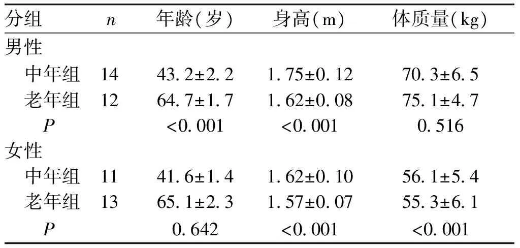
分组 n年龄(岁)身高(m)体质量(kg)男性 中年组 14432±22175±012703±65 老年组 12647±17162±008751±47 P<0001<00010516女性 中年组 11416±14162±010561±54 老年组 13651±23157±007553±61 P0642<0001<0001
2.2 年龄、性别、冠状动脉分支对TAG值的影响
采用手动测量法分析显示,50例受检者中全部女性受检者的TAG值低于全部男性受检者,但差异无统计学意义(P=0.73);50例受检者中老年组受检者的TAG值低于中年组,差异无统计学意义(P=0.28);RCA的TAG值稍高于LAD、LCx,但其差异无统计学意义(P=0.32,见表2)。
表2 年龄、性别、冠状动脉分支对TAG的影响
Table 2 Effect of the age,sex,and different branch of coronary artery on the TAG value
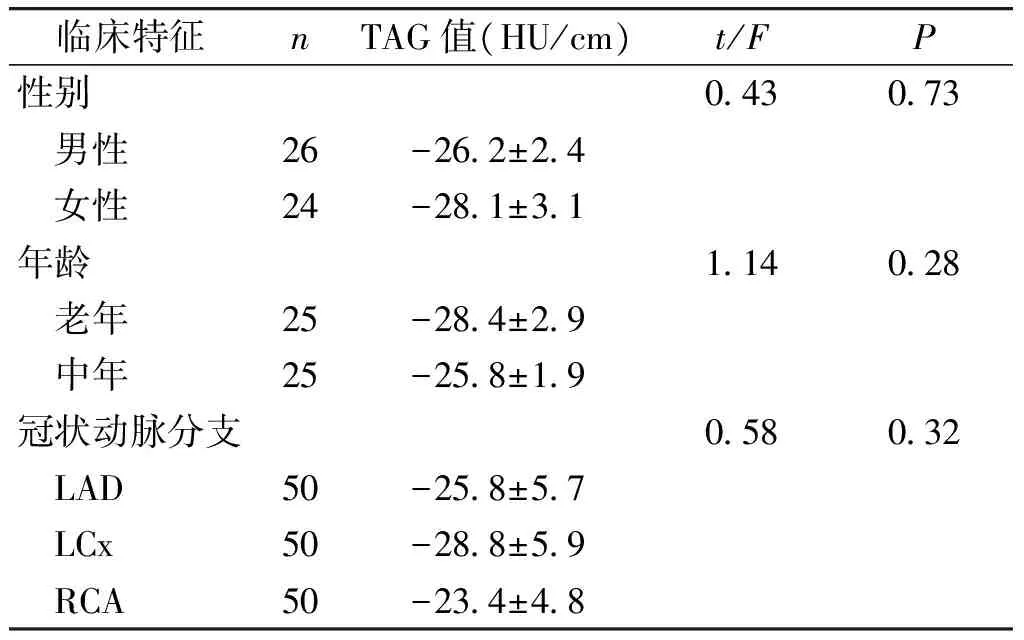
临床特征nTAG值(HU/cm)t/FP性别043073 男性26-262±24 女性24-281±31年龄114028 老年25-284±29 中年25-258±19冠状动脉分支058032 LAD50-258±57 LCx50-288±59 RCA50-234±48
2.3 自动测量法和手动测量法的比较
选取20例受检者,对60支冠状动脉用手动测量法和自动测量法进行测量,自动测量法测量一支冠状动脉的时间比手动测量法少64%[(4.5±0.4)minvs(15.7±0.9)min,P<0.01]。自动测量法的TAG平均值为-29.1 HU/cm,手动测量法的TAG值为-28.2 HU/cm,但差异无统计学意义(P>0.05)。Bland-Altman分析显示自动测量法和手动测量法的差值均数-1.0 HU/cm,95%的一致性界限的上下界为(-19.5)-17.6 HU/cm(见图1)。线性回归分析得线性回归方程为y=1.0x-0.9 HU/cm,显示斜率为1.0,截距较小为0.9,相关系数较高(r=0.93,P<0.001,见图2),进一步说明自动测量法和手动测量法间的一致性高。
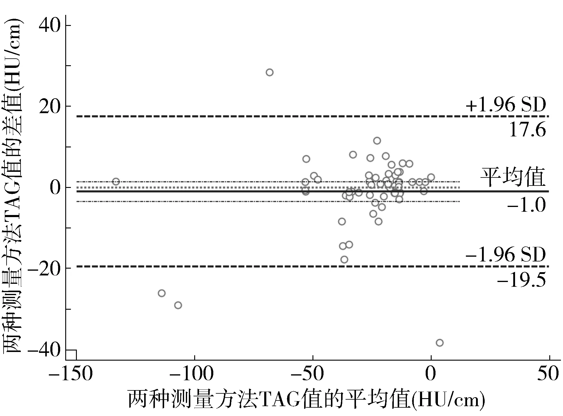
图1 自动测量法VS手动测量法的Bland-Altman分析Figure 1 Bland-Altman plot of automated method versus manual method
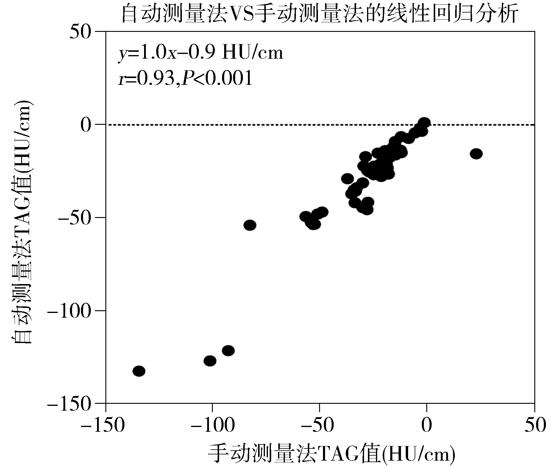
图2 自动测量法VS手动测量法的线性回归分析Figure 2 Linear regression analysis plot of automated method versus manual method
2.4 观察者间一致性比较
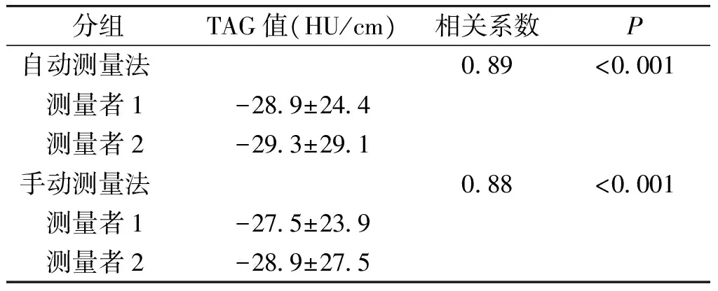
选取20例受检者,对60支冠状动脉采用手动测量法和自动测量法进行测量,无论自动测量法还是手动测量法其相关系数均较高,因此观察者间的一致性较好(y自动法=1.1x+3.8 HU/cm,r=0.89,P<0.001;y手动法=1.1x+2.2 HU/cm,r=0.88,P<0.001,见图3B和4B)。同时Bland-Altman分析显示,与手动测量法相比,自动测量法差值均数小(-0.4 HU/cm),95%的一致性界限窄(-17.5,16.8 HU/cm)(见图3A和4A)。
表3 两种测量方法观察者间一致性比较
Table 3 Inter-observer agreement of automated and manual method
分组TAG值(HU/cm)相关系数P自动测量法089<0001 测量者1-289±244 测量者2-293±291手动测量法088<0001 测量者1-275±239 测量者2-289±275
3 讨论
临床上,对于一个未能确定是否引起心肌缺血的冠状动脉病灶进行支架植入或冠状动脉搭桥手术的血运重建是存在争议的。因此为了避免进行不必要的支架植入或冠状动脉搭桥手术,对于冠状动脉病变进行功能学评估是明智且必要的[7,8]。TAG由CT冠状动脉造影时获得,其反映造影剂通过管腔的下降率,有潜力评估冠状动脉的血流动力学即生理学情况[9,10]。TAG依赖于管腔内衰减的CT值的变化,其准确性受多种因素影响。本研究采用320排CT,其时间分辨率高,在一次心动周期内即可完成全心容积扫描成像,使得各个位置的扫描数据均处于同一时相,相较于回顾性心电门控螺旋扫描和前瞻性心电门控步进(step-and-shoot)扫描,在一定程度上降低了造影剂浓度、注射速率及注射量等因素的影响[11,12]。在此基础上,本研究进一步评估了年龄、性别及不同冠状动脉分支对TAG值的影响。同时,TAG值的测量是一项较为繁琐的工作,所需时间较长,因此本研究进一步评估自动测量软件包测量TAG的准确性,为TAG进一步应用于临床提供依据。
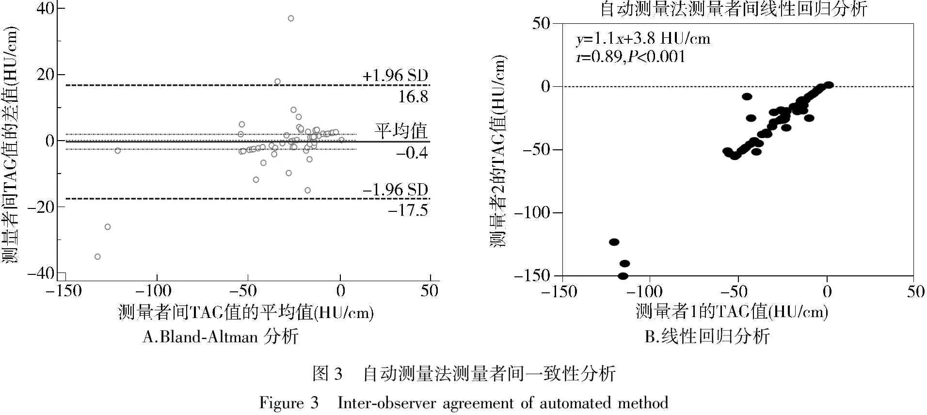
A.Bland⁃Altman分析B.线性回归分析图3 自动测量法测量者间一致性分析Figure3 Inter⁃observeragreementofautomatedmethod
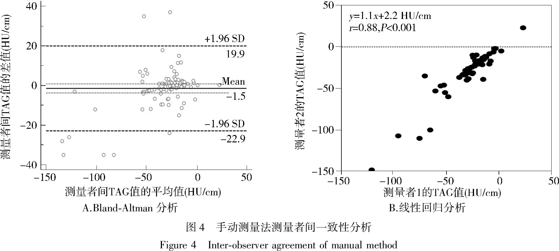
A.Bland⁃Altman分析B.线性回归分析图4 手动测量法测量者间一致性分析Figure4 Inter⁃observeragreementofmanualmethod
本研究发现女性受检者的TAG值低于男性受检者,老年组受检者的TAG值低于中年组,这可能是由于一般健康成年男性在安静状态下的心输出量为4.5-6.0 L/min,女性则比男性低10%左右,且研究发现心输出量是影响TAG值的一个重要因素[6];同样老年组受检者的TAG值低于中年组,这可能与老年人心功能下降,心输出量、射血分数降低有关。RCA的TAG值稍高于LAD、LCx,可能是由于RCA较长,走行比LAD,LCx平缓,LAD多出现心肌桥,LCx走行多迂曲造成的。
本研究证实自动测量法比手动测量法节省时间,且观察者间一致性较高。TAG值的测量从理论上讲简单,但是人工法测量时需要放置30-80个ROI,是项较为繁琐的工作。自动测量法仅需简单的手动进行矫正,TAG值测量所需时间短,且测量值的变异小。此外自动测量法采用每隔1 mm放置ROI(手动测量法每隔5 mm),这减少了狭窄处对TAG值测量的影响,尤其是ROI内存在斑块成分的时候。观察者间一致性高,这也证实了测量者的偏倚不是影响TAG值测量的因素。因此自动测量法可能为测量TAG值的较好方法。
本研究局限性在于,本研究采用320排CT,因此64或256排CT扫描仪、造影剂注射速率等因素对TAG值重复性的影响有待进一步验证。
综上所述,本研究证实自动测量法对TAG值的测量是准确的,可重复的。
[1] Wijns W,Tu S.Diagnostic optimization of coronary CT angiography[J].JACC Cardiovasc Imaging,2011,4(11):1158-1160.
[2] Wong DT,Ko BS,Cameron JD,etal.Transluminal attenuation gradient in coronary computed tomography angiography is a novel noninvasive approach to the identification of functionally significant coronary artery stenosis:a comparison with fractional flow reserve[J].J Am Coll Cardiol,2013,61(12):1271-1279.
[3] Ko BS,Wong DT,Norgaard BL,etal.Diagnostic performance of transluminal attenuation gradient and noninvasive fractional flow reserve derived from 320-detector row CT angiography to diagnose hemodynamically significant coronary stenosis:an NXT substudy[J].Radiology,2016,279(1):75-83.
[4] Norgaard BL,Jensen JM,Leipsic J.Fractional flow reserve derived from coronary CT angiography in stable coronary disease:a new standard in non-invasive testing?[J].Eur Radiol,2015,25(8):2282-2290.
[5] Park EA,Lee W,Park SJ,etal.Influence of coronary artery diameter on intracoronary transluminal attenuation gradient during CT angiography[J].JACC Cardiovasc Imaging,2016,9(9):1074-1083.
[6] Funama Y,Utsunomiya D,Oda S,etal.Transluminal attenuation-gradient coronary CT angiography on a 320-MDCT volume scanner:Effect of scan timing,coronary artery stenosis,and cardiac output using a contrast medium flow phantom[J].Phys Med,2016,32(11):1415-1421.
[7] Pijls NH,Fearon WF,Tonino PA,etal.Fractional flow reserve versus angiography for guiding percutaneous coronary intervention in patients with multivessel coronary artery disease:2-year follow-up of the FAME (Fractional Flow Reserve Versus Angiography for Multivessel Evaluation) study[J].J Am Coll Cardiol,2010,56(3):177-184.
[8] Zhang D,Lv S,Song X,etal.Fractional flow reserve versus angiography for guiding percutaneous coronary intervention:a meta-analysis[J].Heart,2015,101(6):455-462.
[9] Stuijfzand WJ,Danad I,Raijmakers PG,etal.Additional value of transluminal attenuation gradient in CT angiography to predict hemodynamic significance of coronary artery stenosis[J].JACC Cardiovasc Imaging,2014,7(4):374-386.
[10] Ko BS,Wong DT,Cameron JD,etal.320-row CT coronary angiography predicts freedom from revascularisation and acts as a gatekeeper to defer invasive angiography in stable coronary artery disease:a fractional flow reserve-correlated study[J].Eur Radiol,2014,24(3):738-747.
[11] Xu L,Sun Z,Fan Z.Noninvasive physiologic assessment of coronary stenoses using cardiac CT[J].Biomed Res Int,2015,2015:435737.
[12] Yoon YE,Choi JH,Kim JH,etal.Noninvasive diagnosis of ischemia-causing coronary stenosis using CT angiography:diagnostic value of transluminal attenuation gradient and fractional flow reserve computed from coronary CT angiography compared to invasively measured fractional flow reserve[J].JACC Cardiovasc Imaging,2012,5(11):1088-1096.
Accuracy of automated method in computed tomography coronary angiography in the evaluation of transluminal attenuation gradient
LI Jiaojiao*,ZHANG Bin,LIU Di,LI Chuangui,LI Xiaodong,LANG Yingtao
(DepartmentofRadiology,FirstAffiliatedHospitalofHebeiNorthUniversity,Zhangjiakou075000,China;*Correspondingauthor,E-mail:dr_lijiaojiao@163.com)
ObjectiveTo evaluate the accuracy of the automated method for measuring the transluminal attenuation gradient(TAG) value,and to assess the influence of age,sex,and different branch of coronary artery on the TAG value.MethodsTotally 50 patients underwent CT coronary angiography with 320 row CT scanner.The automated method and manual method were applied to measure the TAG value of anterior descending branch(LAD),left circumflex(LCx) and right coronary artery(RCA).The difference of TAG was compared in patients with different age,sex,and branch of coronary artery.The accuracy of the automated method and the inter-observer agreement were assessed by Bland-Altman and linear regression analysis.ResultsTAG was lower in female than in male.TAG was lower in elderly group than in middle-age group.The TAG in RCA was slightly higher than that in LAD and LCx.The analysis time per artery of the automated method was shortened by 64% compared to the manual method.Bland-Altman analysis showed low mean diffe-rence(-1.0 HU/cm) between the automated method and the manual method.Automated method and manual method had high inter-observer agreement.ConclusionAutomated method could reduce the computation time with high accuracy and reproducibility.
320 row CT; CT coronary angiography; transluminal attenuation gradient; Hounsfield units
张家口市科技攻关计划资助项目(1521094D)
李娇娇,女,1988-06生,硕士,住院医师,E-mail:dr_lijiaojiao@163.com
2017-02-21
R541.4
A
1007-6611(2017)06-0566-05
10.13753/j.issn.1007-6611.2017.06.012

