儿童肱骨内髁骨折克氏针贯穿骨折端固定九例报告
王来喜 程富礼 景小博 刘永立 申子龙
儿童肱骨内髁骨折克氏针贯穿骨折端固定九例报告
王来喜 程富礼 景小博 刘永立 申子龙
目的 探讨儿童肱骨内髁骨折的诊断及治疗方法,以期提高儿童肱骨内髁骨折的诊治水平。方法 回顾性分析本院 2 0 1 0~2 0 1 5 年手术治疗的 9 例儿童肱骨内髁骨折患儿的临床资料,其中 K i l f o y l e-I I 型3 例,K i l f o y l e-I I I 型 6 例,2 例合并肘关节脱位。术前详细体格检查了解肘关节的肿胀、压痛及畸形情况,肘后三角的改变情况,本组均行肘关节正侧位 X 线片及 C T 检查,1 例 5 岁患儿并行肘关节 M R I 检查以证实诊断。手术采取切开复位 2~3 枚克氏针贯穿骨折端固定。术后石膏固定 3 周后开始肘关节功能锻炼。结果 本组术后骨折均愈合良好,无肘关节畸形出现。2 例术后肘关节功能活动受限,其中 1 例为陈旧性骨折。无伤口感染、医源性血管神经损伤、内固定松动、骨坏死、骨化性肌炎等并发症。肘关节按 D h i l l o n 评分标准,7 例优,1 例良,1 例可。结论 儿童肱骨内髁骨折容易误诊及漏诊,诊断宜体格检查与影像学相结合。小年龄患儿 X 线片检查时应充分考虑肱骨远端骨骺不显影,必要时行肘关节 M R I 检查。治疗应早期诊断及复位固定。
肱骨;肘关节;骨折;肱骨内髁;肘关节骨折;儿童;治疗
儿童肱骨内髁骨折是较为少见的肘关节损伤,骨折块通常包括肱骨滑车内侧 1 / 2 以上和肱骨内上髁部分,占所有儿童肘部骨折的 1%~2%。由于儿童肱骨内髁骨折属于 S a l t e r-H a r r i s I V 型关节内骨骺骨折,同肱骨外髁骨折一样若处理不当容易出现骨折不愈合。骨骺骨折本身还可能出现骨骺生长发育异常,再加上滑车部分软骨较多,血运相对较差,骨折后软组织血运如果破坏严重可能会出现骨坏死。另外尺神经距离肱骨内髁滑车较近,骨折块移位较大时可能会出现尺神经损伤症状。如果不能及时诊断及正确治疗,儿童肱骨内髁骨折出现后期并发症的可能性会很大,将会对儿童肘关节的发育及功能产生不可挽救的影响[1]。由于儿童肱骨内髁骨折罕见,且我国目前专业从事小儿骨科的医生较少,我国骨科医生目前对于儿童肱骨内髁骨折的认识水平差别很大。2 0 1 2~2 0 1 5 年我院诊治 9 例儿童肱骨内髁骨折患儿,现回顾分析如下。
资料与方法
一、一般资料
本组 9 例,其中男 6 例,女 3 例;左侧 5 例,右侧 4 例;新鲜骨折 8 例,伤后至手术时间 2~7 天,平均 3 天,陈旧性骨折 1 例,伤后至手术时间 2 5 天;年龄 5~1 4 岁,平均 1 1 岁。K i l f o y l e-I I 型3 例,K i l f o y l e-I I I 型 6 例,2 例合并肘关节前脱位。术前详细体格检查了解肘关节的肿胀、压痛及畸形情况及肘后三角的改变情况,所有患儿均行肘关节正侧位 X 线片及 C T 检查,以了解骨折移位的程度及方向。1 例 5 岁患儿并行肘关节 M R I 检查以证实诊断。所有病例均采取切开复位克氏针内固定,屈肘 9 0° 功能位石膏外固定,术后 3~4 周去除石膏外固定开始行主动肘关节伸屈功能锻炼,术后 2~3 个月左右骨折骨性愈合后取出内固定。
二、手术方法
采用全麻或臂丛神经阻滞麻醉,上臂上段止血带下手术,采用肘关节内侧切口,注意保护尺神经。显露骨折端后,清理瘀血及软组织,适当松解前臂屈肌总腱,直视下轻柔解剖复位后以 2~3 枚直径为 1.2 5 m m 或 1.6 m m 的克氏针由内下斜向外上交叉贯穿固定骨折块,术中拍 X 线片示骨折复位良好,克氏针长短合适后,折弯针尾埋于皮下。冲洗缝合切口。
三、随访检查
术后 1 2~1 4 天切口愈合拆线后出院,出院后1 周、3 周、7 周、3 个月来院复查,以后定期随访,随访时间 4~2 8 个月,平均 1 6.9 个月。随访时注意观察切口愈合情况,肘关节外观有无畸形,测量肘关节屈伸活动度,按 D h i l l o n 评分系统进行功能评分[2]( 表 1 )。行 X 线片检查了解骨折愈合情况,有无畸形愈合及内侧过度生长,测量提携角。

表1 Dhillon 肘关节功能评分系统Tab.1 Dhillon elbow function scoring system
结 果
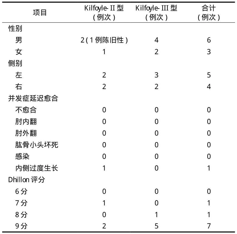
术后无切口感染,所有患儿切口均一期愈合。所有患儿术后骨折均达骨性愈合,骨折愈合时间为6~1 2 周,平均 8.1 周。无肱骨内髁滑车坏死出现。1 例出现内侧过度生长,可见内侧隆突畸形,但无功能受限。2 例肘关节屈伸活动部分受限。术后肘关节功能评分显示优 7 例,良 1 例,可 1 例。具体结果见表 2。

表2 骨折随访并发症及数据统计表Tab.2 Complications and data statistics in the follow-up
讨 论
肱骨内髁骨折较少见,好发于儿童,波及范围包括内上髁与滑车的大部分。受伤后肘内侧和内上髁周围软组织肿胀或有较大血肿形成。临床检查肘关节的等腰三角形关系存在。患儿表现为疼痛,特别是肘内侧局部肿胀、压痛、正常内上髁的轮廓消失。肘关节活动受限,前臂旋前、屈腕、屈指无力。合并肘关节脱位者,肘关节外形明显改变,功能障碍也更为明显,常合并有尺神经损伤症状[3]。
C h a c h a[4]认为肱骨内髁骨折出现在儿童肱骨内髁骨化中心未完全出现的时期,一般常见于 8~1 2 岁。F o w l e s 等[5]报道了 1 8 例儿童肱骨内髁骨折,平均年龄 1 1 岁。P a p a v a s i l i o u 等[6]报道 1 5 例肱骨内髁骨折病例平均年龄 9 岁。B e n s a h e l 等[1]认为肱骨内髁骨折好发的年龄更小,为 5~7 岁。A r a b e l l a 等报道了 2 1 例肱骨内髁骨折患儿,平均年龄 4.7 岁[1]。本研究的 9 例肱骨内髁骨折患儿,年龄 5~1 4 岁,平均 1 1 岁,同以上国外学者所报道的年龄基本一致。但有报道 2 岁患儿也出现肱骨内髁骨折[7],甚至有最小 6 个月患儿发生肱骨内髁骨折的报道[8]。

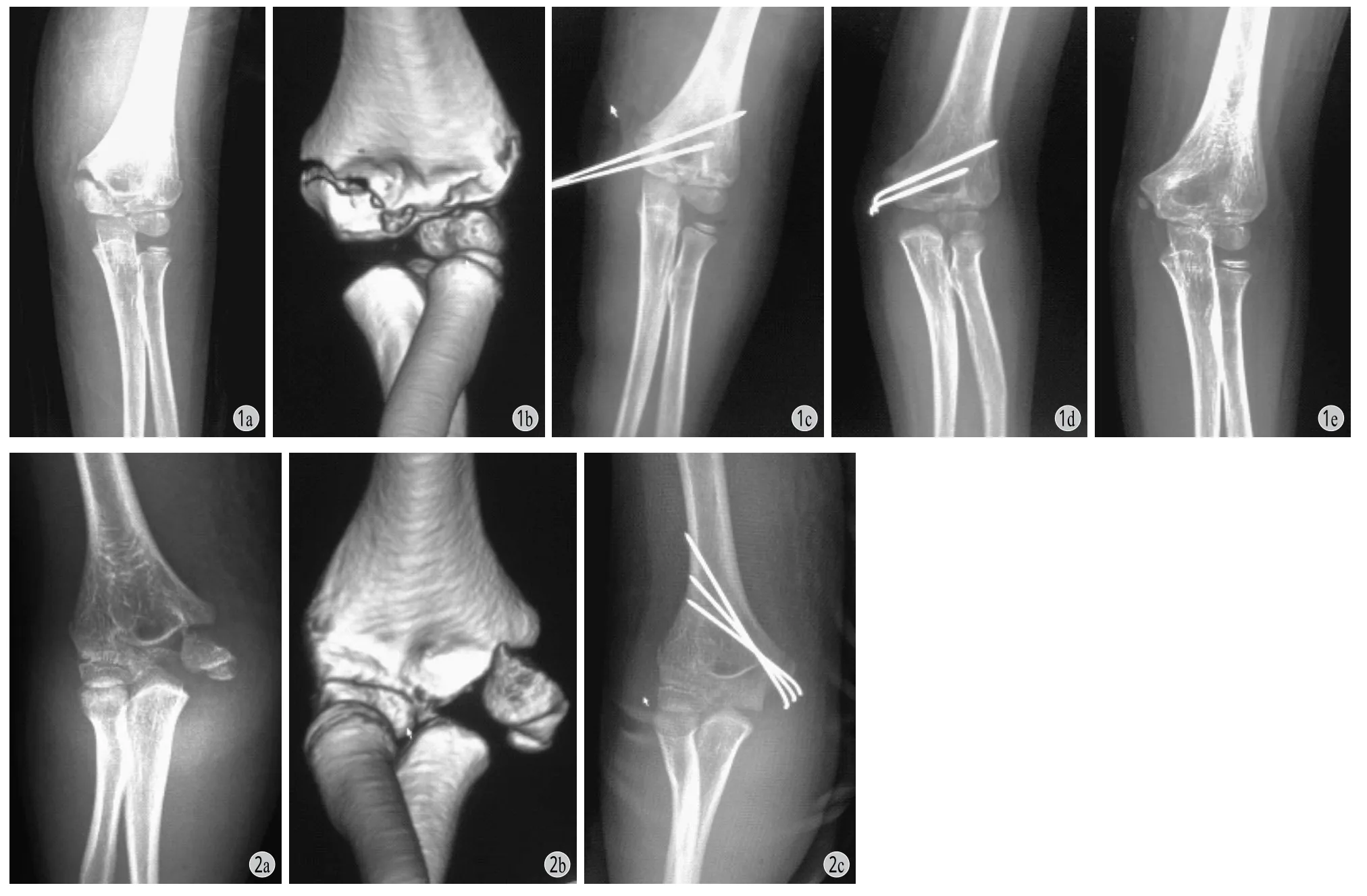
图1 患儿,女,7 岁 a~b:为术前 X 线片及 CT 片提示 Kilfoyle-II 型骨折;c:为术中X 线片显示骨折复位良好;d:为术后 2 个月X 线片显示骨折愈合良好;e:为术后 3 个月及内固定取出后 X 线片,显示有内侧过度生长图 2 患儿,男,14 岁 a~b:为术前 X 线片及 CT 提示 Kilfoyle-III 型骨折;c:为术后X 线片显示骨折复位良好Fig.1 Female, 7y a - b: Preoperative X-ray and CT showed Kilfoyle-type II fracture; c: Intraoperative X-ray showed the fracture was well restored; d: X-ray showed fracture healed well at 2 months after operation; e: X-ray after removal of internal fixation 3 months pastoperatively the showed medial overgrowth
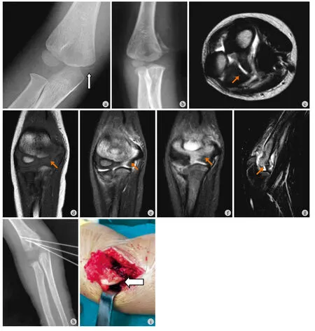
由于肱骨内髁骨折少见,发病率明显低于肱骨外髁骨折,故肱骨内髁骨折的诊断有一定难度。对于大年龄患儿一般通过肘关节标准正侧位 X 线片即能获得明确诊断。本组 6 例年龄较大的患儿均得到了及时的诊断及治疗见图 1~2。但对于小年龄患儿,肱骨内髁骨化中心完全没有出现,肘关节正侧位 X 线片很难做出明确的诊断,本组 1 例 5 岁的患儿由于未能及时诊断,直至伤后 3 周 M R I 检查才提示诊断,最终通过手术确诊 ( 图 3 )。A r a b e l l a 等[1]认为对于年龄小的患儿,可通过肘关节的外翻应力实验判断肱骨内髁骨折的可能性,如果出现外翻时肘关节不稳定应高度怀疑肱骨内髁骨折,此时可通过拍摄肘关节斜位 X 线片或 M R I 检查来明确诊断。
肱骨内髁骨折应与肱骨内上髁骨折和肱骨远端全骨骺骨折相区别。肱骨内髁骨折与肱骨内上髁撕脱性骨折是两个不同范围的损伤。前者骨折波及肱骨滑车大部分和 ( 或 ) 肱骨内上髁,属于关节内骨折,须解剖复位。而后者为关节外骨骺损伤,为前臂屈肌群及旋前肌群猛烈收缩引起的撕脱性骨折,不必须对骨折部位进行解剖复位。对于带有一部分内侧干骺端骨块的肱骨远端全骨骺骨折,由于忽视了肱骨远端骨骺软骨不显影的特点,易被误诊为肱骨内髁骨折。这两种骨折的治疗也存在很大的区别,肱骨远端全骨骺骨折为 S a t e r-H a r r i s I I 型关节外骨骺骨折,大部分可通过手法复位得以治疗。而肱骨内髁骨折为 S a t e r-H a r r i s I V 型关节内骨骺骨折,移位>2 m m 则需切开复位解剖复位内固定治疗[5]。
同肱骨外髁骨折一样,肱骨内髁骨折为关节内骨折,属于儿童骨骺骨折的一种类型,骨折线通过骺板,需要解剖对位以恢复关节面的平整和肱骨远端的正常生长。复位差则骺板处骨桥形成大,鱼尾状畸形大,造成肱尺关节面不适应,发生肘关节半脱位,逐渐发生关节软骨退行性变化,形成创伤性关节炎[3]。为此,大多数病例需要采取切开复位克氏针固定治疗。由于肱骨内髁骨折常包括肱骨滑车的大半部分,手术中直视下观察肱骨滑车是否对合好在复位中至关重要。如肱骨滑车前侧、远侧对合好,一般能达到解剖复位。因此术中拍 X 线片是必不可少的。即使解剖复位和牢固固定,肱骨内髁骨折仍然存在骨折延迟愈合、不愈合,肘内外、翻畸形,鱼尾样畸形,肱骨滑车骨骺无菌性坏死,感染及内侧过度生长及尺神经损伤等诸多并发症[9]。因此,如何恢复关节面的平整和肘关节的功能,尽量减少并发症的发生才是治疗的目的。

图3 患儿,男,5 岁 a:为受伤当时 X 线片,仅正位片显示肱骨远端内侧隐约线状骨折影 ( 箭头所示 ),当时漏诊;b:为伤后 3 周 X 线片,显示肘关节远端内侧骨痂,提示肘关节远端内侧骨折可能;c~g:为伤后 3 周 MRI 检查,显示肱骨内髁损伤;h:为术中复位固定后的 X 线片;i:为术中所见肱骨内髁骨折块 ( 箭头所示 )Fig.3 Male, 5y a: X-ray at the time of injury, only the positive slice showed a slightly fractured line of the distal medial humerus ( arrow ), which was missed at the time; b: 3 weeks after injury, the X-ray showed the distal medial callus of the elbow, suggesting that the distal medial fracture of the elbow might be possible; c -g: MRI performed 3 weeks after injury showed internal humeral condyle injury; h: X-ray after reduction and fi xation; i: Fracture of the medial condyle of the humerus ( arrow ) could be seen in surgery
据统计结果表明通过解剖复位、克氏针张力带坚强内固定及早期功能锻炼可达到骨折愈合及患儿功能恢复的临床治疗效果,而且术后感染、骨折延迟愈合、不愈合、鱼尾样畸形、肱骨小头坏死等并发症无一例出现。尺神经损伤的并发症虽然有文献报道,但本组无一例出现尺神经损伤症状。有 1 例出现内侧过度生长,此前未见报道,因此认为可能与肱骨外髁骨折后出现外侧过度生长的机理类似。有 2 例肘关节伸屈活动部分受限,其中有 1 例为陈旧性骨折,当时未得到及时的诊断及治疗。
综上所述,病例中大部分的肱骨内髁骨折患儿都得到了很好的治疗,伤后得到及时的复位和固定一般都能愈合良好且功能恢复良好。有 1 例因漏诊未及时治疗,最终出现肘关节功能部分受限。因此认为肱骨内髁骨折预后不好的危险因素主要是漏诊、骨折移位不能及时复位,不能得到可靠固定。
[1]Leet AI, Young C, Hoffer MM. Medial condyle fractures of the humerus in children[J]. J Pediatr Orthop, 2002, 22(1):2-7.
[2]Song KS, Ramnani K, Cho CH, et al. Late diagnosis of medial condyle fractures of the humerus with rotational displacement in child[J]. J Orthop Tyaumatol, 2011, 12(4):219-222.
[3]苏琦, 周敏, 廖春来, 等. 儿童肱骨内髁骨折手术治疗[J]. 国际骨科学杂志, 2014, 35(2):129-130.
[4]Chacha PB. Fracture of the medial condyle of the humerus with rotational displacement:report of two cases[J]. J Bone Joint Surg Am, 1970, 52(7):1453-1458.
[5]Fowles JV, Kassab MT. Displaced Fractures of the medial humeral condyle in children[J]. J Bone Joint Surg Am, 1980, 62(7):1159-1163.
[6]Papavasiliou V, Nenopoulos S, Venturis T. Fractures of the medial humeral condyle of the humerus in childhood[J]. J Pediatr Orthop, 1987, 7(4):421-423.
[7]Edmonds EW. How displaced are “nondisplaced” fyactures of the medial humeral epicondyle in children? Result of a threedimensional computed tomography analysis[J]. J Bone Joint Surg Am, 2010, 92(17):2785-2791.
[8]Deboeck H, Casteleyn PP, Opdecam P. Fractures of the medial humeral condyle: report of a case in an infant[J]. J Bone Joint Surg Am, 1987, 69(9):1442-1444.
[9]EI Ghawabi MH. Fractures of the medial humeral condyle of the humerus[J]. J Bone Joint Surg Am, 1975, 57:677-680.
( 本文编辑:李慧文 )
参考文献著录的规范格式
1. 期刊文献著录格式为:
[ 序号 ] 作者 1, 作者 2, 作者 3, 等. 文题[J]. 刊名, 年, 卷( 期 ): 起页码-止页码.
[ 例 1 ] 赵凤朝, 李子荣, 王佰亮, 等. 骨髓水肿与股骨头塌陷及疼痛的相关性研究[J]. 中华骨科杂志, 2 0 0 8, 2 8(8):6 4 5-6 4 8.
[ 例 2 ] K i m Y L, S h i n S I, N a m K W, e t a l. T o t a l h i p a r t h r o p l a s t y f o r B i l a t e r a l l y a n k y l o s e d h i p s[J]. J A r t h r o p l a s t y, 2 0 0 7, 2 2(7): 1 0 3 7-1 0 4 1.
2. 专著文献的著录格式为:
[ 序号 ] 章节作者. 章节名 // 全书主编者. 书名[M]. 版次. 出版地: 出版社. 出版年: 起页码-止页码.
[ 例 1 ] 黄公怡. 肩关节疾病//王澍寰. 临床骨科学[M]. 1版. 上海:上海科学技术出版社. 2 0 0 5: 3 5 5-4 0 7.
[ 例 2 ] S i m o n MA, S p r i n g f i e l d D. S u r g e r y f o r b o n e a n d s o f t t i s s u e t u m o r s[M]. E d 1. P h i l a d e l p h i a: L i p p i n c o t t-R a v e n P u b l i s h e r s. 1 9 8 8: 2 1-4 8.
3. 电子文献著录格式为:
[ 序号 ] 著者. 题名 [ 文献类型标志 / 文献载体标志 ].出版地: 出版者, 出版年 [ 引用日期 ]. 获取和访问路径.
[ 例 1 ] O n l i n e C o m p u t e r L i b r a r y C e n t e r, I n c. H i s t o r y o f O C L C [E B/ O L]. [2 0 0 0-0 1-0 8]. h t t p//w w w.o c l c.o r g/a b o u t/h i s t o r y/d e f a u l t. h t m.
Diagnosis and treatment experience of the humeral condylar fracture in children
WANG Lai-xi, CHENG Fu-li,
JING Xiao-bo, LIU Yong-li, SHEN Zi-long. Department of Pediatric Orthopedics, Zhengzhou Orthopedic Hospital, Zhengzhou, Henan, 450052, China
CHENG Fu-li, Email: chengf l zzgk@163.com
Objective To investigate the diagnosis and treatment of humeral condylar fractures in children to improve the experience. Methods The clinical data of 9 children with humeral condylar fractures treated in our hospital from 2010 to 2015 were retrospectively analyzed, including Kilfoyle-type II in 3 cases, Kilfoyle-type III in 6 cases and combined elbow dislocation in 2 cases. A detailed preoperative physical examination of elbow joint swelling, tenderness and deformity was performed, as well as the change of posterior cubital triangle. All the patients underwent anterioposterior and lateral X-ray and CT examinations of the elbow. One child of 5 years old underwent MRI examination of the parallel elbow to conf i rm the diagnosis. Open reduction was performed with 2 - 3 Kirschner wires penetrating the fixed end. After 3 weeks of cast immobilization, elbow functional exercise was performed. Results All fractures healed well after surgery, without elbow deformity. Two patients had limitation of elbow joint function, among whom 1 patient suffered old fracture. No wound infection, iatrogenic vascular injury, internal fi xation loosening, bone necrosis, myositis ossif i cans or other complications occurred. According to Dhillon scoring standard, there were 7 excellent cases, 1 good case and 1 fair case. Conclusions Misdiagnosis and missed diagnosis of humeral condylar fractures in children often happen. Both X-ray examination and physical examination should be performed. X-ray examination should be paid full attention in younger children when the distal humeral epiphysis is developing. If necessary, the elbow joint MRI examination should also be performed. Early diagnosis and reduction fi xation should be done in the treatment.
Humerus; Elbow joint; Fractures, bone; Humeral condylar; Fracture of elbow joint; Children; Treatment
10.3969/j.issn.2095-252X.2017.07.004
R726.8, R687.3
2017-03-27 )
4 5 0 0 5 2 郑州市骨科医院小儿骨科
程富礼,E m a i l: c h e n g f l z z g k@1 6 3.c o m

