肱骨髁上骨折患儿的外侧小切口切开复位治疗
徐会法 黄鲁豫 雷伟 严亚波 刘峙辰 沙佳 李超 王宏
肱骨髁上骨折患儿的外侧小切口切开复位治疗
徐会法 黄鲁豫 雷伟 严亚波 刘峙辰 沙佳 李超 王宏
目的 探讨切开复位克氏针固定治疗肱骨髁上骨折患儿的疗效。方法 回顾性分析 2 0 0 9 年9 月至 2 0 1 5 年 1 2 月于本院接受切开复位手术治疗的 3 1 例儿童肱骨髁上骨折的临床资料,其中男 1 8 例,女1 3 例,年龄 1 1 个月~1 4 岁,平均 4.5 岁。受伤至手术间隔时间为 1 0 h~1 3 天,平均 1.8 天。均采用肱骨远端外侧入路切开复位,直视下克氏针内固定术,术后支具功能位外固定。术后 3、6、1 2、1 8、2 4、3 0 个月时采用 F l y n n 肘关节评分标准评定疗效。结果 本组均获得随访,资料完整,随访 1 2~3 0 个月,平均 2 4.6 个月。闭合复位失败者 2 3 例;开放骨折者 4 例;合并神经损伤者 4 例。本组病例平均手术时间为 3 8.2 ( 3 0~4 8 ) m i n,术中出血平均 2 0.8 ( 1 5~4 0 ) m l,平均住院日 6.2 ( 4~1 5 ) 天。按照 F l y n n 肘关节临床功能评分标准评定疗效:术后 3 个月优良率为 7 4.2% ( 2 3 / 3 1 ),术后 1 2 个月优良率为 9 6.8% ( 3 0 / 3 1 ),其中 1 例肘关节屈伸活动度减少>1 5°。术后患儿均未出现伤口不愈合、骨筋膜室综合征、血管损伤、神经损伤、骨折不愈合、肘关节内、外翻畸形等并发症。结论 外侧小切口切开复位治疗儿童肱骨髁上骨折疗效确切。该方法操作相对简单,手术风险小、时间短,对患儿创伤小,并发症少,可以有效治疗上述类型的肱骨髁上骨折。
儿童;肱骨骨折;内固定器;骨折切开复位;骨固定针
肱骨髁上骨折指骨折线经过肱骨远端鹰嘴窝中 心的有着特殊解剖基础和损伤机制的骨折。是儿童第二常见的骨折,占儿童所有骨折的 1 7.9%,仅次于桡骨远端骨折 2 0.2%[1],好发年龄为 5~6 岁。其每年的发病率为 1 7 7.3 / 1 0 0 0 0 0[2]。其治疗主要采取闭合复位石膏固定或者经皮穿针固定治疗[3-5],随着手术技术的发展对于难复性,不稳定性,需要多次复位且多次 X 线曝光、多次穿针的病例应行外侧小切口切开复位[6-7],通过回顾性分析 2 0 0 9 年 9 月至 2 0 1 5 年 1 2 月于本院治疗的儿童肱骨髁上骨折患儿,旨在探讨其开放复位的手术指征。
资料与方法
一、一般资料
本组 3 1 例,男 1 8 例,女 1 3 例,年龄 1 1 个月~1 4 岁,平均 4.5 岁。其中闭合复位失败者:2 3 例;开放骨折者:4 例;合并神经损伤者:4 例。受伤至手术间隔时间为 1 0 h~1 3 天,平均 1.8 天。
二、手术方法
全部采用肱骨远端外侧切口,长约 3~5 c m,由肱三头肌与肱桡肌间隙进入,剥离肱骨远端骨膜,显露骨折端,清理骨折端的血肿及嵌顿的软组织,于肘关节前方部分切开肘关节囊,清理肘关节内的积血,直视下复位骨折端。自肱骨小头向内上逆行钻入 1 枚克氏针,再于骨折线近端约 2 c m 的肱骨外侧向肱骨内髁顺行钻入 1 枚克氏针,直径均为2 m m,交叉固定骨折端,剪短针尾,埋于伤口内,放置负压引流管后逐层关闭切口,术后给予支具功能位固定后 1 个月去除支具行肘关节屈伸功能锻炼,术后 6~8 周手术取出克氏针。
三、评价方法
疗效评定标准依照 F l y n n 肘关节临床功能评分标准分为优 :肘关节屈伸活动度减少 5° 内,提携角减少 5° 内;良:肘关节屈伸活动度减少 6°~1 0°,提携角减少 6°~1 0°;一般:肘关节屈伸活动度减少1 1°~1 5°,提携角减少 1 1°~1 5°;差:肘关节屈伸活动度减少>1 5°,提携角减少>1 5°。
结 果
本组均获 1 2~3 0 个月随访,平均 2 4.6 个月。本组病例平均手术时间 3 8.2 ( 3 0~4 8 ) m i n,术中出血平均 2 0.8 ( 1 5~4 0 ) m l,平均住院日 6.2 ( 4~1 5 ) 天。至末次随访,平均年龄 6.6 ( 2.5 ~1 6 ) 岁。术后 4 周均获骨折临床愈合,去掉支具,开始指导主动功能锻炼。术后 6~8 周取出克氏针。按照F l y n n 肘关节临床功能评分标准评定疗效:术后 3 个月优良率为 7 4.2% ( 2 3 / 3 1 ),术后 1 2 个月优良率为 9 6.8% ( 3 0 / 3 1 ),其中 1 例肘关节屈伸活动度减少>1 5°。术后患儿均未出现伤口不愈合、骨筋膜室综合征、血管损伤、神经损伤、骨折不愈合、肘关节内、外翻畸形等并发症。
讨 论
肱骨髁上骨折的首选治疗方法是闭合复位经皮克氏针内固定,已达成共识。而对不可复性肱骨髁上骨折的治疗存在争议。有学者认为此种骨折为避免加重血管神经损伤,应行切开复位[8]。郭源等[9]认为不可复性肱骨髁上骨折行切开复位,手术中的剥离、骨折端的显露极可能会加重肘部的损伤,延长骨折愈合时间,影响肘关节功能。S l o n g o 等[10]认为不可复性肱骨髁上骨折可以通过特殊的手法复位,不需要切开复位。通过这组病例对肱骨髁上骨折切开复位的指征总结如下。
一、闭合复位失败者
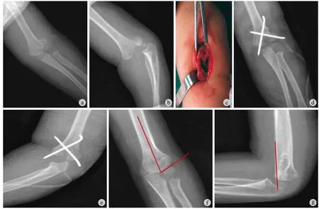
本组有 2 3 例为闭合复位失败后进行切开复位克氏针固定治疗的占 6 0.5%。其中有 3 例受伤至手术间隔时间>1 周,术中手法复位困难。另有 2 例为多方不稳定骨折,术中复位时过度屈曲并能维持复位。有 5 例术中透视不易观察复位情况,虽然肱骨髁上骨折多发于 5~7 岁,但是 1 岁左右的肱骨髁上骨折时有发生,且部分患儿骨折位置较低,肱骨滑车骨骺骨化中心未出现,相关操作经验不足时术中透视不能证实骨折复位 ( 图 1 ),建议切开复位治疗。虽然有学者对这类患儿进行肘关节造影或者 B 超引导下行闭合复位克氏针固定取得成功,但是并没有得到大范围的推广,且有一定的学习曲线,没有相关操作经验者盲目实施可能会对患儿造成不可恢复的并发症。
二、开放骨折者
本组有开放骨折 4 例,占比为 1 0.5%。均为肱骨干骺端的尖端先前刺破皮肤所致。这种骨折一般建议做肘关节前方切口,必要时可向近端内侧及远端外侧延长。但是肘关节存在血管、神经束且软组织覆盖较多,手术损伤较大的风险。本组 4 例开放骨折均采用外侧切口,暴露骨折端,将断端间血肿等软组织彻底清理,使用双氧水及碘伏水冲洗3 次,术后常规使用抗生素,4 例均未发生感染。

图1 患儿,女,15 个月 a~b:术前肘关节正侧位 X 线片,伸直桡偏型肱骨髁上骨折,Gartland III 型;c:术中照片,骨折线从肱骨外侧干骺端经鹰嘴窝至肱骨内髁骺线;d~e:术后肘关节正侧位 X 线片,骨折解剖复位,2 mm 克氏针交叉固定;f~g:术后 18 个月复查,Baumann 角:64°,肱前线通过肱骨外髁骨化中心Fig.1 Female, 15 months old a - b: Anterioposterior and lateral photograph of the elbow joint before the operation, deviation-ulnar type humeral supracondylar fracture, Gartland type III; c: Fracture line from humerus lateral metaphysis through olecranon fossa to entepicondyle humeral epiphyseal line during operation; d - e: Anterioposterior and lateral photograph of the elbow joint post-operatively, fracture anatomical reduction, intersecting fi xation with 2mm Kirschner wire; f - g: Reexamination 18 months later, Baumann angle: 64°, anterior humeral line is in humeral lateral condyle ossif i cation center
三、合并神经损伤者
文献报道 G a r t l a n d I I I 型肱骨髁上骨折合并创伤性神经损伤占比高达 1 2%~2 0%,神经损伤程度与骨折严重程度有关[11]。本组有 2 例合并有桡神经损伤,2 例正中神经损伤,共 4 例。占比 1 0.5%。曾裴等[12]报道闭合复位、经皮穿针内固定治疗儿童闭合性 G a r t l a n d I I I 型肱骨髁上骨折 3 9 8 例,合并神经损伤 3 3 例,术后 3~2 4 周 ( 平均 6 周 ) 神经损伤恢复,可作为首选治疗方法之一。但是前期有1 例 G a r t l a n d I I I 型肱骨髁上骨折合并正中神经损伤,术后神经损伤症状加重,术后 1 年无明显恢复。建议 G a r t l a n d I I I 型肱骨髁上骨折合并神经损伤者切开复位治疗,防止神经卡压于骨折端,导致神经损伤加重。
笔者认为对于肱骨髁上骨折的治疗,闭合复位或者切开复位都有其优缺点。对于肱骨髁上骨折闭合复位经皮穿针固定治疗,最大的缺点就是医源性尺神经损伤,其发生率报道不一,S k a g g s 等[13]报道尺神经损伤的发生率为 5%,L y o n s 等[14]报道尺神经损伤的发生率为 6%,而冯超等[15]报道闭合复位经皮穿针固定治疗 G a r t l a n d I I I 型肱骨髁上骨折4 6 0 例,并无医源性尺神经损伤发生,其认为手术操作经验至关重要。笔者认为闭合复位经皮穿针固定治疗肱骨髁上骨折不能完全避免医源性尺神经损伤,本院闭合复位经皮穿针固定治疗肱骨髁上骨折4 2 8 例,发生医源性神经损伤 5 例,占 1.2%。现在的医学模式为生物-心理-社会医学模式,积极和家属沟通各种术式利弊,要求切开复位者可以给予切开复位治疗。
手术治疗肱骨髁上骨折的报道中曾提到多种手术入路,包括肘前方横切口、肘后切口、前内侧切口、外侧切口、内侧切口以及内外侧双切口等[6-7,16]。其中 We i l a n d 等[17]提出外侧切口,其具有创伤小、住院时间短、并发症少、固定牢固、复位佳等优点。本组病例均采用外侧入路。
综上所述,对于 G a r t l a n d I I I 型肱骨髁上骨折,闭合复位经皮克氏针固定仍为首选治疗方案,但是存在下述情况:闭合复位失败者、开放骨折者、合并神经损伤者,患儿家长要求切开复位者建议采用外侧小切口切开复位克氏针内固定治疗,该方法具有操作简单、风险小、时间短、创伤小、术后并发症少的优点,是治疗 G a r t l a n d I I I 型肱骨髁上骨折直接、有效、安全的方法。
[1]Cheng JC, Ng BK, Ying SY, et al. A 10-year study of the changes in the pattern and treatment of 6,493 fractures[J]. J Pediatr Orthop, 1999, 19(3):344-350.
[2]Ladenhauf HN, Schaffert M, Bauer J. The displaced supracondylar humerus fracture: indications for surgery and surgical options: a 2014 update[J]. Curr Opin Pediatr, 2014, 26(1):64-69.
[3]吴伟平, 李旭, 史强, 等. Gartland III 型儿童肱骨髁上骨折的微创治疗[J]. 南方医科大学学报, 2014, 34(9):1351-1354.
[4]罗冬冬, 张智勇, 刘彩娥, 等. 急诊闭合复位外侧经皮穿针固定治疗儿童 Gartland II 型及 III 型肱骨髁上骨折[J]. 中国骨与关节损伤杂志, 2014, 29(7):723-724.
[5]张成强. 儿童肱骨髁上骨折的治疗选择[J]. 中华小儿外科杂志, 2014, 35(10):798-800.
[6]李玉婵, 陈博昌, 徐蕴岚, 等. 肘内侧进路切开复位治疗Gartland III 肱骨髁上骨折[J]. 中国矫形外科杂志, 2004, 12(3):167-169.
[7]姜锋, 王晓, 张明辉, 等. 两种外侧入路结合张力带钢丝内固定治疗儿童肱骨髁上骨折疗效比较[J]. 中国骨与关节损伤杂志, 2013, 28(3):267-268.
[8]任东, 邢丹谋, 冯伟, 等. 克氏针固定治疗儿童肱骨髁上骨折不同进针方式的比较[J]. 中华手外科杂志, 2011, 27(2): 102-104.
[9]郭源, 王承武, 范源, 等. 儿童“不可复性”肱骨髁上骨折的治疗[J]. 中华小儿外科杂志, 1998, 19(2):67-69.
[10]Slongo T, Schmid T, Wilkins K, et al. Lateral external fixation--a new surgical technique for displaced unreducible supracondylar humeral fractures in children[J]. J Bone Joint Surg Am, 2008, 90(8):1690-1697.
[11]Cramer KE, Green NE, Devito DP. Incidence of anterior interosseous nerve palsy in supracondylar humerus fractures in children[J]. J Pediatr Orthop, 1993, 13(4):502-505.
[12]曾裴, 杨建平. 儿童闭合性 Gartland III 型肱骨髁上骨折合并血管神经损伤的治疗[J]. 中华创伤骨科杂志, 2013, 15(4): 352-354.
[13]Skaggs DL, Hale JM, Bassett J, et al. Operative treatment of supracondylar fractures of the humerus in children. The consequences of pin placement[J]. J Bone Joint Surg Am, 2001, 83-A(5):735-740.
[14]Lyons JP, Ashley E, Hoffer MM. Ulnar nerve palsies after percutaneous cross-pinning of supracondylar fractures in children’s elbows[J]. J Pediatr Orthop, 1998, 18(1):43-45.
[15]冯超, 郭源, 张建立. 克氏针治疗儿童肱骨髁上骨折的穿针方式效果分析[J]. 中华小儿外科杂志, 2008, 29(5):291-293.
[16]Ladenhauf HN, Schaffert M, Bauer J. The displaced supracondylar humerus fracture: indications for surgery and surgical options: a 2014 update[J]. Curr Opin Pediatr, 2014, 26(1):64-69.
[17]Weiland AJ, Meyer S, Tolo VT, et al. Surgical treatment of displaced supracondylar fractures of the humerus in children. Analysis of fi fty-two cases followed for fi ve to fi fteen years[J]. J Bone Joint Surg Am, 1978, 60(5):657-661.
( 本文编辑:李慧文 )
. 会议 ●征文 ●消息 Conference / Call for Paper / News .
本刊被美国化学文摘数据库收录公告
本刊现为中国科技论文统计源期刊。2 0 1 3 年 1 月,本刊经美国化学文摘 ( C h e m i c a l A b s t r a c t s,C A ) 数据库审理委员会审核通过,并从 2 0 1 3 年第 1 期开始,正式被美国化学文摘数据库收录。特此公告!
C A 只收录本刊论著,其它文章不收录。
查询本刊请使用拼音:Z h o n g g u o G u Y u G u a n j i e Z a z h i或本刊标准国际刊号 ( I S S N ):2 0 9 5-2 5 2 X,查询网址:h t t p://c a s s i.c a s.o r g/s e a r c h.j s p。
《中国骨与关节杂志》编辑委员会
An investigation on surgical indications of humeral supracondylar fracture in children: open reduction through small lateral incision
XU Hui-fa, HUANG Lu-yu, LEI Wei, YAN Ya-bo, LIU Zhi-chen, SHA Jia, LI Chao, WANG
Hong. Department of Orthopedics, Xijing Hospital, Fourth Military Medical University, Xi’an, Shaanxi, 710032, China
HUANG Lu-yu, Email: huangly@fmmu.edu.cn
Objective To investigate the surgical indications and use of Kirschner wire in the fi xation of open reduction by small lateral incision on Children’s humeral supracondylar fracture by following up the cases in recent years. Methods From September 2009 to December 2015, 459 cases of children with humeral supracondylar fracture were analyzed retrospectively, in which 31 cases ( 6.8% of all the cases ) were treated in open reduction. There were 31 cases ( 18 male and 13 female cases aged from 11 months to 14 years, 4.5 years on average ), and the time interval from injury being from 10 hours to 13 days ( 1.8 days on average ). All patients were treated with open reduction by lateral incision of distal humerus with Kirschner wires and brace was used in functional position after surgery. Clinical effects were assessed by Flynn standard in 3, 6, 12, 18, 24 and 30 months. Results All the 31 cases were followed up from 12 to 30 months ( 24.6 months on average ) with complete data, of whom 23 cases failed in close reduction, 4 cases were of open fractures, 4 cases had combined nerve injuries. The average surgery time was 38.2 minutes ( range: 30 - 48 minutes ), the average intraoperative blood loss was 20.8 ml ( range: 15 - 40 ml ), and the average hospital stay was 6.2 days ( range: 4 - 15 days ). According to the Flynn standard of joint function, the effects were “good”in 74.2% ( 23 / 31 ) of the cases in 3 months postoperatively, and in 96.8% ( 30 / 31 ) in 12 months postoperatively, in whom the decrease of joint fl ection-extension motion was larger than 15° in 1 case. No unhealed wounds, osteofascial compartment syndrome, vascular injury, nerve injury, fracture disunion, cubitusvarus and valgus deformity or other complications were found in any case. Conclusions Open reduction of small lateral incision is effective on children’s humeral supracondylar fracture with simple operation, less surgical risk, shorter operation time, less trauma and complications.
Child; Humeral fractures; Internal fi xators; Open fracture reductions; Bone wires
10.3969/j.issn.2095-252X.2017.07.003
R726.8, R683
2017-03-27 )
7 1 0 0 3 2 西安,第四军医大学第一附属医院骨科
黄鲁豫,E m a i l: h u a n g l y@f m m u.e d u.c n

