牙本质折断恒牙牙髓预后影响因素logistic回归分析
高云云,黄建静,谢亚伦
(温州医科大学附属第一医院 口腔科,浙江 温州 325015)
牙本质折断恒牙牙髓预后影响因素logistic回归分析
高云云,黄建静,谢亚伦
(温州医科大学附属第一医院口腔科,浙江温州325015)
目的:回顾性分析牙本质折断的恒牙牙髓预后及其影响因素。方法:对2012年2月至2014年3月就诊于我院口腔科的牙本质折断患者进行logistic回归分析,记录患者的治疗方法、年龄、性别、就诊时间、牙根发育情况、冠折近髓程度、松动度及就诊叩痛,分析治疗效果及牙髓预后情况,并分析影响牙髓预后的因素。结果:共收集到305例符合纳入条件的牙本质折断患者,共有患牙325颗。其中有248颗(占76.3%)牙髓存活或钙化,为治疗成功;77颗(占23.7%)发生了牙髓坏死,为治疗失败;牙本质折断行间接盖髓术的成功率为76.3%。冠折近髓程度、就诊叩痛与牙髓坏死率有显著相关性(P<0.05)。患者年龄、性别等因素与牙髓坏死率无显著相关性(P>0.05)。结论:牙本质折断后行间接盖髓术的成功率较高;冠折近髓程度、就诊时叩痛是判断牙髓预后的重要因素。
牙本质折断;间接盖髓术;牙髓预后;影响因素
牙齿外伤发生率约为6%~30%[1-3]。根据Andreasen分类法,牙齿外伤包括牙体硬组织、牙周围组织及牙髓组织损伤[4-7]。牙冠折断是牙外伤的常见类型,而根据牙冠的折断程度可以分为牙本质折断、牙釉质折断和冠折露髓,其中牙本质折断占25%~ 70%[8-9]。牙本质折断,外界细菌及其代谢产物可能进入暴露的牙本质小管并持续刺激牙髓组织导致炎症的发生,从而造成牙髓组织的损伤[10]。目前对于牙本质折断治疗主要采用对牙髓组织无刺激的材料覆盖断面以隔绝外界刺激。本研究选择2012年2月至2014年3月在我院收治的牙本质折断患者进行logistic回归分析,主要对牙本质折断恒牙的牙髓预后影响因素进行评估,为防止牙本质折断后牙髓组织受到损害提供治疗依据。
1 资料和方法
1.1一般资料 选取2012年2月至2014年3月我院收治的牙本质折断患者305例(患牙325颗)进行回顾性分析,其中男199例,女106例;年龄≤10岁的患者170例,11~19岁的48例,20~44岁的49例,≥45岁的38例。其中上颌中切牙311颗(占95.7%),上颌侧切牙10颗(占3.1%),下颌中切牙4颗(占1.2%);170颗牙根发育成熟、根尖孔闭合,155颗牙根未发育成熟、根尖孔未闭合,平均复查时间(48.2±21.1)月。
入选标准:①据李弘毅分类法,确诊为牙本质折断;②进行氢氧化钙盖髓治疗;④复诊时间>2年。排除标准:①外伤并伴有脱臼、挫入等严重牙周组织损伤;③牙本质折断前有牙周炎症或其他牙髓疾患。
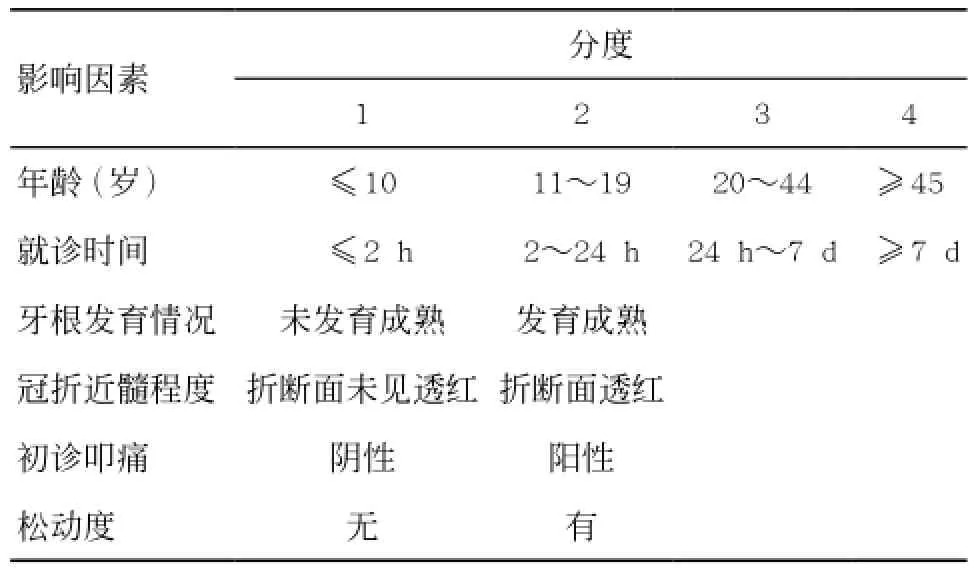
1.2牙髓预后影响因素分度 将可能影响牙髓预后的因素(年龄、就诊时间、牙根发育情况、冠折近髓程度、初诊叩痛、松动度)进行分度,见表1。

表1 牙髓预后影响因素分度
1.3牙髓治疗及预后情况 预后转归可分为牙髓存活、牙髓钙化和牙髓坏死3种:①牙髓存活:牙冠颜色自然正常,电活力、温度测试正常,根尖片无病理改变;②牙髓钙化:牙冠变色,根尖片发现根管影响变窄、消失或模糊;③牙髓坏死:牙冠变色,电活力、温度测试异常,叩诊不适,根尖区可见骨密度减低影[11-13]。根据预后情况,可分为牙髓坏死和非牙髓坏死(包括牙髓存活及牙髓钙化)。将无牙髓坏死、根尖区无低密度影像、无牙根内外吸收的定义为治疗成功;出现牙髓坏死、根尖区显示低密度影像、牙根内外吸收的定义为治疗失败。1.4 统计学处理方法 采用SPSS17.0软件进行统计学分析,对各变量进行单因素x2分析,将得出的显著性变量带入非条件logistic回归模型分析。P< 0.05为差异有统计学意义。
2 结果
2.1牙髓预后情况 325颗患牙中,248颗(占76.3%)治疗成功,即牙髓预后为存活和牙髓钙化(未发现根尖周病变);77颗(占23.7%)治疗失败,即牙髓预后为坏死。牙本质折断后行盖髓术成功率为76.3%。
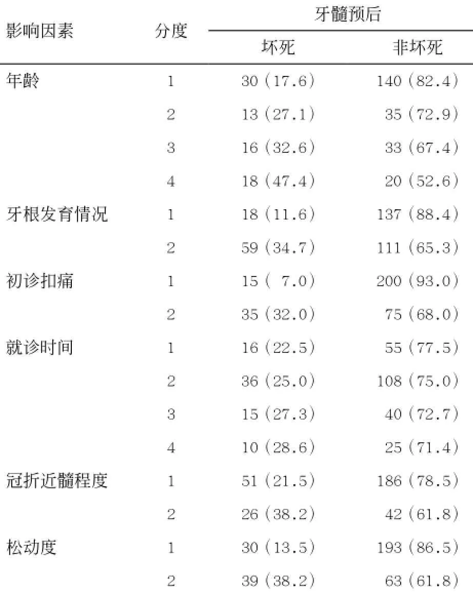
2.2牙髓预后转归的影响因素 患者年龄、初诊叩痛、冠折近髓程度与牙髓组织坏死率呈显著相关,差异有统计学意义(P<0.05),见表2。

表2 不同因素对牙本质折断恒牙的牙髓预后因素分析[n(%)]
2.3根尖孔闭合对牙髓预后的影响 复诊期间发现155颗未发育成熟的恒牙中,18颗发生牙髓坏死,坏死率为11.6%;170颗发育成熟的恒牙中,59颗发生牙髓坏死,坏死率为34.7%,两者的牙髓坏死率差异无统计学意义(P>0.05),见表3。
2.4影响牙髓预后因素的logistic分析 冠折近髓程度、初诊叩痛与牙髓坏死率有显著相关性(P<0.05),冠折近髓程度、初诊叩痛为判断牙髓预后情况重要指标。牙本质折断近髓的患牙发生坏死的风险性为未近髓患牙的4.45倍(OR=5.8),初诊叩痛阳性患牙发生坏死的风险性为初诊叩痛阴性患牙4.48倍(OR=5.2),见表4-5。

表3 根尖孔闭合对牙髓预后的影响

表4 各不同因素不同分度下的牙髓预后[n(%)]

表5 冠折近髓程度、初诊叩痛对牙髓组织预后影响的logistic回归分析
3 讨论
本研究据文献调研[14-16],分析可能影响牙髓组织预后的6个临床相关因素(性别、年龄、冠折近髓程度、初诊叩痛、外伤后就诊时间、是否接受急诊处理)。牙髓位于牙本质内部的牙髓腔内,通过根孔尖与周围组织相连接,无侧支血液循环,故牙髓损伤之后很难恢复。因此,当牙本质折断后,对牙髓的保护就显得尤为重要,如若不及时治疗就可能导致牙髓坏死。有研究报道牙本质折断后牙髓的存活率为60%~90%[17-18],但由于不同研究纳入病例的标准不统一,所得出的结果差异性也较大。本研究共收集305例符合纳入条件的牙本质折断病例,涉及患牙325颗。研究显示,牙本质折断行间接盖髓术的成功率为76.3%。此外,有研究[11]表明,即使是简单冠折但伴有严重牙周组织损伤时,牙髓坏死率也会显著提高,尤其对于根尖闭合患者,坏死率会更高。简单冠折并伴有移位时,牙髓坏死率由3%提升至67%[19]。故本研究在病例筛选时排除了伴有严重牙周组织损伤的病例。
有研究[10]发现,当牙本质折断后,外界细菌及其代谢产物可能进入暴露的牙本质小管并持续刺激牙髓组织导致炎症的发生,从而造成牙髓组织的损伤。目前对于牙本质折断治疗主要采用对牙髓组织无刺激的材料覆盖断面以隔绝外界刺激,可用氢氧化钙制剂进行有效封闭。对于牙本质折断后的治疗方法,有研究[20]认为,当露髓孔径较小时可以行盖髓术。但由于露髓断面处的牙髓可能存在充血、出血或污染等问题,这使得行盖髓术的成功率降低。而且一旦感染扩散,会导致牙髓的广泛炎症。故对于恒牙冠折露髓的病例,应该提倡初诊时就行活髓切断术以提高活髓的保存率,使得牙根继续发育,为远期修复治疗创造条件。
本研究表明:恒牙牙本质折断后行盖髓术的成功率较高;冠折近髓程度及叩痛程度与牙髓坏死率呈现显著性相关;患者年龄、性别等因素是牙髓坏死的非相关因素;冠折近髓程度及初诊时叩痛程度是判断牙髓预后的重要指标。
[1]GOETTEMS M LTORRIANI D DHALLAL P Cet al.Dental traumaprevalence and risk factors in schoolchildren[J].Community Dent Oral Epidemiol201442(6)581-590.
[2]HAMILTON F AHILL F JHOLLOWAY P J.An investigation of dentoalveolar trauma and its treatment in an adolescent population.Part 1The prevalence and incidence if injuries and the extent and adequacy of treatment received [J].Br Dent J201018(3)91-95.
[3]王岐麟, 黄山娟, 陈洁, 等.年轻上颌切牙冠折露髓的预后及影响因素回顾分析[J].华西口腔医学杂志, 2011, 29 (6): 622-625.
[4]ANDREASEN J OANDREASEN F M.Textbook and coloratlas of traumatic injuries to the teeth[M].4th ed.CopenhagenMunksgaard2007.
[5]秦满.儿童恒牙外伤后牙髓预后评估及其影响因素[J].中国实用口腔科杂志, 2010, 26(2): 182-185.
[6]FELICIANO K MD E FRANÇA CALDAS A J R.A systematic review of the diagnostic classifications of traumatic dental injuries[J].Dent Traumatol201422(2)71-76.
[7]明洪菊, 刘长春.双期矫治恒牙初期骨性安氏III类错(牙合)[J].四川医学, 2013, 34(6): 826-827.
[8]黄山娟, 王岐麟, 陈洁, 等.242颗牙本质折断恒牙牙髓预后及其影响因素的回顾性分析[J].华西口腔医学杂志, 2013, 31(3): 275-278.
[9]MARTIN I GDALY C GLIEW V P.After-hours treatment of anterior dental trauma in Newcastle and western Sydney:A four-year study[J].Aust Dent J201135(1)27-31.
[10]OLSBURGH SJACOBY TKREJCI I.Crown fractures in the permanent dentitionPulpal and restorative consideration [J].Dent Traumatol201318(3)103-115.
[11]ROBERTSON AANDREASEN F MANDREASEN J O,et al.Long-term prognosis of crown-fractured permanent incisors.The effect of stage of root development and associated luxation injury[J].Int Paediatr Dent201110(3)191-199.
[12]CVEK MLUNDBERG M.Histological appearance of pilps after exposure by a crown fracturepartial pulpotomyandclinical diagnosis of healing[J].J Endod20119(1)8-11.
[13]宋春林.80例年轻恒牙外伤后牙髓处理分析[J].中外医疗, 2012, 26(1): 35-37.
[14]上官索奕, 郭向晖, 柳静, 等.12岁人群恒牙龋病抽样调查分析研究[J].医学研究杂志, 2012, 41(5): 121-123.
[15]WILSON SSMITH G APREISCH Jet al.Epidemiology of dental trauma treated in an urban pediatric emergency department[J].Pediatr Emerg Care201213(1)12-15.
[16]常亮.浅谈牙髓病临床表现及诊断[J].中国伤残医学, 2014, 22(4): 155-157.
[17]OZCELIK BKURANER TKENDIR Bet al.Histopathological evaluation of the dental pulps in crown-fractured teeth[J].J Endod201226(5)271-273.
[18]CASTRO J CPOI W RMANFRIN T Met al.Analysis of the crown fractures and crown root fractures due to dental trauma assisted by the integrated clinic from 1992 to 2002 [J].Dent Traumatol201121(3)121-126.
[19]HECOVA HTZIGKOUNAKIS VMERGLOVA Vet al.A retrospective study of 889 injured permanent teeth[J].Dent Traumatol201026(6)466-475.
[20]FLORES M TANDERSSON LANDREASEN J Oet al.Guidelines for the management of traumatic dental injuries.I.Fractures and luxations of permanent teeth[J].Dent Traumatol200723(2)66-71.
(本文编辑:吴彬)
Retrospective study of the pulp prognosis and influence factors of dentin-fractured teeth
GAO Yunyun,HUANG JianjingXIE Yalun.Department of Stomatologythe First Affiliated Hospital of Wenzhou Medical UniversityWenzhou325015
ObjectiveThe dental pulp prognosis and influential factors of dentin-fractured permanent teeth were analyzed retrospectively.MethodsRetrospective analysis was performed on the dentin-fractured patients treated in the Department of Stomatology of our hospital during the period from February 2012 to March 2014.The associative information of patients such as sexagethe treatment time and methodthe teeth-root developmentthe extent of fracture close to dental pulpthe tooth mobility and treatment pain were recorded in detail.The therapeutic effectprognosis of dental pulp and the factors influencing the prognosis of dental pulp were analyzed by retrospective analysis.ResultsTotally 305 patients who met the inclusion criteria and 325 diseased teeth were collected.Among all the dentin-fractured teethpulp necrosis occurred in 77 teeth (23.7%) and pulp survived or calcification occurred in 248 teeth (76.3%).The success rate of indirect pulp capping on the dentinfractured teeth was 76.3% and had a certain statistical significance.The extent of fracture close to dental pulp and the percussion pain of treatment were highly significantly correlated to the pulp necrosis rate (P<0.05)contrarily,there is no significant correlation between the pulp necrosis rate and the factors such as age and sex of patients (P>0.05).ConclusionThe indirect pulp capping on the dentin-fractured teeth can get a higher success rate.The extent of fracture close to dental pulp and the percussion pain of treatment can be the important factors to judge the prognosis of dental pulp.
dentin fracture; indirect pulp capping; pulp prognosis; influential factors
R781.2
ADOI10.3969/j.issn.2095-9400.2016.06.010
2015-12-09
国家自然科学基金资助项目(81000461);浙江省公益性技术应用研究计划(2016C33179)。
高云云(1981-),女,浙江温州人,住院医师,硕士。

