
Bibliometric analysis and mapping knowledge domain of pterygium: 2000-2019
2021-06-11 00:49YuChiWangFangKunZhaoQianLiuZiYanYuJingWangJinSongZhang
Yu-Chi Wang, Fang-Kun Zhao, Qian Liu, Zi-Yan Yu, Jing Wang, Jin-Song Zhang,
1Department of Ophthalmology, the Fourth Affiliated Hospital of China Medical University, Eye Hospital of China Medical University, the Key Laboratory of Lens in Liaoning Province,Shenyang 110000, Liaoning Province, China
2Cataract Centre, Shenyang Aier Excellent Eye Hospital,Shenyang 110000, Liaoning Province, China
3Aier School of Ophthalmology, Central South University,Changsha 410000, Hunan Province, China
Abstract
● KEYWORDS: pterygium; bibliometric analysis; mapping knowledge domain; VOSviewer
INTRODUCTION
Pterygium, which is commonly regarded as a proliferatively disorder for its growth and propensity for recurrence, is a noncancerous growth of fibrovascular tissue, characterized by wing‐shaped outgrowth of the conjunctiva over the cornea.The growth of pterygium can cause foreign body sensation and dryness, limit eye movement, and impair vision[1]. Vision impaired typically results from visual axis involvement,astigmatism and tear film rupture. In view of the paucity of studies using bibliometrics and visualization method to conduct deep mining and reveal a panorama in this field, we adopted the method in the form of mapping knowledge domain(MKD) to reveal the status, topics of intense focus, and trend of emerging research in pterygium.
Bibliometric analysis is a statistical analysis of literature,and represents an evaluation of the research literature in all forms of written publications, and information based on co‐citation and co‐occurrence data of keywords[2]. VOSviewer is a software tool for visual analysis of network data. The program can construct a bibliometric map of co‐citation data or co‐occurrence data and explore maps. Using this program, one can view the inner association of publications in a graphical presentation of the map[3]. MKD is a series of images in which a larger quantum of items can be contained. Knowledge domain map output from VOSviewer consists of items together with the links between these couples. Through the use of labels, colors, circles, lines to describe items, it is possible to visually present the complex associations of every item to the others, as well as an overview of the characteristics of each[4].Although bibliometric analysis has been conducted in certain area of ophthalmology[5‐6], no analysis of pterygium has been published before. In present study, to provide scholars,researchers, and ophthalmologists with structured information on the status and hotspots of pterygium research, we performed bibliometric analysis and citation analysis in the field of pterygium during 2000‐2019.
MATERIALS AND METHODS
Database and Searching ProcessLiterature data sources were queried online through the Web of Science Core Collection (WoSCC) database on May 26, 2020 by using advanced search strategy. By searching for the term:TS=[pterygium NOT (pterygium‐syndrome)] for a period ranging from Jan 1, 2000 to Dec 31, 2019, documents of the type ‘article’ were retrieved. In order to get more information,no language limitation was placed for articles. Because every publication in this database must at least include English title, abstract, keywords and author information. The basic information in each publication was collected including title,journal, author, country, institution, abstract, keywords, and citation references. The authors Wang YC and Liu Q conducted the process of data screening, extraction, and verification to ensure authenticity and avoid duplication.
Data Collection and Analysis MethodsCollected data were saved as “plain text” with the basic information. The VOSviewer v.1.6.14. (www.vosviewer.com) program uses the mapping technique of visualization of similarities (VOS)to construct bibliometric map and obtain the strength of association between publications through calculate similarity matrix. Then, the data retrieved from WoSCC were used by VOSviewer to generate tables and knowledge domain maps of relevant components such as countries, institutes,journals, authors, co‐citation references, and co‐occurrence keywords in a certain field, for a better view of the structure.We analyzed maps from the following visualization include network visualization and density visualization. In the network visualization, it mainly focused on the label, size and color of each items. Meanwhile, the distribution density and distance between the items were also the focus of the analysis. Each item was connected with others by links, which could reflect relationships between every item. In the density visualization,it paid attention to the color and distribution of each point. The denser the area, the higher the frequency of occurrence of this item. It is useful to get an overview of the general structure of a map and to draw attention to the most important areas in a density map. Due to problems of overlapping labels in map visualization, every bibliometric analysis result was also tabulated.
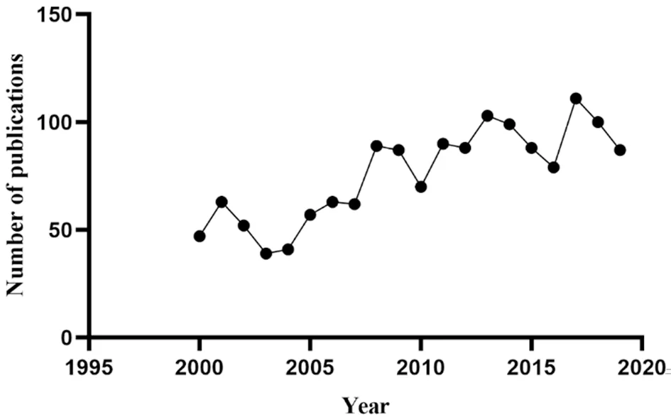
Figure 1 Annual publication outputs for pterygium from 2000 to 2019.
RESULTS
Annual Number of Publications in PterygiumUsing the retrieval strategy mentioned above, a total of 1516 records were collected from WoSCC for the period 2000‐2019. The overall amount of literature published was on the rise, but it fluctuated a little as shown in Figure 1. In 2000‐2005, the number of publications dropped to the first trough in 2003 (40 publications). The period of 2000‐2005 is the low production stage of the past two decades. Followed a sustained increase for 5y, the volume of publications suddenly dropped the second trough with 70 publications in 2010. But on the whole,the number of publications was on an increasing trend. The most articles published in 2017, 111 publications.
Distribution of Countries in Pterygium ResearchMost of the 1516 publications concerning pterygium were contributions from 83 active countries, among which the United States recorded the most publications, citations, and links with other countries. As shown in Table 1, the top ten countries in terms of literature volume accounted for 75.07%of the relevant research, with 1138 publications. Specifically,the United States submitted the most publications (252,16.62%), followed by China (198, 13.06%), and Turkey (138,9.10%). From VOSviewer analysis in Figure 2, each node represents a country and lines between the nodes indicate the strength of relation between countries. Although countries like Germany, England, Iran, and Spain in blue node had relatively low research output, their lines’ strength indicated strong connection with others and great impact on other countries’research. To the contrary, although China’s publications ranked second, its link strength was relatively low. Even so,considering all the countries, the dispersed and low‐density distribution indicated that studies in pterygium are ongoing around the world.
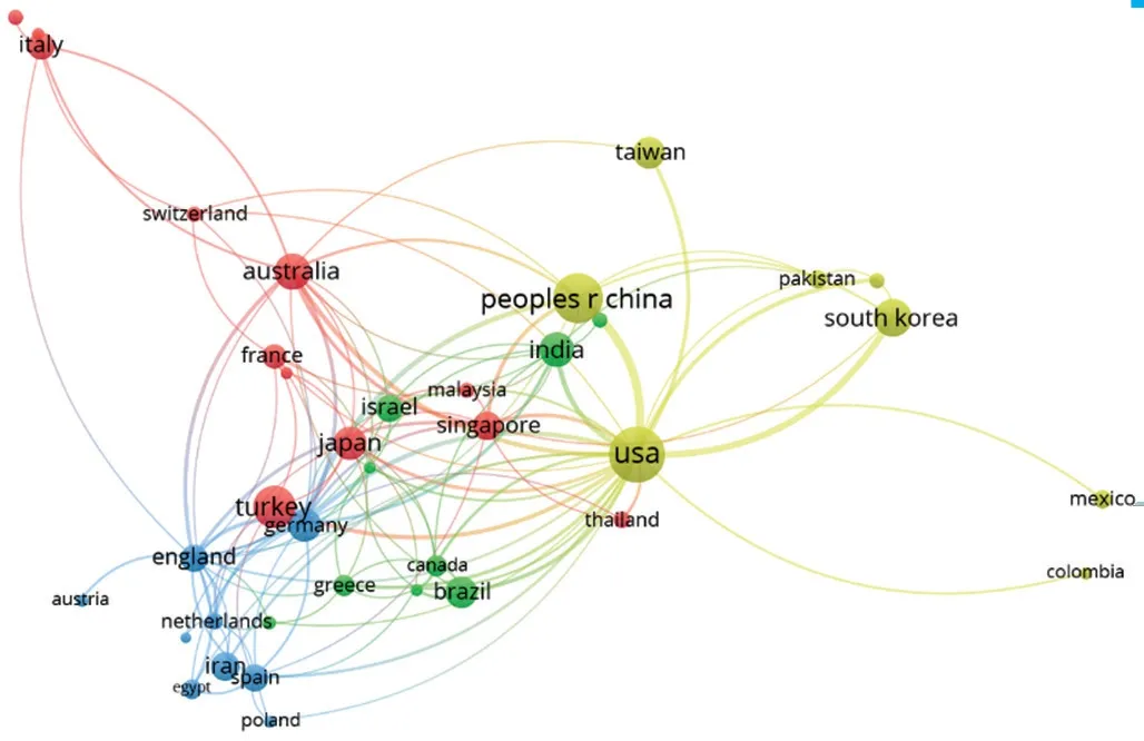
Figure 2 Network map of countries reporting pterygium research Totally 38 countries that met the threshold of a minimum of 5 publications related to pterygium are included. Each node represents a country; the links represent the association between countries; the color and distance between items represent the similarity between countries.

Table 1 Ranking of the top 10 countries with the most publications in pterygium research, during 2000-2019
Distribution of Institutions in Pterygium ResearchRanking by output of publications, the top most five institutions were arranged in order, including Singapore National Eye Center(52 publications), National University of Singapore (47 publications), The University of New South Wales Sydney(40 publications), Tel Aviv University (28 publications),Chung Shan Medical University (27 publications). The top most ten prolific institutions, with a combined total of 321 publications, accounted for 21.17% of the entire published output, were listed in Table 2. Among them, two institutions were located in Singapore (Singapore National Eye Center,National University of Singapore), a finding coincident with whole network visualization analysis where Singaporean organizations formed tightly connected clusters, as shown by the blue nodes in Figure 3. Figure 3 displays the knowledge map of 25 institutions that met the threshold of least eight publications and linked with each other. As shown in the map,the University of Melbourne linked the most, followed by the Chinese University of Hong Kong, and Singapore Eye Research Institute. However, some organizations with a high number of publications, due to lack of citations, have reached the threshold, are not shown in the network visualization.Meanwhile, the small links between institutions and low map density of the knowledge domain map demonstrate the broad lack of collaboration.
Distribution of Journals in Pterygium ResearchIn terms of journal distribution, as displayed in Table 3,Corneahad the highest outputs and the most citations, which were, both, much higher than other journals. Pterygium‐related publications inCorneanumbered 221 in the past decade, almost four times that of the second most popular journal,Investigative Ophthalmology & Visual Sciencewith 56 publications,followed byThe British Journal of Ophthalmologywith 54 publications,Ophthalmologywith 53 publications, andEyewith 52 publications.
Distribution of Authors in Pterygium ResearchAs shown in Table 4, the ten most productive authors ranked by output of publications included Coroneo MT (28 publications), Tsai YY(24 publications), Tseng SCG (22 publications), Cheng YW(21 publications), and Tan DTH (21 publications),et al. Figure 4 shows the knowledge map of nine authors with a minimum of five publications in pterygium research and connected with each other. The authors Coroneo MT, di Girolamo N and Wakefield D were in green node, and they are all Australian writers, while the authors represented by the red node are more Italian authors. It is found that the authors in the same country are closely related. Meanwhile, the low map density and few lines among authors reflected the absence of any influential core research group in this field.
Distribution of Co-citation References: Background of Pterygium ResearchSetting the minimum citations’ count at 30, 155 publications were selected from 20 854 cited documents and formed the knowledge map of co‐citation references, which revealed the basic knowledge structure and background of pterygium research. Through a series of complex algorithms in VOSviewer software, including calculating the co‐citation of cited references’ similarity matrix and mapping similarity matrix, references with similar topics were close together and formed clusters in the Figure 5, denoted in color red, blue, and green. Cluster #1 in red contained the most references with 74 publications, occupied 47.74% of the map, broadly centered around the theme of pterygium treatment. References in this cluster chiefly discussed surgical options and adjuvant drugs, in large part proven by designed clinical trial experiments. Cluster #2 in gre en contained 53 publications, accounting for 34.19%, mostly focused on the topics of pathogenesis, morphological changes in pterygium,and novel options for its treatment. Cluster #3 in blue contained 28 nodes (18.06% of total 155 co‐citation publications)concentrated on epidemiology, especially the prevalence rate of and risk factors for pterygium. Table 5 lists the top 20 cited references from all the lists match the threshold; the detail content is discussed later.
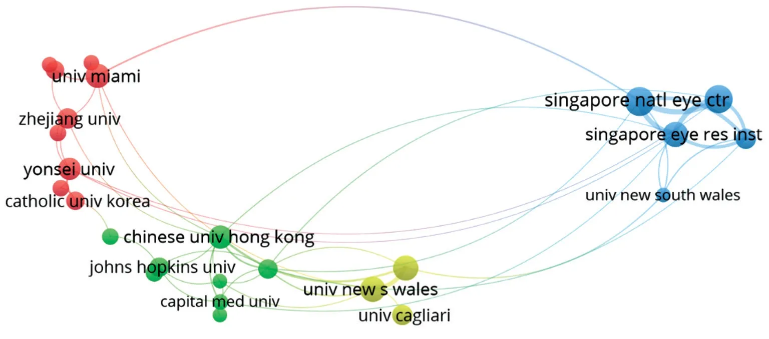
Figure 3 Network map of institutions involved in pterygium research Totally 51 institutions met the threshold of a minimum of 8 publications related to pterygium; only 25 of them are linked to each other. Each node represents an institution; the links represent the association between institutions; the color and distance between items represent the similarity between institutions.
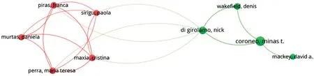
Figure 4 Network map of authors in pterygium research Totally 91 authors met the five publications threshold; only 9 of them are linked to each other. Each node represents an author; the links represent the association between authors; the color and distance between items represent the similarity between authors.
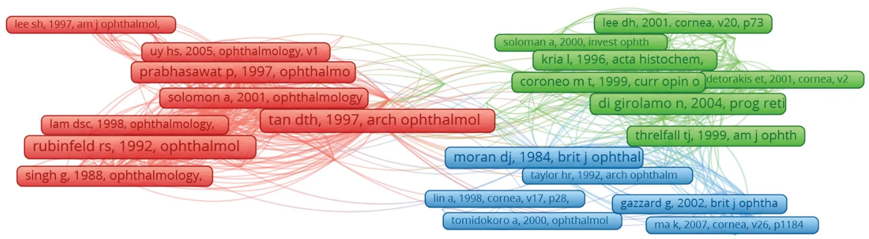
Figure 5 Co-citation analysis network map of references in pterygium research Totally 155 publications met the threshold of 30 citations.Each node represents a publication; the links represent the association between cited publications; the color and distance between items represent the similarity between cited publications.
Analysis of Co-occurrence of Keywords: Topics of Pterygium ResearchFrom the retrieved publications, keywords were extracted and co‐occurrence frequencies calculated. Totally 67 keywords met the requirement of appearing at least 30 times among all 4258 keywords. Based on difference between co‐occurrence matrix of keywords, three clusters containing keywords in analogous topics were separated by VOSviewer.Figure 6 shows the network map of the three keyword clusters expressed in color red, green, and blue. The three clusters correspond with the following research fields: from the density map displayed in Figure 7, colors range from blue to green to yellow. The yellow area is the current research hotspots,while the blue area is relatively low. Meanwhile, it is easy‐to‐interpreting the general structure of pterygium research from this density view. Especially the excision and pathogenesis areas turn out to be important topics. These areas with dense density indicated overall the keywords in these areas received a lot of citations. The top 20 high‐cycle occurrence keywords are listed in Table 6.

Table 2 Ranking of the top 10 institutions with the most publications in pterygium research during 2000-2019

Table 3 Ranking of the top 10 journals with the most publications in pterygium research during 2000-2019
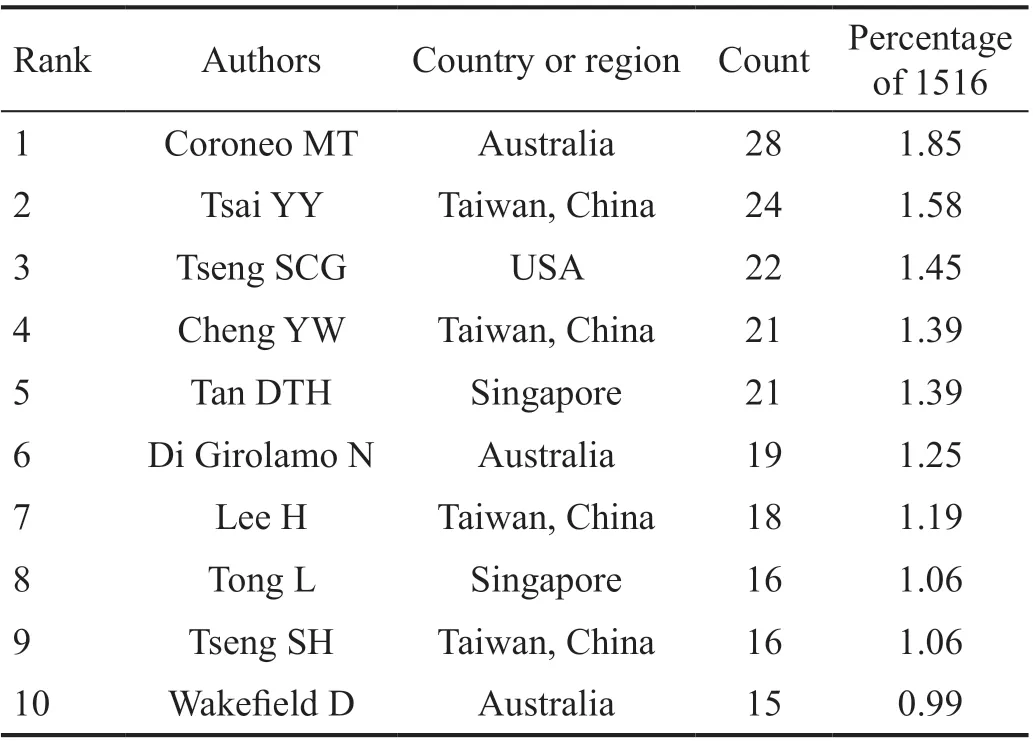
Table 4 Ranking of the top 10 authors with the most publications in pterygium research during 2000-2019
DISCUSSION
By performing bibliometric analysis and drawing knowledge domain, our results revealed a retrospective and current view of the pterygium research. In terms of output in pterygium research, the number of annul publications reflect the change of subject knowledge level and is also an important indicator of the development trends in this field. Judging from the number of publications each year, the overall publication is on the rise, but fluctuates a little. The number of publications in recent years has reached its peak. Although the overall publication is on the rise, there is no blowout growth, so it could explain why not much progress has been made and/or nothing novel has been discovered in this field in the past two decades. In addition, it can be interpreted as since pterygium affects only humans, resulting in difficulties in establishing an animal model[7]. In the analysis of countries (Table 1), the United States has the most publications, citation frequency and connection with others, reflecting its dominant position and international influence in the field of pterygium. China and Turkey ranked high in the publication lists which indicate that significant progress has been made in these countries during the past two decades. Furthermore, some European countries,such as Germany, England, and Spain have relatively low publications but high links with other countries indicating their core status and the value of their publications. The distribution of institution vis‐a‐vis publications and citations reflect research power and influential within the groups. As described above (Table 2), the prominent position of Singaporean institutions, such as the National University of Singapore and the Singapore National Eye Center, suggest that these institutions are prolific and excel in the field of pterygium research. The analyzed results provide valuable information for scholars interested in pterygium research to identify and approach appropriate institutions to cooperate with. On the other hand, the overall sparse and loose distribution of institutions represents inter‐institution cooperation ought to be reinforced, and more combined research should be focused on pterygium. In terms of journal distribution,Corneahad the most citations and publications and was considerably ahead of other journals in pterygium research.Corneais an ophthalmologic journal mainly publishing articles related to the cornea and external ocular structures, thus its published research in pterygium is profound and has a wide influence.Table 3 provides researchers and relevant staff useful information to search articles and to submit achievements.By analyzing the distribution of authors, Coroneo MT, Di Girolamo N, and Wakefield D were included in both, the list of publications and the network visualization, which means their publications are of high quality and are widely recognized.Combining with publication year analysis, the high citation rate of scholars may be because their earlier research in pterygium was significant and reliable and has formed the base knowledge structure in this domain. In the analysis of authorship network map (Figure 4), insufficient collaboration among authors indicates the need for strengthening mutual cooperation. On the premise that high‐quality publications are more likely to be extensively cited, the quote parameter is used to select appropriate publications with commonly accepted theories and results. By exploring the field of highly cited references and keywords, the knowledge structure and research hotspots can be revealed. Furthermore, high frequency cited references could express the basis and background of knowledge. Thus,the cited references (Figure 5) were divided into three clusters by VOS technique, and these three themes will be clarified in the following keyword clusters. As shown in Figure 6, the co‐occurrence keywords with similar characteristics were divided into three clusters, every cluster reflecting a different research topic. Ingeniously, these three clusters are the same as the clusters of the co‐citation clusters. Figures 5 and 6 further illustrate that the background of pterygium research basically revolves around these three themes.

Figure 6 Co-occurrence network map of keywords in pterygium research Totally 67 keywords met the threshold of a minimum of 30 occurrences. Each node represents a keyword; the links represent the association between keywords; the color and distance between items represent the similarity between keywords.

Figure 7 Co-occurrence density map of keywords in pterygium research.
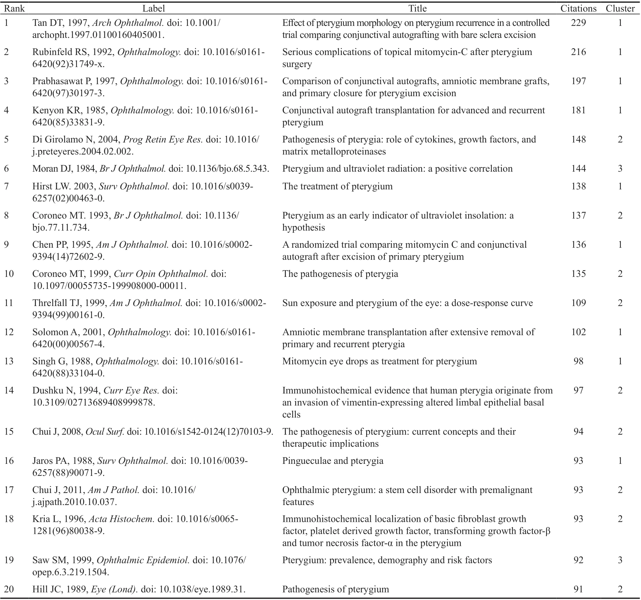
Table 5 Ranking of top 20 co-citation references in pterygium research during 2000-2019
Cluster #1 represents surgical therapeutic techniques and adjuvant for pterygium, reporting keywords such as “excision”,“recurrent pterygium”, “mitomycin‐C”, “conjunctival autograft”, “transplantation”, “complications”, “fibrin glue”,“amniotic membrane”. The topic is consistent with the topic described by Cluster #1 in the co‐cited references. Pterygium excision is the only effective treatment to prohibit growth onto the cornea, but pterygium recurrence is a common post‐operative complication, and a tough problem to overcome. A top co‐citation research in 1990s has reported that recurrence rate after bare‐sclera excision of pterygium could be as high as 88% after the mean followed up for 9.3mo[8]. There is still no definitive treatment proven to reduce recurrence rate to zero reliably, even though several modified strategies have been described, such as graft transplantation and adjuvants’application. Graft material commonly used include amniotic membrane and conjunct membrane. Amniotic membrane contains with abundant growth factors, anti‐angiogenic factors, anti‐inflammatory proteins, and with multiple functions in ocular surface, which can be used in ocular surface reconstruction and conjunctival reconstruction[9‐10].From a Cochrane systematic review, amniotic membrane transplants (AMT) have higher recurrence rates (6.4%‐42.3%)than conjunctival autograft (3.3%‐16.7%), thus it can only be beneficial in the situation of large conjunctival defects and conjunctival scarring[11]. Conjunctival autograft remains the preferred surgical techniques for primary and recurrence pterygium patients: they do not require tissue allograft and have a lower incidence of recurrence than AMT. The risk ratios between conjunctival autograft and AMT were 0.53(95%CI 0.33 to 0.85). No significant complication was mentioned in either surgery techniques. A comparative study has found that AMT and conjunctival autograft resulted in consequential reduction in astigmatism, which are a better surgical method than the bare sclera[12]. Limbal stem cells have the potential to differentiate into corneal epithelial cells and can inhibit the migration of conjunctival epithelial cells to the corneal surface[13]. Limbal conjunctival autograft can promote corneal epithelialization, rapidly tissue repair, and effectively prevent postoperative recurrence[14]. In a high citation study conducted in Spain in the 1990s, patients with recurrent pterygium had no recurrences in the 14mo follow‐up after performing limbal‐conjunctival autograft transplantation[15]. To preserve conjunctiva tissue as much as possible, Hernández‐Boganteset al[16]combined amniotic membrane (AM) and conjunctival graft material. AM was used to replace part of the conjunctiva and limbal epithelial cells were transplanted to repair the exposed sclera, and promising results were obtained. In order to preserve the structure and barrier function of healthy conjunctiva, Yoshitomi and Oshika[17]introduced a new method for the treatment of primary pterygium, which does not require excision or incision, but inverts the growth direction of pterygium. It was found that the recurrence rate was as low as 2.4% at one year, and most cases restored conjunctival physiological vascular loop and the palisades of Vogt. Although the results of these studies are encouraging,more research and observations are needed to validation in the future. Mitomycin C, an alkylating agent, could be used as chemotherapeutic to inhibit synthesis of RNA/DNA/protein. Applying mitomycin C as adjunctive therapy has proven effective in minimizing recurrence rate when compare with non‐mitomycin C surgery[18]. A ten‐year follow‐up randomized controlled trial found that recurrence rate in primary pterygium of mitomycin C group was much higher than limbal conjunctival autograft group, in which mitomycin C group was applied 0.02% mitomycin C intraoperatively in bare‐sclera excision surgery[19]. However, in a 15‐year follow‐up study, the presence or absence of mitomycin C could not influence the recurrence rate of limbal conjunctival autograft for recurrence pterygium[20]. Considering that higher doses of mitomycin C pose an increasing risk of serious and sight‐threatening complications like corneoscleral melt and necrosis,the administration indicators for mitomycin C must be carefully evaluated. Reducing postoperative inflammation and vascular formation may play a significant role in preventing recurrence. To clarify the efficacy of anti‐vascular endothelial growth factor (VEGF) as adjuvant in bare‐sclera excision of pterygium, Elgouharyet al[21]injected bevacizumab subconjunctival after surgery, then they found less recurrence rate than control after 7 to 15mo follow up. VEGF inhibitor can halt angiogenesis and fibroplasia and therefore, suppress the growth of pterygium. In one clinical trial, 5‐fluorouracil(antimetabolites) combined with Avastin (anti‐VEGF) to treat pterygium, 89% present of patients had mean 29 µm reduction of pterygium thickness as evidences by optical coherence tomography, but 67% had astigmatism increased after injection[22]. In most studies, anti‐VEGF had great safety profile. However, the application of anti‐VEGF in pterygium is controversial for the lack of conclusive determination of rouse and dosages and method of surgery. Comparing with suture used in pterygium surgery, fibrin glue as a common alternative to suture: it could be applied in damage tissue gluing, which is better in timesaving, less postoperative pain and less inflammation. In a Meta‐analysis, including 14 randomized controlled trials applied conjunctival autograft, fibrin glue had less recurrence rate and higher incidence of complications(graft loss, granuloma,etc.)[23]. In addition, autologous serum or another sutureless and fibrin‐free approach has not been widely applied for immature technology and unknown complications.

Table 6 Top 20 high co-occurrence keywords in pterygium research during 2009-2018
Cluster #2 broadly covers the occurrence process and pathogenesis of pterygium; keywords in this cluster include“expression”, “pathogenesis”, “conjunctiva”, “fibroblasts”,“epithelial‐cells”, “endothelial growth‐factor”, “angiogenesis”,“apoptosis”, “p53 expression”,etc.
It’s a common sense that pterygium arise from limbal stem cells and characteristic of epithelial cell proliferation,extracellular matrix changes, inflammatory infiltrating,angiogenesis and fibrosis in stroma. The high proliferation potential of limbal stem cells can inhibit the invasion of conjunctival epithelial cells and prevent the limbus from growing of conjunctival blood vessels. In a study published in 2000, it was found that pterygium tissue is potentially equipped with proliferative and invasive power, with evidence of co‐expression of putative limbal stem cell markers CK‐15,CK‐19, and p63α. Meanwhile, Coroneo MT, the author with the largest publications participated in this study[24]. Upon the cumulative stimulate of UV exposure or other environmental factors, the specific zones of limbal stem cell are destroyed.Mediated by cytokines, matrix metalloproteinases and growth factors, altered limbal stem cell may migrate along the cornea and dissolve Bowman’s membrane, followed by conjunctival tissue invasion. Many pro‐inflammatory cytokines[25], such as interleukin‐1 (IL‐1), IL‐6, IL‐8 and tumor necrosis factor‐α (TNF‐α), can promote the proliferation of conjunctival fibroblasts and neovascularization[26]. Therefore,a UV‐blocking contact lens was designed to prevent cellular damage and secretion of pro‐inflammatory cytokines in limbal epithelial cells, which may be beneficial in preventing recurrence[27]. Moreover, cytokines can stimulate production of matrix metalloproteinases (MMPs), which play an important role in the promote cell invasion and metastasis. Abundant of MMPs could potentially be responsible for the matrix collagen remodeling. In a study, the expression of MMP9 was stronger in progressive pterygium than normal and quiescent pterygiumviaimmunohistochemistry. To explore the mechanism, it was found that RNA binding protein human antigen R (HuR) was induced under the stimulation of IL‐1β, interacted with and regulated the stability of MMP9 at posttranscriptional level[28]. TNF‐α is secreted from corneal and conjunctival epithelial cells after being stimulated by UV, which can directly activate NF‐κB pathway and promote cell proliferation and differentiation. Recently, it has been found that TNF‐α can activate hypoxia‐inducible factor (HIF)‐1α, which can not only recruit progenitor cells to promote the progression of pterygium, but also induce the expression of downstream growth factors, such as VEGF‐A and VEGF‐C[29]. VEGF‐A was found highly expressed in epithelial and vascular endothelial cells in pterygium and was positively correlated with vessel density. In addition, VEGF‐C was secreted from pterygium fibroblasts, which may promote endothelial cell migration[30]. Meanwhile, the upregulation of VEGFR‐2 in pterygium fibroblasts also increases susceptibility to VEGF, resulting in higher recurrence rate[29,31]. Since both HIF‐1a and VEGF are overexpressed in pterygium, and anti‐VEGF treatment can only get a transient effect; one study found angiopoietin‐like 4 (ANGPTL4), as an HIF‐regulated angiogenic factor, is overexpressed in pterygium epithelial.Knockout of ANGPTL4 can inhibit the tubule formation in conjunctival epithelial cellsin vitro. It is speculated that ANGPTL4 may be involved in the formation of pterygium neovascularization and may become a similar target like VEGF[32]. Besides, there is a novel target receptor, low‐density lipoprotein receptor (LDLr), which over‐expressed after exposure to VEGF in the subconjunctival fibroblasts in pterygium, correlating with cellular proliferation[33]. However,a definite etio‐pathogenic mechanism remains elusive. Apart from UV exposure, genetic component could play a role in pterygium pathogenesis. A multigenerational family reporting little UV exposure but affected by pterygium presented with missense variants in CRIM1 gene. Wild‐type CRIM1 plays a protective role by reducing proliferation as well as increasing apoptosis, but variants in CRIM1 are unable to participate in protecting against pterygium formation[31]. Besides, the increased expression of p53 in altered limbal cell of pterygium did not cause apoptosis, suggesting the probability of p53 gene mutation, which may be a genetic factor[34]. In 2013,differential expressed mRNA and microRNA were obtained through gene chip microarray technology. Among them, the expression of genes in the miRNA‐200 family was consistently down‐regulated, and these genes can inhibit fibronectin (FN1)in the epithelial‐mesenchymal transition (EMT) pathway[35].Performing RNA sequencing technology in pterygium and normal conjunctival tissue; it was found that 200 differentially expressed genes were up‐regulated, mainly enriched in extracellular matrix and cell migration, while the down‐regulated 139 differentially expressed genes were related to endocrine and inflammation. Through the weighted gene co‐expression network analysis (WGCNA), five hub genes were identified from complex molecular network. Three out of them,including ECM1, IQGAP2 and FN1, were associated with cell adhesion and extracellular remodeling. Although lack ofin vivoandin vitroexperiments, this data provided key biological activities of pterygium for research[36]. Genome‐wide circRNA expression profiling was analyzed to explore differential expressed circRNA between primary pterygium and normal conjunctival tissue.In vitro, circ_0085020 (circ‐LAPTM4B)influence phenotype of pterygium fibroblast and epithelial cell, and may be used as a biomarker of disease[37]. Besides common histological features (angiogenesis, fibrogenesis,and inflammatory factor infiltration), there are few examples of neoplasms appearing in pterygium[24]. Besides, studies showed that the expression of K‐ras oncogene mutation was significantly up‐regulated in pterygium specimens compared with normal conjunctiva, supporting the tumoral correlation of pterygium[38]. At present, there is no complete system for the pathogenesis of diseases, but as the second cluster, research on pterygium mechanism is also a current hot topic.
Cluster #3 mainly include keywords concerning the epidemiology and etiology of pterygium, like “prevalence”,“risk‐factors”, “population”, “epidemiology”, “dry eye”,“Singapore”, and “ultraviolet‐radiation”. In a large population epidemiological study conducted in north‐western Spain,it was found that the prevalence of pterygium was 5.9% in people≥40y[39]. According to a recent Meta‐analysis, the average prevalence rate of pterygium was 12% in the total global population, varying by lifestyle (alcohol consumption,wearing sunglasses), environment factors (UV exposure),and geographical (outdoor occupation, living in rural environment)[40]. Many studies have found that increased sun exposure is associated with a higher incidence of pterygium in lifestyle variables such as outdoor activities, rural dweller, and gender[41]. Strong epidemiological data indicate that ultraviolet ray (UVR) could cause a destructive effective in the long term,including DNA damage, induced inflammation, and increased expression of VEGF[27]. Older age is a commonly recognized risk factor in many publications[39]. Wearing sunglasses and cigarette smoking are protective factors. Smoking decreasing the incidence of pterygium can possibly be explained by decreased inflammation, antibody secretion, and changes in tear composition[42]. In anin vitroexperiment, it was confirmed that after the treatment with a mixture of nicotine and cotinine, the proliferation and migration of pterygium cells were reduced. The related biomarker of migration, such as Snail, α‐smooth muscle actin protein, MMP1 and MMP9 were decreased[43]. However, the role of smoking is controversial due to its role in cancer formation and cardiovascular diseases.In addition, attention is also paid to risk factors such as tear film change, viral infection, cytokines, and growth factors imbalance. Tear film instability or dry eye is common in pterygium patients. Meibomian gland dysfunction has been identified as a cause of the tear function instability in pterygium patients[44]. The association between viruses and pterygium is not clear, but research has shown that HPV appears to function as a pathogenic co‐factor, along with UVR and HIV co‐infection[45]. Better understanding of epidemiology and risk factors can help ophthalmologists in improving treatment method and clarifying research trends.
In the process of searching publications through WoSCC, there is a certain gap with the PubMed database because the search strategies of the two databases are different. In the WoSCC database, publications were searched by subject words, which were searched in the title, abstract, or keywords of the paper.The raw data may bring false‐positive results. However, when we used VOSviewer to analyze the raw data, every step with a threshold could exclude the data with a low occurrence rate,so this tool shows high reliability. Due to the limitations of this database, unpublished articles and academic papers were not included, which affected the recall rate of final data. Even though we only download data from WoSCC, the results of this study were stable and highly reproducible, since this study covers the vast majority of references from 2000 to 2019,and the latest published articles are unlikely to affect the final results[3]. Besides, we set no language restriction while we conduct an advanced search, but it didn’t prevent the titles and abstracts of the retrieved 1516 publications from being displayed in English. Even though English brings unavoidable language bias[46], English is the universal language nowadays,and it is no doubt that a publication published in a language other than English must at the very least include English title and abstract. In the analysis of authors, institutions, and countries, all authors were included without ranks on the contribution of authors, which will the limit first author and correspondence authors’ contributions. In consistence with previous studies[6,47], the United States is the country with the largest number of publications, and China also occupies a certain position, probably as a consequence of the high resources and population of these countries. In co‐citation analysis, the number of citations is an important factor in evaluating the quality of the publication. However, the earlier the articles, the easier it is to be cited[48]. Therefore, we applied VOSviewer to conclude the connection of publications and used cluster analysis to draw knowledge structure, for better analysis the research trends of the pterygium research.
Several bibliometric studies have been published in the field of ophthalmology, such as dry eye[48], keratoconus[47], and retinal vein occlusion[6]. To the best of our knowledge, this is the first bibliometric analysis in pterygium research and the first study using VOSviewer to visualize the network of countries,institutions, journals, and authors in the field of pterygium. The retrieved data from WoSCC database objectively and clearly show the topics of current intensive research and predict frontiers using the tools of VOSviewer, which allow a scientific assessment of the clinical and academic research from the information provided by publications, including the academic value of the literature and relevant authors, institutions, and research output level. Treatment and prevention of recurrence are currently the research focus of pterygium. Since the only effective treatment is excision, it is hopeful that after a comprehensive study of epidemiology and pathogenesis,effective drugs can be developed to replace surgical excision.
ACKNOWLEDGEMENTS
Authors’ contributions:Wang YC: conceptualization,methodology, writing original draft; Zhao FK: methodology,resources; Liu Q: validation, data curation; Yu ZY: visualization;Wang J: project administration, writing, review, and editing;Zhang JS: supervision, funding acquisition.
Foundation:Supported by the National Natural Science Foundation of China (No.81870644).
Conflicts of Interest: Wang YC,None;Zhao FK,None;Liu Q,None;Yu ZY,None;Wang J,None;Zhang JS,None.
 International Journal of Ophthalmology2021年6期
- International Journal of Ophthalmology的其它文章
- A decrease in macular microvascular perfusion after retinal detachment repair with silicone oil
- Surgical outcomes in acute dacryocystitis patients undergoing endonasal endoscopic dacryocystorhinostomy with or without silicone tube intubation
- Dr. Father Wacław Szuniewicz, a forgotten pioneer in refractive surgery and his work in China
- Deterioration of Avellino corneal dystrophy in a Chinese family after LASlK
- A mutated CRYGD associated with congenital coralliform cataracts in two Chinese pedigrees
- Autophagy dysregulation mediates the damage of high glucose to retinal pigment epithelium cells