Borassus flabellifer L.crude male flower extracts alleviate cisplatin-induced oxidative stress in rat kidney cells
Ornanong Tusskorn, Kanoktip Pansuksan, Kwanchayanawish Machana
1Chulabhorn International College of Medicine, Thammasat University, Pathum Thani 12121, Thailand
2Department of Pharmacognosy, Faculty of Pharmaceutical Sciences, Burapha University, Chon Buri 20131, Thailand
ABSTRACT
KEYWORDS: Borassus flabellifer L.; Cisplatin; Oxidative stress;Mitochondrial dysfunction
1.Introduction
Borassus
flabellifer
(B
.flabellifer
) L.(Arecaceae), commonly known as palmyra palm, is broadly distributed in various Southeast Asian countries including Thailand.The fruit pulp ofB
.flabellifer
is widely consumed and the sap from the flower is used as a source of palm sugar[1].Moreover, many parts of the plant are used in traditional medicine as antidiabetic, antimicrobial, antiinflammatory, and antioxidant[2-4].The male inflorescence shows a significant anti-inflammatory activity and analgesic property[5].In addition, flowers ofB
.flabellifer
have been investigated for their antipyretic effects[6-8], and immunosuppressant properties[9].It has been reported that 2,3,4-trihydroxy-5-methyl acetophenone, isolated from the palm syrup has potent radical scavenging activity[2].Furthermore, murine experiments have shown antidiabetic activity of crude methanol extracts from male flowers ofB
.flabellifer
[10].Seeds and rhizomes have been reported to have antioxidant activity[11,12], but to our knowledge, none from male flowers.Therefore, this warrants investigation of the male flower for its antioxidant and cytoprotective properties.Cisplatin is a platinum compound approved for use as a single drug and in combination with other drugs, in the treatment of various types of cancers[13].However, its clinical application is limited because of the high toxicity that it generates in kidney cells,which is its most common and severe side effect.Cisplatin-induced nephrotoxicity is associated with oxidative stress, cellular redox disturbance, and mitochondrial dysfunction[14].Thus, protective strategies are essential in reducing cisplatin-induced nephrotoxicity.This study aimed to evaluate the antioxidant potential ofB
.flabellifer
,and its cytoprotective effects on cisplatin-induced oxidative stress in kidney cells.2.Materials and methods
2.1.Plant collection and extraction
Male flowers ofB
.flabellifer
were obtained from Chai Nat,Thailand and authenticated by Miss Tapewalee Kananthong, a botanist of the Royal Forest Department.A sample was deposited in the Forest Herbarium, Royal Forest Department, Bangkok, Thailand(BKF No.193907).B
.flabellifer
was extracted with ethyl acetate, and methanol and the powdered crude extract was stored at –20 ℃.2.2.Cell culture
A rat kidney cell line (NRK-52E) was purchased from ATCC(Manassas, Virginia, USA) and grown in Dulbecco’s Modified Eagle Medium (DMEM) (Sigma Aldrich, St.Louis, Missouri, USA)supplemented with 4 mML
-glutamine, 1.5 g/L sodium bicarbonate,100 U/mL penicillin, 100 μg/mL streptomycin sulfate and 10% fetal bovine serum.The cells were maintained under 5% COin air at 37 ℃ and subcultured every 2-3 days using 0.25% trypsin ethylenediaminetetraacetic acid, and the medium was changed after overnight incubation.2.3.2,2-Azino-bis-(3-ethylbenzothiazoline-6-sulfonic acid) (ABTS) assay
A modified method of Taiet
al
.[15] was used to evaluate ABTS radical cation (ABTS) indicated by reduced antioxidants.Briefly,7 mM ABTSwas produced by reacting ABTS stock solution with 2.45 mM potassium persulfate (KSO), allowing the mixture to store in the dark at room temperature for 12-16 h before use.The ABTSsolution was then diluted with saline phosphate buffer (PBS,pH 7.4) at an absorbance level of 0.70 (±0.02).After adding diluted ABTSsolution to each extract, or Trolox standard, the reaction mixture was measured after 15 min of initial mixing.Reduced absorbance was recorded at 734 nm using a UV spectrophotometer.Antioxidant activity of each extract was measured using the ICthreshold defined as the concentration required to cause 50% inhibition of radical scavenging activity.2.4.2,2-Diphenyl-1-picrylhydrazyl (DPPH) free radical scavenging assay
Free radical scavenging activity was assessed according to the method of Vongsaket
al
.[16] with some modifications.Twenty μL of the extract and a standard Trolox control at varying concentrations(10-1 000 μg/mL) were mixed with 180 μL of 1 mM DPPH in ethanol.Then, the solution was incubated at 37 ℃ for 30 min, and reduced DPPH free radicals were evaluated with a microplate reader at an absorbance of 520 nm.Antioxidant activity ofB
.flabellifer
extracts was presented as ICwhich is defined as the concentration of extract required to cause a 50% decrease in the initial absorbance of free radical scavenging activity.2.5.Ferric reducing antioxidant power (FRAP) assay
The FRAP assay of Al-Mansoubet
al
.[17] was used to determine the total antioxidant potential of each extract.In brief, a FRAP reagent was prepared by mixing 300 mM acetate buffer pH 3.6, with 10 mM 2,4,6-tripyridyl-s-triazine in 40 mM HCl, and 20 mM FeClat ratio of 10:1:1 (v/v).Twenty μL of the sample solution at 1 mg/mL was mixed thoroughly with the FRAP reagent and incubated for 30 min in dark.The absorbance level of theB
.flabellifer
samples and Trolox control was measured at 600 nm.A lower level of iron is indicated by increased absorbance of the reaction with the results expressed in mg Trolox equivalent antioxidant capacity (TEAC)/100 g extract.2.6.Total polyphenolic content (TPC)
TPC was measured using Folin-Ciocalteu colorimetric method described by Vongsaket
al
[18].Twenty μL of the plant extracts were mixed with Folin-Ciocalteu reagent and incubated at room temperature.The mixture was added with 7.5% sodium carbonate,and then incubated for 30 min at room temperature.Total polyphenols were determined by a microplate reader at a wavelength of 765 nm.A resulting absorbance of blue color indicates substantial polyphenol content.A standard curve of gallic acid was used for quantitation and the results were represented as g GAE of 100 g extract.2.7.3-(4,5-dimethylthiazol-2-yl)-2,5-diphenyltetrazolium bromide (MTT) assay
Cytotoxicity was assessed by the MTT assay, which quantifies cell proliferation and viability.NRK-52E cells were seeded onto 96-well culture plates at a density of 10 000 cells/well.After overnight culture, cells were plated in fetal bovine serum (FBS)-free medium and treated with varying concentrations of ethyl acetate and methanolic extracts ofB
.flabellifer
(10-50 μg/mL) or cisplatin (1-100 μM), and cell viability was examined 24 h posttreatment.The cytoprotective effect was also evaluated after exposure toB
.flabellifer
(1 and 10 μg/mL) and cisplatin (5 and 10 μM), or combinations of both.Then, the medium was removed after treatment, and the cells were incubated in complete medium (with FBS) containing 0.5 mg/mL MTT dye.After 2 h, the FBS medium was removed, and the cells were solubilized with dimethyl sulfoxide.Absorbance was measured using a microplate reader with a filter at a wavelength of 570 nm.Culture medium served as a negative control.The concentration ofB
.flabellifer
and cisplatin required to inhibit 50% growth of the NRK-52E cells (ICvalues) was calculated by analyzing the relationship between concentrations and percent(%) inhibition using GraphPad Prism 6 version 6.01 for Windows(GraphPad Software, La Jolla, CA, USA).2.8.Glutathione (GSH) assay
The antioxidant activity was evaluated by the GSH assay, which measures cell survival and cellular function.GSH is a primary regulator of cellular redox.Both reduced GSH and glutathione disulfide (GSSG) were assayed using thiol green, a fluorescent probe that detects thiol compounds, according to a method described by Tusskornet
al
[19].Redox stress, as indicated by the decrease of GSH/GSSG ratio, is involved in cellular dysfunction and cell death[20].Cultured cells treated for 6 h were trypsinized and washed with cold PBS.Afterward, aliquots of cell suspensions were made to react with 1-methyl-2-vinyl pyridinium trifluoromethanesulfonate for detection of GSSG and another aliquot for detection of total GSH and protein content.Samples used for assays of GSSG and total GSH were deproteinized with meta-phosphoric acid before performing the enzymatic assay.Protein concentration was evaluated by the Bradford dye-binding assay.2.9.Determination of cellular reactive oxygen species (ROS)by 2’,7’-dichlorofluorescin diacetate (DCFDA) assay
Cisplatin-induced intracellular ROS was measured by staining kidney cells with a cell-permeable fluorescent probe, DCFDA.In brief, 20 000 NRK-52E cells were seeded in a 96-well black microplate and cultured overnight.After treatment ofB
.flabellifer
and cisplatin in FBS-free medium for 3 h, the medium was replaced with 100 μL of 25 μM DCFDA in PBS.The NRK-52E cells were incubated for 45 min, after which the dye was replaced with 100 μL of PBS.A microplate reader was used to measure fluorescence signal at excitation and emission wavelengths of 485 and 535 nm,respectively.2.10.Measurement of mitochondrial transmembrane potential (ΔΨm)
The detection of ΔΨm changes in cells is a key indicator of mitochondrial function and cell death.The collapse of ΔΨm is considered as an early event towards mitochondrial damage and apoptosis[21].It is more straightforward than the detection of mitochondrial DNA or protein.In order to measure the change in ΔΨm, NRK-52E cells were seeded onto a 96-well black microplate at a density of 20 000 cells/well, cultured overnight, and then treated withB
.flabellifer
and cisplatin in FBS-free medium for 6 h.The assay was performed using the cationic, lipophilic JC-10 dye according to a modified method described by Tusskornet
al
[22].In brief, a cultured plate was centrifuged at 1 000 rpm for 5 min at room temperature.The cultured media was removed, and cultured cells were loaded with JC-10 dye, then incubated for 30 min.The ΔΨm was measured with a fluorescent plate reader, and its differential images were captured by fluorescence microscopy.Accumulation of JC-10 in the mitochondrial matrix of healthy cells produces red fluorescent J-aggregates.However, in apoptotic and necrotic cells,as ΔΨm decreases, JC-10 monomers are generated, resulting in a fluorescent shift to green[23].Fluorescent intensity in NRK-52E cells was quantified by the ratio of J-aggregates with monomers.2.11.Statistical analysis
Normal distribution of the continuous data warranted descriptive expressions of central tendency and dispersion in terms of mean ±standard deviation (SD).Control and treated groups were statistically compared using the Student’st
-test (two groups) or one-way analysis of variance with Duncan multiple rangepost
-hoc
test as appropriate.The level of significance was set atP
<0.05.
Table 1.Antioxidant capacities and total phenolic contents of Borassus flabellifer extract.
3.Results
3.1.Antioxidant activity of B.flabellifer
The results of four assays (ABTS, DPPH, FRAP, TPC) regarding antioxidant activity are shown in Table 1.B
.flabellifer
methanolic extract showed more prominent antioxidant activity than its ethyl acetate extract with lower ICvalues in ABTS and DPPH assays and higher FRAP value and TPC.The antioxidant activity of Trolox, as a standard control, was comparable toB
.flabellifer
methanolic extract in ABTS assay, but significantly higher in DPPH assay (P
<0.05).3.2.Cytotoxic effects of B.flabellifer in NRK-52E cells

Figure 1.Cytotoxicity of B.flabellifer and cisplatin in NRK-52E cells.Cells were treated with (A) BFE or (B) BFM (10-50 μg/mL) or with (C) cisplatin (1-100 μM).Each value represents mean ± SD of 3 experiments.BFE and BFM represent the ethyl acetate and methanol extracts of B.flabellifer, respectively.

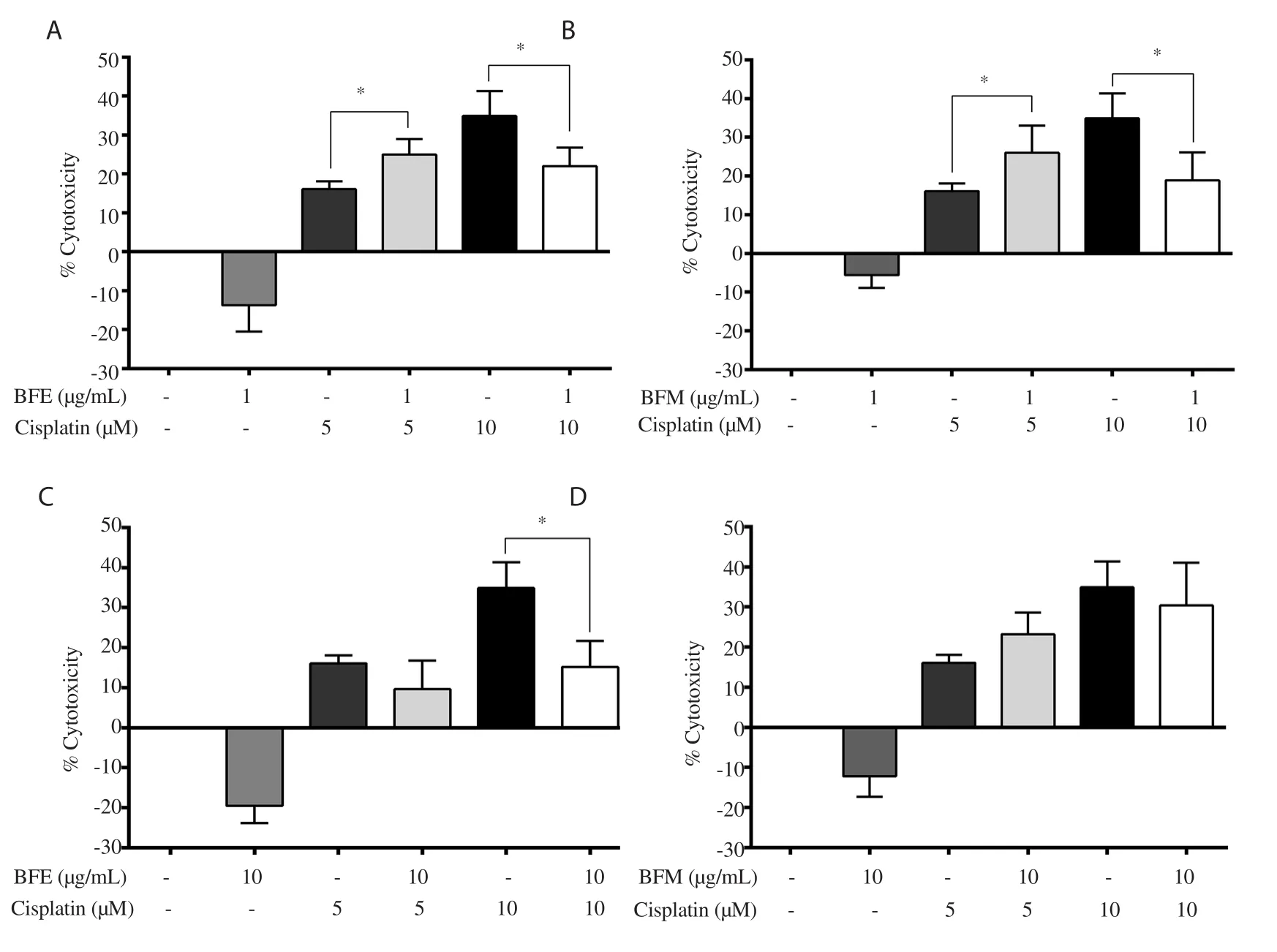
Figure 2.Cytoprotective effect of B.flabellifer extracts on cisplatin-induced cytotoxicity.(A) BFE (1 μg/mL) + Cisplatin (5/10 μM); (B) BFM (1 μg/mL) +Cisplatin (5/10 μM); (C) BFE (10 μg/mL) + Cisplatin (5/10 μM); (D) BFM (10 μg/mL) + Cisplatin (5/10 μM).Cytotoxicity was assessed by MTT assay.Each bar represents mean ± SD of 3 experiments.*P<0.05.
A screening protocol was used to determine acceptable levels of cytotoxicity of ethyl acetate and methanolic extracts ofB
.flabellifer
using NRK-52E cells.The similarity of dose-response curves between the ethyl acetate and methanolic extracts ofB
.flabellifer
and cisplatin are shown in Figure 1A-C with ICvalues of (26.2±1.8)μg/mL, (35.8±2.4) μg/mL and (34.6±6.7) μM, respectively,demonstrating that relative safety of the extracts was likely to be below IClevels.For cytoprotective effect of the ethyl acetate and methanolic extracts in NRK-52E cells, cells were employed at concentrations below the ICvalues,i
.e
.1 and 10 μg/mL against cisplatin (5 and 10 μM).Cytoprotective effect of both extracts was observed in combination with cisplatin at a high concentration (10 μM) (Figure 2A-C).The methanolic extract ofB
.flabellifer
at 10 μg/mL had a tendency of the protective effect (Figure 2D).Cisplatin at a low concentration(5 μM) conferred small cytotoxicity (about 15%), where both ethyl acetate and methanolic extracts did not show protection against the cytotoxic effect.
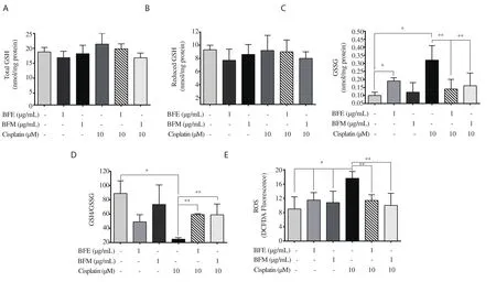
Figure 3.Effect of B.flabellifer extracts and cisplatin on cellular GSH contents and ROS production.(A) Intracellular total GSH, (B) reduced GSH, (C) GSSG,(D) the ratio of GSH/GSSG, and (E) ROS production.Each bar represents mean ± SD of 3 experiments.Values with asterisks indicate significance (P<0.05); *and ** indicate comparisons with the control and cisplatin group, respectively.GSH: glutathione; GSSG: oxidized glutathione disulfide; ROS: reactive oxygen species.

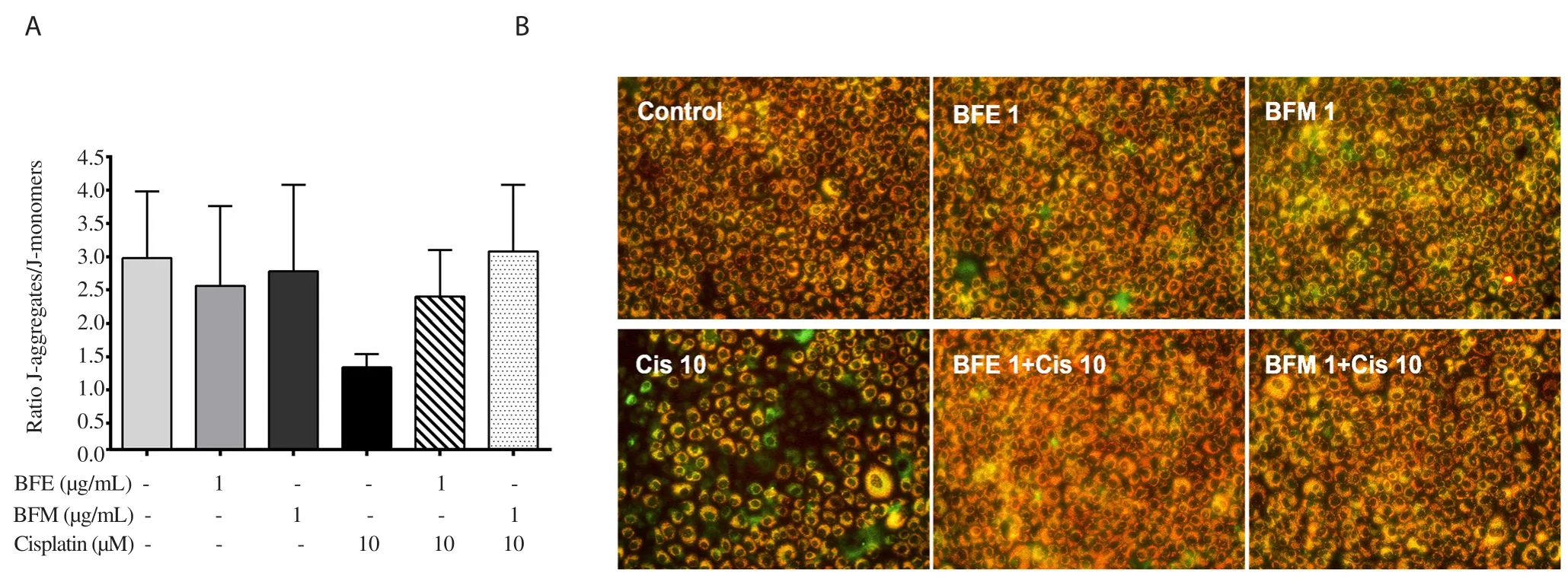
Figure 4.Effect of B.flabellifer extracts and cisplatin on mitochondrial transmembrane potential (ΔΨm).JC-10 fluorescent signals were measured and the ratio of J-aggregates and J-monomers was calculated as indicative of ΔΨm (A).Each bar represents mean ± SD.(B) Images of JC-10 staining in treatment groups are shown.
3.3.Effect of B.flabellifer in combination with cisplatin on cellular redox stress and ROS formation
To understand the mechanisms of the cytoprotective activity ofB
.flabellifer
ethyl acetate and methanolic extracts, we evaluated the effects of these extracts (1 μg/mL) along with cisplatin (10 μM)on oxidative status (redox).Total GSH and reduced GSH were unchanged in all groups (Figure 3A and 3B).However, treatment with both extracts prevented cisplatin-induced increased oxidized form of GSSG, the level of which was comparable to the control group (Figure 3C).GSH/GSSG ratio, an indication of cellular redox status, was markedly decreased by cisplatin while increasing by treatment with both methanolic and ethyl acetate extracts (Figure 3D).In ROS analysis, cisplatin caused a significant elevation of ROS formation, whereasB
.flabellifer
ethyl acetate and methanolic extracts completely normalized the levels to the control (Figure 3E).3.4.Effects of the drug combination on ΔΨm
Since mitochondria are vulnerable to oxidative stress which leads to cell damage, effects of the extracts on ΔΨm were examined.Figure 4A shows a decrease in the JC-10 ratio, indicative of the loss of ΔΨm in the cisplatin group.Treatment with both methanolic and ethyl acetate extracts prevented cisplatin-induced decrease, which was further confirmed in images of JC-10 dye staining (Figure 4B).Cisplatin induced an apparent green fluorescence in NRK-52E cells representative of the depolarized ΔΨm, while cells treated withB
.flabellifer
extracts showed orange fluorescence similar to control cells.4.Discussion
Thisin
vitro
study focused on two areas of investigation where we used a number of laboratory tools to examine antioxidant activity and cytoprotective effect ofB
.flabellifer
.The four chemical assays used for this purpose favored the methanolic extract over the ethyl acetate one.Cytoprotective activity of theB
.flabellifer
extracts was demonstrated in NRK-52E cells.The effect was associated with prevention of oxidative stress and mitochondrial dysfunction.Studies of herbs with antioxidant properties are important in expanding the biomedical evidence for use in populations that can fully utilize their value[24].Studies have presented data underpinning the role of dietary phytochemical antioxidants in preventing cancer,cardiovascular diseases, diabetes, and other chronic diseases related to oxidative stress[25-27].
Our findings onB
.flabellifer
extracts agree with another study that found potentin
vitro
antioxidant potential from theB
.flabellifer
ethanol extract[28].The strong antioxidant property ofB
.flabellifer
might be of use as a nutraceutical product[29].It is noted that the methanol extract ofB
.flabellifer
shows stronger antioxidant activity in chemical assays than ethyl acetate extract.However, the prevention of oxidative stress by both extracts in the cell system is comparable.This demonstrates some disparity between the chemical assay and cell-based assay, suggesting the ethyl acetate and methanolic extracts exert a high antioxidant activity at the cellular level by scavenging ROS and preserving redox status.The potential to develop health products from herbs warrants investigation of safety for human use.In our study, we investigated the safe use ofB
.flabellifer
by examining cytotoxicity in renal cells,whichB
.flabellifer
ethyl acetate extract significantly prevented.Phenolic compounds such as gallic and tannic acids have been shown to reduce cisplatin-induced functional and histological renal damage by suppressing ROS formation, lipid peroxidation, and oxidative stress[30].Free radicals, especially ROS, are considered to cause the emergence and development of cell injury as well as various degenerative diseases[31].Cisplatin-induced ROS in renal cells causes damage to cellular biomolecules such as nucleic acids, lipids, and proteins resulting in cell dysfunction[32].Cisplatin conjugation with GSH forming cisplatin adducts induces mitochondrial oxidative stress through lipid peroxidation[33].This, in turn, leads to dysregulation of the endogenous antioxidant system[34].We have shown that these oxidative stress situations in the kidney cells were ameliorated with an increase in the GSH redox ratio and a decline in GSSG to the levels that were not significantly different from control levels.This result suggests that ROS formation and GSH redox stress may cause mitochondrial dysfunction[35].Reduced GSH and its redox ratio have been shown to regulate various redox-sensitive enzymes and transcription factors in maintaining the operation of cellular function and mitochondria[36].We have demonstrated thatB
.flabellifer
prevented the cisplatin-induced loss of ΔΨm by increasing antioxidant activity and reducing oxidative stress.The present study showed that the ethyl acetate and methanolic extracts ofB
.flabellifer
significantly protected renal cell death induced by cisplatin at a high concentration (10 μM).However,cisplatin at a low concentration (5 μM) exerted only marginal cytotoxicity (below 15%), where treatment withB
.flabellifer
added on a small cytotoxic effect.Since both extracts themselves also induce small oxidative stress, as indicated by the slightly declined GSH ratio, this may explain additional cytotoxicity to the low concentration of cisplatin.However, cisplatin at a high concentration induced a clear cytotoxic effect with marked oxidative stress,indicated by the GSH redox ratio and ROS formation.The ethyl acetate and methanolic extracts ofB
.flabellifer
showed a preventive effect in association with cellular antioxidant activity,i
.e
.prevention of ROS formation and maintenance of GSH redox ratio.This finding suggests a beneficial effect ofB
.flabellifer
on oxidative stressinduced cell injury.A multitude of cytoprotection mechanisms ofB
.flabellifer
may be present in the kidney cells that protect against cisplatin-induced cytotoxicity.The effects could be attributed to cellular antioxidant activity, as shown in the GSH and ROS assays, and inhibition of cisplatin uptake into the cells.However, cytoprotective effect ofB
.flabellifer
is strongly associated with cellular antioxidant activity.Moreover, ifB
.flabellifer
inhibited uptake of cisplatin,B
.flabellifer
could have shown some cytoprotection especially at a low concentration of cisplatin.Definitive mechanisms ofB
.flabellifer
on cisplatin uptake may need further investigation, as cisplatin is transported by several ubiquitously expressed transporters, such as copper transporter1, 2, ATP7A and ATP7B, and organic cation transporters OTC1-3[37].Our study provides evidence of antioxidant potential ofB
.flabellifer
and its cytoprotective effects on cisplatin-induced redox stress and mitochondrial dysfunction.B
.flabellifer
extracts prevent cytotoxicity in association with the maintenance of cellular redox status.Our findings indicate that the male flower ofB
.flabellifer
has good antioxidant properties.Therefore, it is reasonable to consider the male flower for food and nutraceutical applications in the promotion of health.More studies using thein
vivo
approach may confirm or modify our findings.Conflict of interest statement
The authors declare that there are no conflicts of interest.
Funding
This work was supported by the Agricultural Research Development Agency (Public Organization), Thailand (Contract No.CRP5905021020).
Authors’ contributions
OT and KM contributed to the design and performance of the experiments, and also drafted the manuscript.OT, KM and KP contributed to the final version of the manuscript.OT supervised the project.
 Asian Pacific Journal of Tropical Biomedicine2021年2期
- Asian Pacific Journal of Tropical Biomedicine的其它文章
- Anti-inflammatory, anti-oxidative and anti-apoptotic effects of Heracleum persicum L.extract on rats with gentamicin-induced nephrotoxicity
- Screening of phytocompounds, molecular docking studies, and in vivo antiinflammatory activity of heartwood aqueous extract of Pterocarpus santalinus L.f.
- Anti-senescence and anti-wrinkle activities of 3-bromo-4,5-dihydroxybenzaldehyde from Polysiphonia morrowii Harvey in human dermal fibroblasts
- Corchorus olitorius aqueous extract attenuates quorum sensing-regulated virulence factor production and biofilm formation
- 9-Hydroxy-6,7-dimethoxydalbergiquinol suppresses hydrogen peroxide-induced senescence in human dermal fibroblasts through induction of sirtuin-1 expression
