Formononetin alleviates diabetic cardiomyopathy by inhibiting oxidative stress and upregulating SIRT1 in rats
Manisha J.Oza, Yogesh A.Kulkarni
1Shobhaben Pratapbhai Patel School of Pharmacy & Technology Management, SVKM's NMIMS, V.L.Mehta Road, Vile Parle (W), Mumbai 400056,India
2SVKM's Dr.Bhanuben Nanavati College of Pharmacy, Vile Parle (W), Mumbai 400056, India
ABSTRACT
KEYWORDS: Cardiomyopathy; Formononetin; Type 2 diabetes;SIRT1; Creatinine kinase; Cardiac hypertrophy
1.Introduction
Diabetic cardiomyopathy is characterized as disordered myocardial structure and function due to cardiomyocyte dysfunction, lipid deposition, left ventricular hypertrophy and fetal gene activity[1].Heart failure is the main cause of the death of diabetics.Framingham's study reported that the risk of heart failure is 2 to 8-fold higher in type 2 diabetic patients.An increase in 1 percent of glycated hemoglobin is linked with a 30 percent increase in the risk of heart failure[2,3].Hyperglycemia, hyperinsulinemia and oxidative stress are associated with altered signaling pathways in diabetic cardiomyopathy[4].The other risk factors of diabetic cardiomyopathy include obesity, hypertension, smoking, dyslipidemia, inflammation,cardiac fibrosis, and coronary artery disease.
Formononetin is a phenolic compound with estrogenic activity.It was initially isolated from legume plants like Cicer arietinum and Trifolium pratense and also present in genus Iris, Dalbergia,and Amorpha[5-7].Formononetin can lower blood pressure by regulating endogenous nitric oxide synthase expression and down-regulating 1-adrenoceptors and 5-HT2A/1B[8].Its antihypertensive and vasorelaxant activity is also associated with the endothelium-dependent pathway by blocking Ca2+channel in the thoracic aorta[9,10].Sulphonated derivative of formononetin has cardioprotective and neuroprotective effects against acute myocardial infarction and cerebral ischemia-reperfusion in rats by reducing cell apoptosis and improving angiogenesis[11,12].Formononetin and its analogs could reduce lipid as PPARγ agonists[13,14].The cardioprotective potential of formononetin is attributed to its antioxidant effect and inhibitory effect on MAP kinase activity in aortic smooth muscle cells[15,16].Formononetin also protects cardiomyocytes by reducing ROS and increasing phosphorylation of GSK-3β[17].Furthermore, formononetin has antidiabetic effects on the animal model of type 1 & type 2 diabetes[18,19].It protects pancreatic β-cells against the inflammatory and apoptosis process via inhibiting nuclear factor-kappa β activity[20].Formononetin could ameliorate diabetic nephropathy via silence information regulator 1 (SIRT1) activation and reduce inflammatory processes via upregulation of SIRT1[21,22].SIRT1 is most abundantly expressed histone deacetylase in heart and protects cardiac tissue against oxidative stress-induced damage.However, the effect of formononetin on type 2 diabetic cardiomyopathy is still unclear.Thus, this study intended to evaluate the effect of formononetin on type 2 diabetic cardiomyopathy.
2.Materials and methods
2.1.Chemicals
Chemicals were purchased from Sigma Aldrich, USA.Formononetin was procured from Tokyo Chemical Industry Co.,Ltd.(TCI), Japan.
2.2.Experimental animals
Male Sprague Dawley rats (n=36) weighting 160-170 g were procured from the National Institute of Biosciences, Pune, India.The animals were acclimatized for one week in humidity and temperature-controlled room.
2.3.Induction of type 2 diabetes
Type 2 diabetes was induced as the methods described by Oza et al.and Srinivasan et al using high-fat diet and streptozotocin (35 mg/kg,intraperitoneally)[22,23].Formononetin was given for 16 weeks after confirmation of diabetes.
The rats were divided into six groups containing six in each.Group 1: Normal control, rats received 0.1% carboxymethylcellulose solution for 16 weeks; Group 2: high-fat diet control, rats received high-fat diet; Group 3: Diabetic control received no drug treatment.Group 4, 5 and 6 received 10, 20 and 40 mg/kg dose of formononetin, respectively[18].All rats in group 2, 3, 4, 5 and 6 received high-fat diet till the end of the study[24].At the end of each week, body weight was measured for each animal.
2.4.Measurement of biochemical parameters
The blood sample was collected from retro-orbital plexus.Blood glucose, total cholesterol, low-density lipoprotein, triglyceride,high-density lipoprotein, creatinine kinase-MB (CK-MB), aspartate aminotransferase (AST) and lactate dehydrogenase (LDH) were measured at the end of the study by Erba Chem-7 biochemistry analyser (Germany) as per manufacturers’ protocol.
2.5.Measurement of ECG and hemodynamic parameters
After 16 weeks of treatment, ECG was performed through a needle electrode (+ve, -ve and neutral) using the data acquisition system (AD Instruments, Australia).Change in the QT interval was recorded.
Hemodynamic parameters such as heart rate, left ventricular enddiastolic pressure (LVEDP), diastolic blood pressure (DBP) and systolic blood pressure (SBP) were recorded by cannulating left carotid artery using data acquisition system (AD Instruments,Australia).Heparinized saline was used to fill cannula and attached to a pressure transducer[25].
2.6.Relative organ weight
Animals were sacrificed and heart tissue was harvested from each animal.The relative organ weight was determined by using the following formula:
Relative organ weight=(Heart weight)/(Body weight)×100
2.7.Determination of oxidative stress parameters in heart tissue
Parts of the heart sample were used for measurement of oxidative stress parameters and the rest were kept in 10% buffered formalin solution for immunohistochemical and histopathological study.
Heart tissue homogenate was prepared using polytron homogenizer in phosphate buffer (pH 7.4).The total homogenate was separated into post-nuclear supernatant and post mitochondrial supernatant as described by Oza and Kulkarni[22].Malondialdehyde (MDA) was measured in the total homogenate as described by Ohkawa et al[26];reduced glutathione was measured (GSH) in total homogenate by the Ellman method[27]; catalase (CAT) was measured in post-nuclear supernatant as described by Luck[28]; superoxide dismutase (SOD)was estimated in post mitochondrial supernatant as described by Paolettie and Mocali[29].
2.8.Immunohistochemical study of heart tissue
The heart tissue sections were prepared as described by Oza and Kulkarni[22].The heart sections were treated with mouse antirat SIRT1 (Santa Cruz Biotechnology, Inc., USA) and visualized in a microscope using diaminobenzidine as the visualizing agent and DPX as the mounting agent.Images were taken using photomicroscope and optical density was measured for SIRT1 expression using ImageJ1.51a software.
2.9.Western blotting analysis of heart tissue
Expression of SIRT1 in heart tissue was quantified by Western blotting analysis using anti-SIRT1 antibody (mouse; 1:1 000)(Santacruz Biotechnology, Inc.USA)[30].The quantification of SIRT1 was carried out as described by Oza and Kulkarni[22].Briefly, the heart tissue homogenate was prepared using radioimmuno-precipitation assay buffer.Total proteins were isolated and protein concentration was estimated as per the Lowry method[31].Sodium dodecyl sulfate-polyacrylamide gel electrophoresis was used to separate the protein and transferred on polyvinylidene difluoride membrane by electrophoresis (Bio-Rad, California,USA).Immunoblots examination was performed using anti-SIRT1 antibody (mouse; 1:1 000), β-actin (goat; 1:2 000) and HRP-conjugated secondary antibodies (anti-rabbit: 1:2 000).The enhanced chemiluminescence method was used to detect immunoblots.The results were expressed as SIRT1/β-actin using ImageJ1.51a software.
2.10.Histopathological study of heart tissue
Heart tissue fixed in formalin solution was cut longitudinally,processes, rehydrated, cleaned in xylene and embedded in paraffin.The sections were prepared and stained with Masson trichome stain and hematoxylin-eosin stain as described by Oza and Kulkarni[22].The sections were observed under a photomicroscope (Motic,Canada).The deposition of collagen in heart tissue was determined using ImageJ1.51a software.
2.11.Statistical analysis
The data were expressed as mean ± SEM and analyzed by GraphPad Prism ver.5.00.Differences among groups were analyzed by analysis of variance test followed by Dunnett's multiple comparison.P-value less than 0.05 was considered to be significant.
2.12.Ethical statement
The study protocol was approved by the Institutional Animal Ethics Committee (CPCSEA/IAEC/P-53/2017) of Shri Vile Parle Kelavani Mandal’s animal facility, Mumbai.
3.Results
3.1.Measurement of plasma glucose & lipid profile
As shown in Supplementary Table 1, formononetin at 10, 20 and 40 mg/kg significantly reduced the levels of glucose, triglycerides,cholesterol and LDL in plasm (P<0.001), which were significantly increased in diabetic rats (P<0.001).Besides, formononetin at 20 and 40 mg/kg significantly increased HDL level (P<0.01 or 0.001).
3.2.Measurement of cardiac markers
As shown in Supplementary Table 2, diabetic rats exhibited significant elevation (P<0.001) in levels of CK-MB, LDH and AST;while formononetin significantly reduced these levels (P<0.01).
3.3.Measurement of ECG and hemodynamic parameters
As shown in Figure 1A, diabetic rats showed significant reduction(P<0.001) in heart rate as compared with the normal rats; while formononetin at 10, 20 and 40 mg/kg significantly increased heart rate (P<0.05).Formononetin significantly decreased systolic and diastolic blood pressure (P<0.05) (Figure 1B and C), and the blood pressure of formononetin at 40 mg/kg group was comparable with that of the normal control group.Diabetic rats exhibited significantly prolonged (P<0.001) QT interval comparing with normal rats; while formononetin significantly shortened (P<0.001) QT interval (Figure 1D).
As shown in Figure 2, as compared with normal rats, diabetic rats showed increased LVEDP, -dp/dt and decreased +dp/dt (P<0.001).Formononetin at three doses significantly reduced LVEDP and -dp/dt (P<0.05), but only improved +dp/dt significantly (P<0.01) at 20 and 40 mg/kg.
3.4.Measurement of cardiac hypertrophy
Body weight of diabetic rats was significantly declined while relative heart weight was significantly increased (P<0.001) compared with normal rats.Formononetin significantly increased body weight(P<0.001) and significantly decreased relative heart weight at three doses (P<0.001) (Figure 3).
3.5.Measurement of oxidative stress parameters in heart tissue
As shown in Figure 4, diabetic rats showed significant increase in MDA level and decrease in GSH, SOD and CAT levels in heart tissue (P<0.01).Formononetin significantly declined (P<0.05) MDA level at 20 and 40 mg/kg and significantly increased GSH and SOD levels (P<0.05) at three doses.CAT was significantly increased at 20 and 40 mg/kg (P<0.05).
3.6.Immunohistochemical study of heart tissue
Immunohistochemical result showed significant reduction (P<0.05)in SIRT1 expression in heart tissue of diabetic rats demonstrated by decrease in optical density.Formononetin significantly increased SIRT1 (Figure 5 and 6B) at 40 mg/kg.
3.7.Western blotting analysis of heart tissue
Figure 6A shows significantly reduced SIRT1 expression in heart tissue of diabetic rats (P<0.05).Formononetin at 40 mg/kg significantly increased (P<0.05) SIRT1 expression in heart tissue.
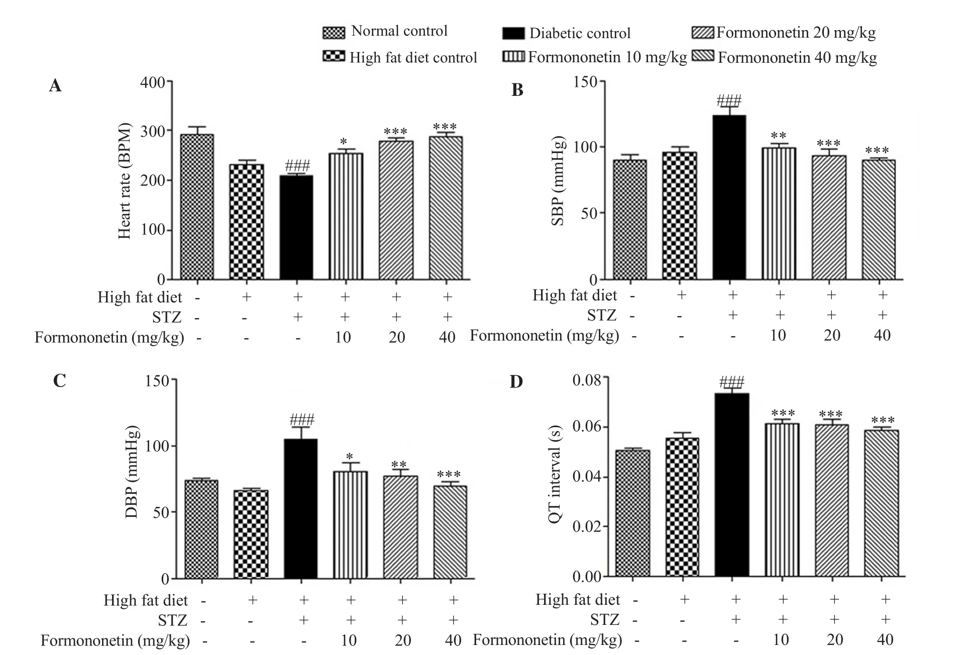
Figure 1.Effect of formononetin on blood pressure and ECG.A: Heart rate, B: systolic blood pressure (SBP), C: diastolic blood pressure (DBP) and D: QT interval.Values are expressed as mean ± SEM (n=6) *P<0.05, **P<0.01, ***P<0.001 when compared with the diabetic control.###P<0.001 when compared to the normal control.STZ: streptozotocin.
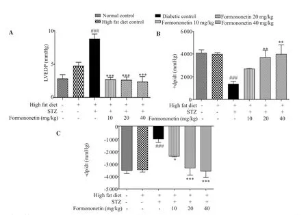
Figure 2.Effect of formononetin on hemodynamic parameters.A: left ventricular end-diastolic pressure (LVEDP), B: +dp/dt and C: -dp/dt.Values are expressed as mean ± SEM (n=6).*P<0.05, **P<0.01, ***P<0.001 when compared with the diabetic control.###P<0.001 when compared to the normal control.STZ: streptozotocin.
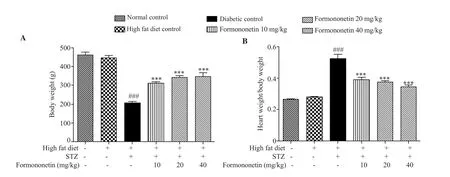
Figure 3.Effect of formononetin on body weight (A) and cardiac hypertrophy (B).Values are expressed as mean ± SEM (n=6).***P<0.001 when compared with the diabetic control.###P<0.001 when compared to the normal control.STZ: streptozotocin.
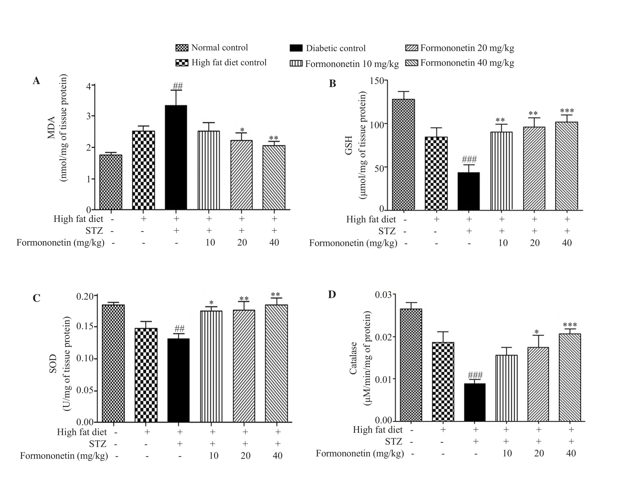
Figure 4.Effect of formononetin on oxidative stress parameters in heart tissue.A: malondialdehyde (MDA), B: reduced glutathione (GSH), C: superoxide dismutase (SOD) and D: catalase.Values are expressed as mean ± SEM (n=6).*P<0.05, **P<0.01, ***P<0.001 when compared with the diabetic control.##P<0.01, ###P<0.001 when compared to the normal control.STZ: streptozotocin.
3.8.Histopathological analysis of heart tissue
Supplementary Table 3 shows significantly higher level of collagen deposition, interstitial fibrosis, inflammatory and degenerative lesions in diabetic rats.Formononetin significantly reduced collagen deposition and interstitial fibrosis at three doses (Figures 7 and 8).
4.Discussion
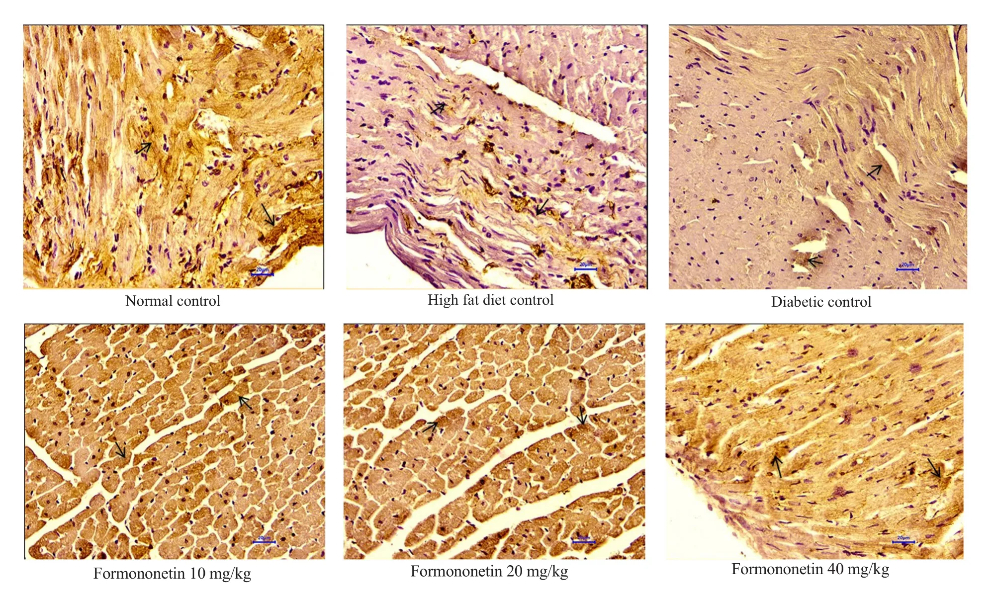
Figure 5.Effect of formononetin on SIRT1 expression in heart tissue (Immunohistochemical staining).All sections were observed at 400× magnification(Arrow indicates SIRT1 expression).Scale bar: 20 μm.
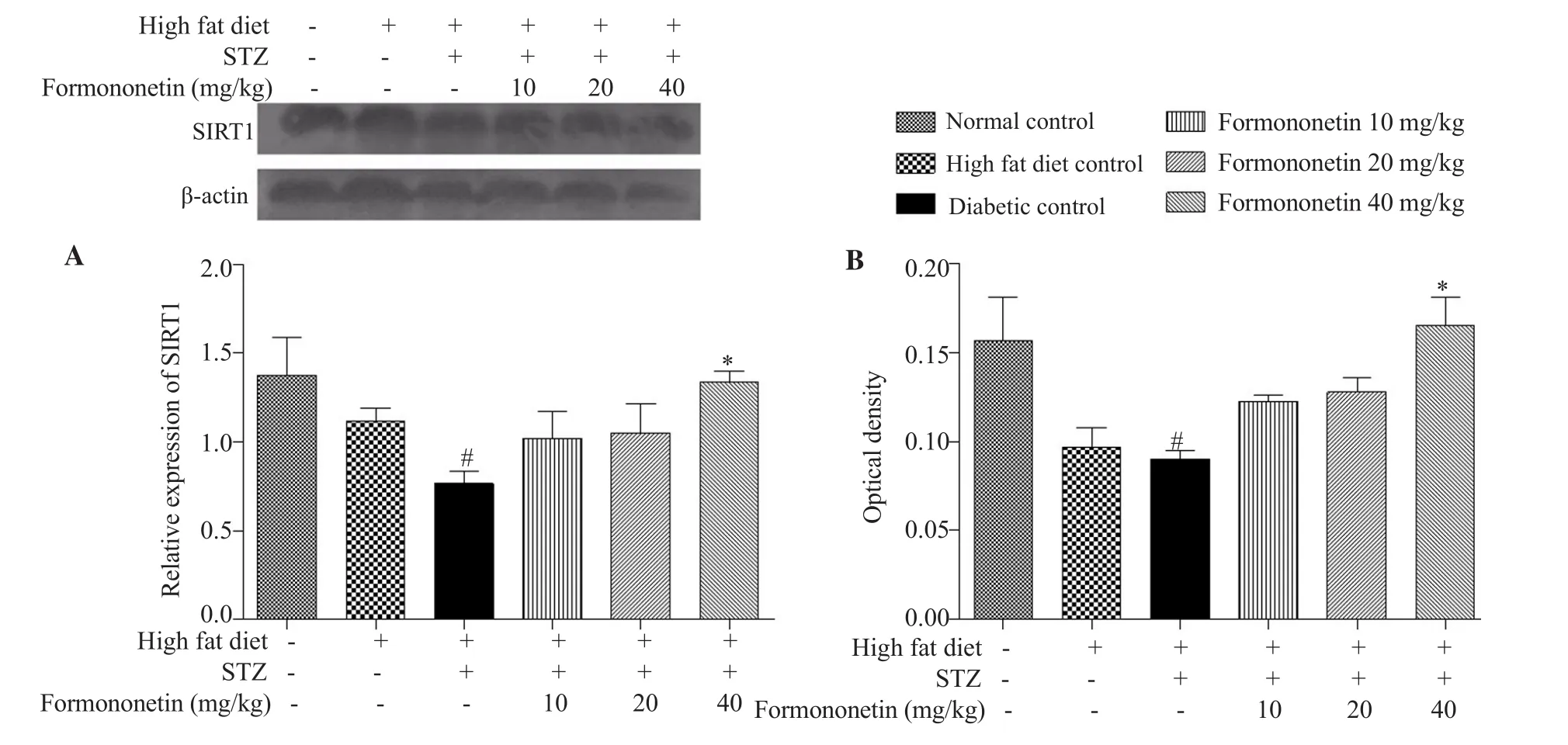
Figure 6.Effect of formononetin on relative expression of SIRT1 in heart tissue.A: Relative expression of SIRT1 in heart tissue by Western blotting method.B:Relative expression of SIRT1 in heart tissue in immunohistochemical staining method.Values are expressed as mean ± SEM (n=6).*P<0.05, when compared with the diabetic control.#P<0.05 when compared to the normal control.STZ: streptozotocin.
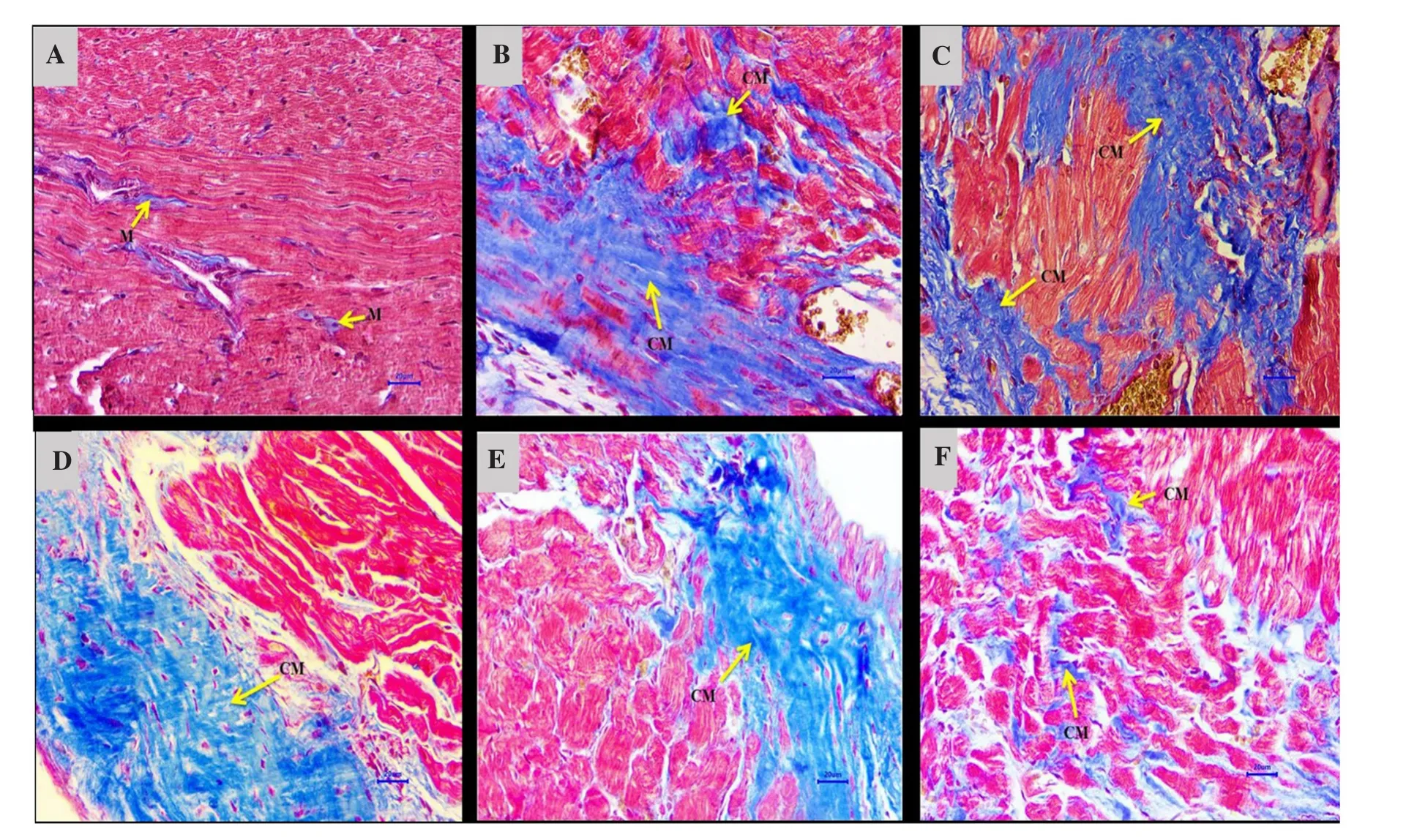
Figure 7.Effect of formononetin on collagen deposition and fibrosis in heart tissue.A: Normal control, B: High fat diet control, C: Diabetic control, D:Diabetic + formononetin (10 mg/kg), E: Diabetic + formononetin (20 mg/kg), F: Diabetic + formononetin (40 mg/kg).(Arrow indicates collagen stained region).All sections were stained with Masson’s trichome stain and observed at 400 × magnification.(M: normal deposition of collagen in myocardium; CM:Increased deposition of collagen in myocardium).Scale bar: 20 μm.
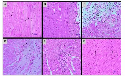
Figure 8.Effect of formononetin on histopathology of heart tissue.A: Normal control, B: High fat diet control, C: Diabetic control, D: Diabetic + formononetin(10 mg/kg), E: Diabetic + formononetin (20 mg/kg), F: Diabetic + formononetin (40 mg/kg).All sections were stained with hematoxylin and eosin stain and observed at 400 × magnification.(M: myofiber; L: lymphocytic infiltration; D: degeneration of myofiber; A: angiogenesis at myofiber).Scale bar: 20 μm.
Diabetic cardiomyopathy is a type of heart diseases due to insulin resistance in heart tissue, chronic hyperglycemia, and compensatory hyperinsulinemia.Persistent hyperglycemia induces glycosylation reaction and augmented advanced glycation end products formation,which is directly associated with fibrosis, crosslinking of connective tissue, cardiac stiffness, and disturbed ventricular functions[32].In the present study, diabetic rats showed persistent hyperglycemia,hyperinsulinemia and insulin resistance (data not shown), while formononetin alleviated hyperglycemia condition.Dyslipidemia is also associated with cardiovascular complications in type 2 diabetes.The chronic high fat diet increases triglyceride, LDL cholesterol,and total cholesterol along with a reduction in HDL cholesterol[33].Diabetic rats in this study showed a disordered lipid profile and formononetin corrected these disorders at the middle and high dose.
The elevation of CK-MB in serum is specifically associated with injury to the myocardium[34].In the present study, hyperglycemic condition for 16 weeks results in cardiomyopathy indicated by elevated CK-MB in the serum of diabetic rats which was reduced by formononetin.LDH is an important indicator of various heart diseases and is elevated under cardiomyopathy condition[35].In the present study, the elevated level of LDH in diabetic rats was reduced by formononetin.The serum AST level is also abnormal in animals with heart problems[36].We found formononetin reduced increased AST level in the serum of diabetic rats.
Hypertrophy in cardiac tissue leads to increased left ventricular end-diastolic volume and increased left ventricular end-diastolic pressure.Therefore, left ventricular end-diastolic pressure is used to measure left ventricular function[37].In this study, the administration of streptozotocin after the high-fat diet resulted in increased LVEDP and -dp/dt along with the reduction in +dp/dt,which were mitigated by formononetin.Changes in the heart rate,blood pressure and QT interval are also important indicators of cardiomyopathy[38] and indicate hypertrophy in cardiac muscles and impaired cardiac function.Our study showed a reduced heart rate along with increased systolic and diastolic blood pressure in diabetic rats.QT interval was also elongated in diabetic animals.Formononetin reduced systolic and diastolic blood pressure and improved heart rate and QT interval to protect cardiac muscles from hyperglycemia-induced damage.Echocardiography is considered as the gold standard test to measure structural changes in cardiac tissue.It provides imperative information regarding structural abnormalities in diabetic cardiomyopathy such as dysfunctional diastolic filling and left ventricular hypertrophy[39].However, it is our limitation that we were not able to perform echocardiography.It is well known that fibrosis in the myocardium is another crucial indicator of diabetic cardiomyopathys[2].Our study showed formononetin mitigated remarkably fibrosis and hypertrophic condition in myocardium.Histopathological examination showed increased collagen deposition, degenerative lesions, and fibrosis in the heart tissues of diabetic rats.Formononetin improved the structure of cardiac tissue with the reduction in collagen deposition and fibrosis.Hypertrophy in the cardiomyocyte is an early indication of ventricular damage since cardiomyocyte hypertrophy results in increased thickness of the left ventricle and expansion of the extracellular matrix[40].Thus, cardiomyocyte size is important to measure structure damage of the left ventricle.However, we could not measure this parameter in this study, which is another limitation of our study.The imbalance between neutralization and production of free radicals results in increased oxidative stress in cardiac tissue and the remodeling of left ventricular functions which is responsible for heart failure in diabetics[41].We found decreased SOD, catalase and reduced glutathione level in cardiac tissue and increased lipid peroxidation in diabetic rats which were improved by formononetin.Recently, it has been reported that SIRT1 activity is linked with the pathogenesis of diabetic cardiomyopathy.SIRT1 affects the activity of various transcription factors responsible for the pathogenesis of cardiomyopathy in diabetics such as nuclear factor kappa-B,matrix metalloproteinase-9, FOXO3a, p300, etc by removing acetyl groups.SIRT1 also increases the activity of peroxisome proliferatoractivated receptor γ coactivator-1α, endothelial nitric oxide synthase,and adenosine'-monophosphate-activated protein kinase which are associated with the reduction in hyperglycemia, insulin resistance and cardiac damage in diabetics.Therefore, SIRT1 might be an important target to improve the cardiac function of diabetic cardiomyopathy patients[42].In the present study, formononetin improved reduced expression of SIRT1 in cardiac tissues of diabetic rats.This indicates that SIRT1 might alleviate hyperglycemiainduced damage in cardiac tissue via various pathways in diabetes.Our study shows that formononetin can protect cardiac tissue against hyperglycemia-induced damage via increasing SIRT1 and reducing oxidative stress in cardiac tissue.Thus, formononetin can be considered as a potential drug candidate for diabetic cardiomyopathy.
Conflict of interest statement
We declare that there is no conflict of interest.
Authors’ contributions
YAK and MJO designed experiments.MJO conducted the experiments, interpreted the results.YAK and MJO wrote and finalized the manuscript.
 Asian Pacific Journal of Tropical Biomedicine2020年6期
- Asian Pacific Journal of Tropical Biomedicine的其它文章
- Enzyme-treated date plum leave extract ameliorates atopic dermatitis-like skin lesion in hairless mice
- Leishmania tropica: The comparison of two frequently-used methods of parasite load assay in vaccinated mice
- Moringa oleifera leaf ethanol extract ameliorates lead-induced hepato-nephrotoxicity in rabbits
- Standardized extract of Centella asiatica ECa 233 inhibits lipopolysaccharide-induced cytokine release in skin keratinocytes by suppressing ERK1/2 pathways
- Response surface methodology-based optimization of ultrasound-assisted extraction of β-sitosterol and lupeol from Astragalus atropilosus (roots) and validation by HPTLC method
