
Emerging Techniques for Cardiovascular PET
2019-05-11 06:01AustinRobinsonMDandJamiesonBourqueMDMHS
Austin A.Robinson, MD and Jamieson M.Bourque, MD, MHS ,2
1 Cardiovascular Division and the Cardiovascular Imaging Center, Department of Medicine, University of Virginia Health System, Charlottesville, VA, USA
2 Department of Radiology, University of Virginia Health System, Charlottesville, VA, USA
Abstract
Keywords: positron emission tomography (PET); absolute blood flow; metabolic imaging; novel tracers; coronary artery disease (CAD); coronary microvascular dysfunction; cardiac infection; congestive heart failure
Introduction
The application of positron emission tomography(PET) to cardiac disease has yielded tremendous developments in the evaluation of coronary artery,myocardial, and valvular heart disease over the past several decades.These advances have included the development of new radiotracers, incremental technological improvements, and coupling of PET with other noninvasive cardiac imaging modalities.The current era has seen rapid, successive and wideranging advances in PET myocardial perfusion and metabolic imaging.This review addresses emerging techniques in cardiovascular PET, including the measurement of absolute myocardial blood flow(MBF), use of novel tracers, and other advances in heart failure, infection imaging, and valvular disease.
Coronary Artery Disease Evaluation:Absolute Flow Quantification
A chief contribution of PET to cardiovascular medicine is in the area of myocardial perfusion imaging(MPI).The favorable kinetics of the primary PET perfusion radiotracers,82Rb and13NH3, and high temporal resolution have enabled the quantification of regional and global MBF [1].The incorporation of MBF into clinical decision-making has yielded substantive improvements in the clinical management of chest pain and coronary artery disease(CAD) and has yielded new data on coronary physiology with far-reaching clinical significance.
Epicardial CAD
Quantification of MBF by PET addresses a shortcoming of single-photon emission computed tomography (SPECT) MPI; in SPECT, the images must be normalized to the region with the highest uptake of radiotracer.This reliance on relative perfusion differences renders SPECT MPI poorly sensitive to global reductions in perfusion in the setting of high-risk multivessel obstructive CAD–termedbalanced ischemia–because abnormal areas are inappropriately scaled to the normal range [2].Global reductions in absolute flow provide a straightforward means to identify balanced ischemia.Moreover, measurement of MBF, coupled with the higher spatial resolution of PET, also facilitates evaluation of complex epicardial CAD by allowing accurate simultaneous assessment of the functional significance of multiple coronary lesions[3].MBF can be compared against visual perfusion defects to more reliably distinguish artifacts from true perfusion defects.In addition to its advantages in diagnosis, measurement of MBF has prognostic importance as well.In patients with abnormal perfusion, an abnormal myocardial perfusion reserve(MPR) portends a higher risk of death and major adverse cardiovascular events [4].
Coronary Microvascular Dysfunction
Beyond improving the diagnosis of epicardial obstructive CAD, MBF quantification during PET MPI has opened new possibilities in the diagnosis,evaluation, and management of coronary microvascular dysfunction (CMD).MPR, calculated as the ratio of hyperemic MBF to resting MBF, has been used as a surrogate for coronary flow reserve [5].An abnormally low MPR in the absence of obstructive epicardial CAD provides evidence of CMD, giving PET MPI a key role in the diagnosis and monitoring of this disease [6].PET MPR evaluation has application in obese and diabetic patients and has an emerging role in monitoring patients for chemotherapy-related cardiotoxicity [7, 8].Furthermore,PET MPR analysis can be used over time to noninvasively monitor efficacy of therapies suggested for the treatment of CMD, a critical step for ongoing research and maturation of the fleld [9].
Coronary Allograft Vasculopathy
Evaluation of coronary allograft vasculopathy(CAV) has historically relied on invasive angiography and intravascular ultrasonography.Traditional noninvasive testing options such as dobutamine stress echocardiography and SPECT MPI have demonstrated inadequate specificity for CAV, only moderate sensitivity for obstructive CAV, and inadequate sensitivity for early or nonobstructive CAV[10].Emerging data suggest that PET MPI with MBF quantification may be able to effectively assess this disease process noninvasively.In 40 posttransplant patients in whom invasive evaluation was used as a gold standard,82Rb PET-derived stress MBF,MPR (adjusted for the rate-pressure product), and increased coronary vascular resistance (calculated as the stress systolic blood pressure divided by stress MBF) all improved the detection of CAV.With use of diagnostic cutoffs of MPR less than 2.9, stress MBF less than 2.3 mL/g/min, and coronary vascular resistance greater than 55 mmHg/mL/g/min,the presence of any two parameters demonstrated a diagnostic specificity of 97% and sensitivity of 56– 68%, depending on the set of parameters [11].While these flndings await necessary validation in larger studies, they certainly give reason for optimism regarding the ability of PET to aid in monitoring heart transplant recipients for CAV.
Obesity with High Resting Flows
Because of the increased count sensitivity, robust attenuation correction, and MBF analysis, PET MPI has decreased susceptibility to soft-tissue-related attenuation artifacts and has become the MPI modality of choice for patients with body mass index (BMI)greater than 40 kg/m2[12].This referral pattern has encouraged careful evaluation of the relationship of MBF with elevated BMI.A study of 827 patients without obstructive epicardial CAD who underwent PET MPI found that MPR is inversely correlated with BMI above a BMI cutoff of 30 kg/m2, suggesting that CMD worsens with increasingly severe obesity [8].Importantly, MPR in this population was an independent predictor of adverse cardiovascular events, discriminating risk better than BMI and traditional cardiovascular risk factors.Further studies into obesity-specific idiosyncrasies of myocardial perfusion, such as the subset of obese patients with high resting MBF, may lead to better risk classifi-cation and improved monitoring and potentially allow improved therapy development in a heretofore understudied patient population.
Diabetes Mellitus
PET MPI is also of value in evaluating another high-risk patient subset: patients with diabetes mellitus.This is of particular interest because of the higher risk of epicardial CAD as well as coronary endothelial dysfunction [13, 14].In a large retrospective cohort of patients under going PET MPI,an impairment in MPR (below the median) in diabetic patients was associated with an increased risk of cardiac death, similar in magnitude to the presence of CAD in nondiabetic patients [15].Given the prognostic information added by MBF quantiflcation and added diagnostic accuracy for balanced ischemia, PET MPI may provide added bene flt over techniques for flrst-line ischemic testing in patients with diabetes mellitus.Consequently, a trial evaluation comparing initial ischemic testing strategies for diabetic patients should be contemplated.
CAD Evaluation: Other Advances
New Tracers: Flurpiridaz
Although they are significantly improved over existing SPECT tracers,82Rb and13NH3have limitations.Both82Rb and13NH3have suboptimal myocardial extraction at flow rates at the higher end of the physiologic spectrum, leading to a misestimation of MBF ( Figure1 ).Moreover, their short halflives create a need for specialized production procedures.13NH3is created in a cyclotron, which must be locally situated because of the 10-minute halflife of this tracer.82Rb has a half-life of 82 seconds and must be eluted from a82Sr generator while the patient is in the PET scanner [16].These limitations have spurred development of18F-BMS-747158-02(18F-flurpiridaz), a radiotracer with high mitochondrial binding that can be used for PET MPI [17].Importantly, the longer half-life of the18F moiety eliminates the requirement for an onsite cyclotron,allowing a marked expansion of the number of stress laboratories capable of using this tracer.Moreover,the longer half-life permits delayed imaging after injection, a prerequisite for combination with exercise stress testing.In addition to these benefits,18F-flurpiridaz maintains myocardial uptake at high coronary flow rates with less roll-off compared with82Rb and13NH3tracers.18F-Flurpiridaz is under evaluation in the ongoing AURORA study(NCT03354273), a phase 3 trial evaluating the diagnostic performance of18F-flurpiridaz PET compared with SPECT and invasive coronary angiography.If the results are consistent with prior data with the tracer,18F-flurpiridaz may well be positioned to address the three most significant limitations of current PET myocardial perfusion tracers: tracer availability, uptake fldelity at high flow rates, and compatibility with exercise stress testing.
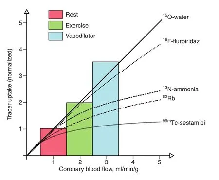
Figure1 Positron Emission Tomography (PET) and Single-Photon Emission Computed Tomography (SPECT)Uptake as a Function of Coronary Blood Flow.
An interesting conundrum arising from the use of18F-flurpiridaz is its compatibility with exercise stress testing and ability to provide absolute flow quantification–but not both simultaneously.The time between the end of exercise and the start of scanning prevents reliable measurement of absolute MBF.The inability to perform both exercise stress testing and absolute flow assessment forces a choice between two important diagnostic and prognostic parameters.Exercise stress testing provides exercise capacity (the most powerful stress prognostic marker), hemodynamic response, and potential for symptom reproduction while increasing the diagnostic accuracy of the ST analysis.Absolute flow measurement provides rich data from resting MBF, hyperemic MBF, and MPR.Rather than omitting one or another important data set, we propose a hybrid protocol in which exercise stress testing is performed to gather stress ECG, hemodynamic,and symptom data followed by on-table vasodilator stress testing to allow absolute blood flow quantifi-cation ( Figure2 ).
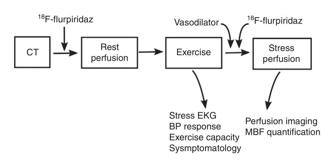
Figure2 Proposed hybrid exercise-vasodilator stress algorithm which, when paired with the use of 18 F-flurpiridaz,could enable the collection of both exercise stress information and myocardial blood flow (MBF) with one clinical study.BP, blood pressure; CT, computed tomography; EKG,electrocardiography.
Plaque Characterization
The utility of cardiac PET in CAD extends beyond the evaluation of myocardial ischemia.Cardiac PET with molecular imaging tracers allows functional interrogation of vascular biology and characterization of atherosclerotic plaques.
18 F-Fluorodeoxyglucose
18F-Fluorodeoxyglucose (18F-FDG) activity correlates with arterial inflammation due to uptake by metabolically active macrophages [18].However,examination of the coronary arteries for inflammation with18F-FDG is hampered by the proximity of coronary arteries to the myocardium, which may have concurrent18F-FDG uptake.While introduction of a low-carbohydrate, high-fat diet may be helpful in suppressing myocardial18F-FDG uptake [19],distinguishing coronary from patchy myocardial uptake remains difficult, even with effective myocardial suppression [20].Underscoring these technical concerns,18F-FDG has demonstrated a low diagnostic performance, with an inability to identify culprit coronary lesions in nearly half of a group of patients presenting with an acute coronary syndrome [21].
Sodium 18 F-Fluoride
The difficulties with using18F-FDG for noninvasive plaque characterization have led to the pursuit of alternative tracers.Sodium18F-fluoride (18F-NaF)has been identified as a marker of valvular and vascular calcification, including the microcalcifications present in the necrotic core of vulnerable, thin-cap flbroatheromas ( Figure3 ) [22].In an early clinical study,18F-NaF outperformed18F-FDG in localizing coronary signal and in distinguishing culprit from nonculprit coronary plaques [20].18F-NaF is the subject of an ongoing, prospective outcomes study to validate its utility in predicting high-risk plaques(NCT02278211).However, the question of the ideal PET radiotracer to characterize vascular plaques is not yet settled.
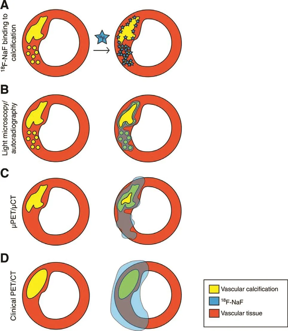
Figure3 18 F-NaF for Noninvasive Characterization of Coronary Plaques.
68 Ga-(1,4,7,10-Tetraazacyclododecane-N, N′, N″ , N″′ -tetraacetic acid)- D-Phe 1,Tyr 3-octreotate
Another promising radiotracer for the evaluation of plaque inflammation has emerged in68Ga-(1,4,7,10-tetraazacyclododecane-N,N′,N″,N″ ′-tetraacetic acid)- DPhe1,Tyr3-octreotate (68Ga-DOTATATE).68Ga-DOTA TATE binds with high affinity to type 2 somato statin receptors, whose expression is upregulated on the surface of activated tissue macrophages [23].In an early clinical study,68Ga-DOTATATE demonstrated fairly good performance in the identification of culprit plaques after acute coronary syndrome, with a sensitivity of 87.5%, specificity of 78.4%, and receiver operating characteristic area under of the curve for diagnostic accuracy of 0.86 [24].Furthermore,68Ga-DOTATATE signal was higher in nonculprit plaques with high-risk morphologic features (spotty calcification, low attenuation, or positive remodeling).In addition to clinical trials, an important next step for plaque-focused radiotracers would include prospective cohort studies, with comparison against other risk stratifiers such as coronary calcium scoring to evaluate reclassification potential.
Heart Failure
Risk Stratification by Viability Assessment
Cardiac PET plays a major role in the evaluation and management of heart failure.Both SPECT and PET MPI are well-established methods for detecting ischemia as a cause of heart failure [25].However,PET can provide additional risk stratification for patients with heart failure through absolute flow quantification and metabolic imaging.Ghannam et al.[26]examined a cohort of 159 patients with implantable cardioverter defibrillators (ICDs) who underwent82Rb PET MPI.They found that PET stress MBF, but not summed rest or stress scores, predicted the risk of ventricular tachycardia or ventricular flbrillation.However, it is the combination of perfusion information with metabolic imaging that brings cardiac PET to the forefront of heart failure evaluation.Metabolic assessment by18F-FDG PET is widely regarded as the gold standard for viability over other modalities that draw conclusions on the basis of resting perfusion uptake [25].The presence of viable myocardium on PET predicts functional improvement after revascularization [27].Additionally, there is evidence that PET-guided care may improve outcomes.In the PARR-2 randomized trial of PET viability-guided care versus standard care for evaluation of severely reduced left ventricular ejection fraction (LVEF),the subgroup of patients in the PET arm whose care adhered to the PET-guided revascularization recommendations experienced a lower rate of composite adverse events [28].
Risk Stratification by Sympathetic Denervation: 11 C-Hydroxyephedrine
Risk stratification is increasingly important for nonischemic heart failure in addition to CAD.Recent trial data indicate that LVEF of 35% or less is an insufficient marker for predicting the risk of sudden cardiac death in patients with nonischemic cardiomyopathy, underscoring the need for additional prognostic markers in this large patient population[29].While there is likely a role for late gadolinium enhancement on cardiac magnetic resonance imaging (MRI), accumulating data suggest that PET has predictive power regarding sudden cardiac death[30].The use of11C-hydroxyephedrine (11C-HED),an analog of norepinephrine, has allowed noninvasive evaluation of cardiac sympathetic innervation.11C-HED binds selectively in the heart to presynaptic sympathetic nerve terminals [31].Regions of cardiac denervation and dysautonomia demonstrate reliably reduced11C-HED uptake compared with controls [32].In a prospective trial of patients with ischemic heart failure with reduced ejection fraction, sudden cardiac arrest was predicted by the amount of sympathetically denervated myocardium but not LVEF or infarct size [33].
Delineation of Inflammatory/Infiltrative Cardiomyopathies
Cardiac Amyloid: 11 C-Pittsburgh compound B, 18 F-Florbetapir,18 F-Florbetaben
Cardiac PET has shown promise in the delineation of nonischemic cardiomyopathy as well.Among nonischemic cardiomyopathies, the identification of cardiac amyloidosis is particularly important because of the aggressive course of the disease,difficulties in noninvasive evaluation, and new effective treatments [34–36].Moreover, traditional diagnostic methods, such as electrocardiography and two-dimensional echocardiography, are inadequately sensitive [37, 38].By contrast, cardiac PET, like99mTc SPECT, offers the ability to directly diagnose amyloid deposits.There is early evidence that hybrid PET and cardiac MRI allows distinction between transthyretin and light-chain amyloid variants (see the section entitled “ Hybrid Imaging”).
Modifications of the amyloid-binding dye thioflavin T led to the identification of11C-Pittsburgh compound B (11C-PIB), structurallyN-methyl-[11C]2-(4′-methylaminophenyl)-6-hydroxybenzothiazole.Although it was originally developed for detection of brain amyloid deposition in patients with Alzheimer’ s disease, it became apparent that11CPIB lent itself to the identification of cardiac amyloid involvement as well [39].11C-PIB myocardial PET appears to identify myocardium infiltrated by both light-chain and transthyretin amyloid with great specificity [40].Other radiotracers have also shown promise in identifying cardiac amyloid flbrils:18F-florbetapir and18F-florbetaben [41, 42].Like11C-PIB,18F-florbetapir and18F-florbetaben were also originally developed to bind brain amyloid.
In the aggregate, cardiac PET for cardiac amyloid appears to have a remarkably robust diagnostic performance.A recent meta-analysis of cardiac PET for the diagnosis of amyloid, aggregating data on 98 participants across six studies (four studies of11C-PIB, one study of18F-florbetapir, and one study of18F-florbetaben), estimated a pooled sensitivity of 0.95 and specificity of 0.98 [43].While promising,much work needs to be done, including analysis in larger groups with broader cardiac disease profiles and development of standardized semiquantitative parameters and reference ranges, before PET can be adopted into clinical practice for patients with suspected cardiac amyloidosis.Additionally, the integration of molecular imaging with new amyloid-targeted therapeutics remains a largely unanswered question.For example, in the pivotal clinical trial of tafamidis for patients with transthyretin amyloid cardiomyopathy, much of the benefit seemed clustered in the subgroup of patients with less severe heart failure according to symptomatic classification [35].These results suggest there may be different disease stages with differing degrees of treatment susceptibility.In such a situation, it would be reasonable to expect targeted molecular imaging with PET to provide flner stratification of disease profiles to guide treatment selection.
Cardiac Sarcoid: 18 F-FDG
Its central role in cardiac sarcoidosis is a chief example of how cardiac PET can be used to diagnose and monitor nonischemic cardiomyopathies [44].Performed in the setting of a low-carbohydrate, highfat diet to suppress physiologic myocardial uptake,18F-FDG PET can identify abnormal glucose uptake indicative of inflammation within the myocardium;in the appropriate clinical scenario, this is consistent with active cardiac sarcoidosis [45].When paired with MPI using a conventional perfusion tracer such as13NH3or82Rb to assess myocardial scar from ongoing severe active inflammation or prior disease,cardiac PET offers the ability to provide a comprehensive assessment of the extent of cardiac sarcoid involvement.
Already a cornerstone of sarcoid diagnosis,18FFDG PET continues to undergo technical improvements to increase diagnostic accuracy.One ongoing development is the addition of18F-FDG activity quantification to visual assessment.A common semiquantitative technique used in PET uses standardized uptake values (SUVs), which are calculated by division of the tracer uptake (corrected for decay) by the weight-based dose of the tracer [46].Common research approaches have involved measuring the maximum SUV and mean SUV throughout the myocardium [47, 48].Another quantitative approach has been developed on the basis of oncologic PET,wherein the aggregate volume of18F-FDG-positive myocardium is quantified [49].This volume, calledcardiac metabolic volume(CMV), is analogous to metabolic tumor volume in oncology.Calculation of CMV allows determination of the volume-intensity product, dubbedcardiac metabolic activity(CMA),which is analogous to total lesion glycolysis in oncology.In a small study comparing CMA with CMV and maximum SUV, CMA had the best diagnostic performance for18F-FDG PET and showed the potential for reclassification [49].As with all new quantitative measures, the thresholds, methods of automation, and processing techniques need to be optimized and standardized.
Infection
The ability of18F-FDG PET to visualize inflammatory metabolic signal has been used to identify infections involving vascular prostheses [50].More recently, this technique has been applied in infection imaging related to the heart as well.
Pacemakers and ICDs
It appears that there is increased18F-FDG uptake in the region of infected ICDs and permanent transvenous pacemaker cans and leads [51].A small prospective study suggests favorable test characteristics for pocket infections, with a sensitivity of 86.7% and specificity of 100% [52].The performance of cardiac PET for device-related endocarditis was much less robust, with a sensitivity of 30.8%and specificity of 62.5%; there were a large number of false negatives (9 of 13 participants).Previous antimicrobial treatment was the presumed explanation for eight of the nine false negatives.The ideal role for18F-FDG PET in diagnosing pocket infections has yet to be fully determined.Accordingly,current recommendations by the Heart Rhythm Society provide a class IIb indication for18F-FDG PET to facilitate the diagnosis of device infection when it cannot be confirmed by other methods [53].Future studies should provide comparisons between relevant patient groups (e.g., antibiotic na ï ve) and address cost-effectiveness.
Left Ventricular Assist Devices
Similarly to the uptake seen with pacemakers and ICDs, other intracardiac and device-related infections appear to show increased18F-FDG signal on PET.For instance, the identification of prosthesis infection can be critical in the case of left ventricular assist devices (LVADs), where the patient is fully dependent on the device for survival and extraction is risky and fraught with complications.Given this importance, it is promising that a study of 35 LVAD patients with suspected device infection(a reasonable population size given the rate nature of this condition) showed that18F-FDG PET added substantially to the diagnostic workup.Among 35 individuals,18F-FDG PET identified potential infection in 28 patients, compared with only four patients on the basis of computed tomography (CT)results alone [54].Additionally, PET was able to establish whether the pattern of infection was limited to peripheral components or involved the central apparatus.Cardiac PET in LVAD patients was found to be prognostically important.None of the patients with no evidence of infection on PET died over a mean follow-up of 23 months, compared with 50% of patients with evidence of infection(P= 0.03).
Infective Endocarditis
Application of infection imaging in infective endocarditis is challenging because of the intricate nature of the criterion-based diagnosis.One creative approach has been to include18F-FDG uptake at the site of a prosthetic valve as a major criterion in the Duke infective endocarditis classification scheme.In a prospective study, inclusion of18F-FDG PET in the updated scoring system increased the sensitivity to 97% from 73% without PET in the classification scheme [55].In a meta-analysis of 13 studies that included prosthetic and native-valve infective endocarditis, the pooled sensitivity and specificity of hybrid PET and CT for infective endocarditis were 76.8 and 77.9%, respectively [56].Concerns persist about limitations in the specificity of this technique, however, as increased18F-FDG signal can result from postoperative inflammatory changes after recent surgery, vasculitis, active thrombi, cardiac tumors/metastasis, and foreign body reactions[57].A reasonable next step is a larger-scale trial of patients without postoperative confounders and cost-effectiveness analysis.
Valvular Disease
In the realm of valvular heart disease, cardiac PET has been applied beyond the assessment of infective endocarditis.The properties that have allowed use of18F-NaF as a marker of calcification within atherosclerotic vascular plaques have also led to its use in the evaluation of calcific heart disease.In a prospective study of 81 participants with echocardiographically diagnosed aortic stenosis, 91% demonstrated increased18F-NaF uptake.Tracer uptake correlated with the severity of aortic stenosis [46].In a 1-year follow-up study with hybrid PET and CT, isolated18F-NaF uptake in a segment predicted the subsequent development of calcification [58].This development suggests that18F-NaF uptake can track the development of valvular processes before development of irretrievable damage, which has implications for disease monitoring and evaluation of preventive therapies.Accordingly,18F-NaF uptake as observed by PET is being used to track progression of aortic stenosis in an ongoing clinical trial of bisphosphonates and inhibitors of receptor activator of nuclear factor κ B ligand (NCT02132026).It follows from these data that18F-NaF could also potentially be applied in the evaluation of mitral annular calcification, an understudied and increasingly frequent cause of mitral valve disease [59].
Technical Advances
Hybrid Imaging
A complete discussion of the uses of hybrid PET and anatomic imaging is beyond the scope of this work,but hybrid imaging has become a critical component of many of the techniques discussed, including MPI, plaque characterization, and infection imaging.Combination of noncontrast CT with PET MPI allows attenuation correction and enhanced risk stratification with coronary artery calcium scoring [60].The addition of PET MPI to coronary CT angiography markedly increases diagnostic specificity for obstructive CAD (from 66 to 93%,according to a recent meta-analysis) [61].Likewise,cardiac MRI and PET provide synergistic information.By colocalizing areas of18F-FDG uptake by PET with subepicardial late gadolinium enhancement and T2 mapping by cardiac MRI, hybrid PET and MRI can provide refinements in the evaluation of myocarditis [62].Whether this approach can improve on the performance of established diagnostic criteria for myocarditis remains to be seen [63].The addition of MRI to PET perfusion and metabolic imaging can provide more refined cardiac sarcoid staging than with either modality alone,but the relevance of this for functional outcomes needs to be further defined [64].Finally, a recent study has suggested that hybrid PET and MRI may allow distinction of cardiac amyloid subtypes.The flndings suggest that colocalized18F-NaF and late gadolinium enhancement is observed in myocardial regions infiltrated by transthyretin amyloid but not light-chain amyloid [65].
Machine Learning
Hybrid imaging, with its roots in combining and integrating different data sets, is an area ripe for benefit from machine learning.Typically applied to numerical data sets, machine learning is positioned to aid in processing the inherent complexity of hybrid imaging techniques, as well as demographic and clinical information within electronic health records [66].Further, machine and deep learning are expected by some investigators to allow fully automated quantification and image interpretation[67].This has implications for artifact identification, workflow streamlining and efficiency, and minimizing interreader variability.
Total-Body PET
A pending innovation with far-reaching implications for myocardial PET assessment is the current effort to develop a total-body PET scanner, consisting of detector rings that span the length of the body.Total-body PET has the potential to provide dramatic technical improvements by allowing signal detection from regions outside the traditional scanner fleld of view.Further, it allows collection of substantially more signal from a region of interest such as the heart.In current PET scanners, only 3– 5% of available signal from the region of interest intercepts with the limited detector rings [68].Use of total-body PET is expected to yield a roughly 40-fold increase in the sensitivity for whole-body imaging, but less for the heart itself.This can enable proportionate reductions in the required radiation doses, which can in turn broaden application of cardiac PET techniques to younger populations and provide greater tolerance for serial scanning when necessary.The pending development of the flrst total-body PET scanner for human use is highly anticipated.
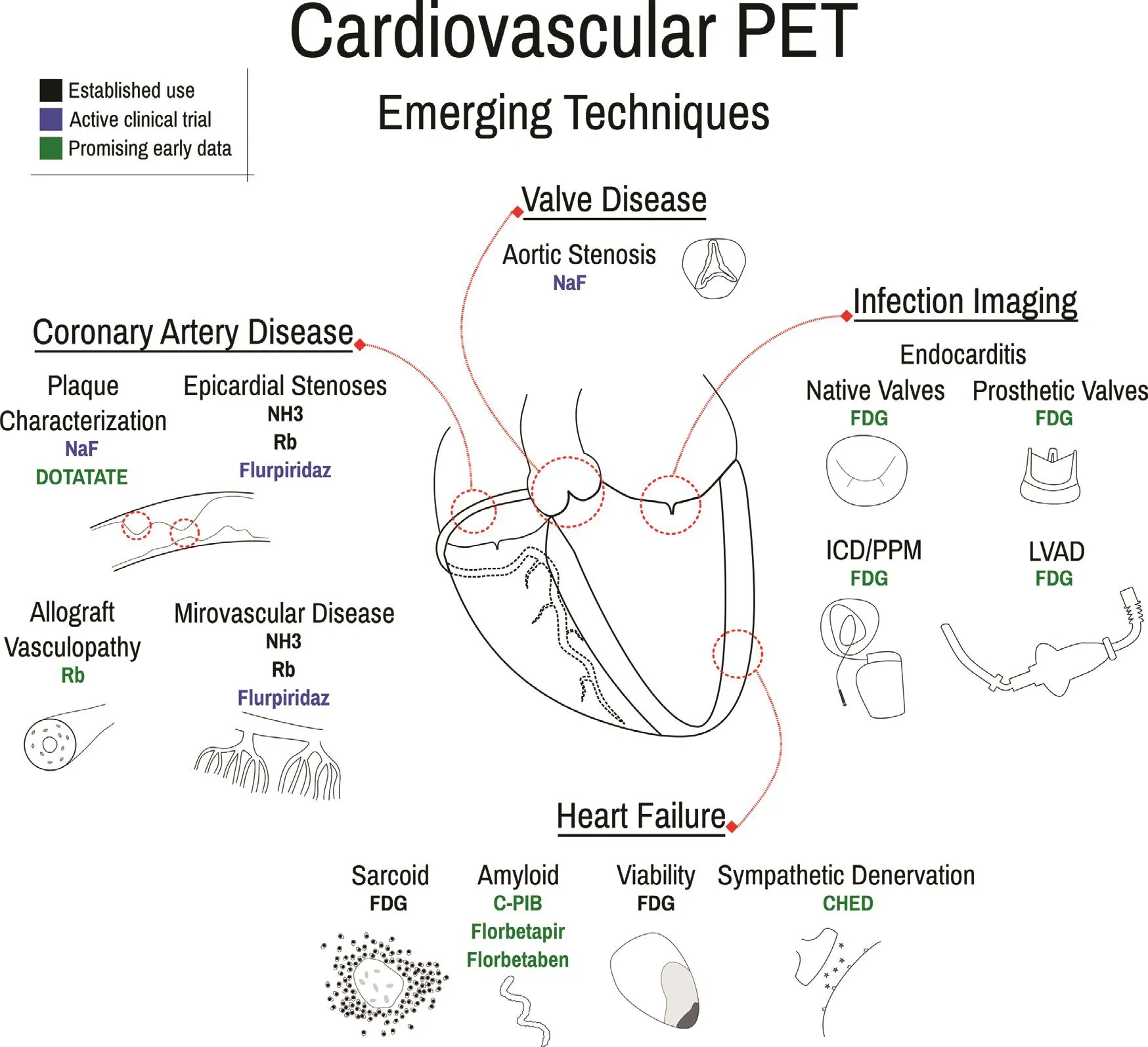
Figure4 Emerging Techniques of Cardiovascular Positron Emission Tomography (PET), Including for Valve Disease Evaluation, Coronary Artery Disease Characterization, Heart Failure Assessment, and Infection Imaging.
Conclusion
From its origin in diagnosing coronary artery stenoses, cardiovascular PET has grown into a staple of contemporary cardiovascular care, with impending advances touching all corners of the fleld, including new tracers and absolute flow assessment in CAD and CMD and characterization of lesions in vascular plaques, cardiac valves, the myocardium,and implanted devices ( Figure4 ).In addition to its breadth of application, cardiovascular PET offers critical diagnostic information, informs prognosis,and has the potential to impact care.Given these strengths and ongoing advances, the present use of cardiovascular PET is bright and its future potential is enormous.
Conflict of Interest
Austin A.Robinson: Work is supported by NIH T32 EB003841.No other conflicts of interest.Jamieson M.Bourque: Work is supported by NIH 5K23HL119620-02; Dr.Bourque does consulting for Pfizer.No other conflicts of interest.
 Cardiovascular Innovations and Applications2019年2期
- Cardiovascular Innovations and Applications的其它文章
- Quality Improvement in Cardiovascular Imaging
- Imaging Beyond the Angiogram in Women with Suspected Myocardial Infarction and No Obstructive Coronary Artery Disease
- Practical Clinical Application of Cardiac Computed Tomography– Derived Fractional Flow Reserve
- Evaluation of the Patient with Incidental Left Ventricular Hypertrophy on Echocardiography
- Using 3D-Printed Models to Advance Clinical Care
- Differential Impact of Appropriate Use Criteria on the Association between Age and Abnormal Stress Myocardial Perfusion SPECT