CHI3L1通过激活TGF-β信号通路促进肝细胞肝癌进展及分子机制
李晓文 王后红 黄海 刘殿雷


[摘要] 目的 观察CHI3L1对肝细胞肝癌Huh7增殖、迁移、侵袭的影响及其分子机制。 方法 通過脂质体转染,观察CHI3L1对Huh7增殖、迁移、侵袭的影响。通过染色质免疫共沉淀、Western blot观察CHI3L1对TGF-β信号通路的作用机制。 结果 转染CHI3L1后,Huh7细胞增殖率显著高于空载组及空白对照组,差异有统计学意义(P<0.05)。转染CHI3L1后,迁移、侵袭至下层细胞数均显著多于空载组、空白对照组,差异有统计学意义(P<0.05)。转染CHI3L1后,TGF-β刺激后形成的Smad2/3/4复合物增加,CHI3L1促进TGF-β诱导下Smads复合物的形成。转染CHI3L1后的Huh7细胞对TGF-β刺激上调p53非依赖的细胞周期抑制因子p21、p15,下调c-myc,抑制Rb磷酸化,上调NOX4;TGF-β抑制剂LY364947可抑制此诱导效应。 结论 CHI3L1可促进肝细胞肝癌增殖、迁移、侵袭,这种作用可能通过激活TGF-β信号通路实现。
[关键词] CHI3L1;TGF-β;增殖;迁移;侵袭
[中图分类号] R735.7 [文献标识码] A [文章编号] 1673-9701(2019)34-0031-04
Study on CHI3L1 promoting the progression of hepatocellular carcinoma by activating TGF-β signaling pathway and the molecular mechanisms
LI Xiaowen1 WANG Houhong2 HUANG Hai1 LIU Dianlei1
1.Department of First Surgery, Hangzhou Hospital of Traditional Chinese Medicine, Hangzhou 310006, China; 2.Department of General Surgery, the Second Affiliated Hospital of Zhejiang University, School of Medicine, Hangzhou 310006, China
[Abstract] Objective To observe the effect of CHI3L1 on the proliferation, migration and invasion of hepatocellular carcinoma Huh7 and its molecular mechanism. Methods The effect of CHI3L1 on the proliferation, migration and invasion of Huh7 were observed by lipofection. The mechanism of CHI3L1 on TGF-β signaling pathway was observed by chromatin immunoprecipitation and Western blot. Results After transfection of CHI3L1, the proliferation rate of Huh7 cells was significantly higher than that of the empty vector group and the blank control group, and the difference was statistically significant(P<0.05). After transfection of CHI3L1, the number of migration and invasion to the lower layer was significantly higher than that of the empty vector group and the blank control group, and the difference was statistically significant (P<0.05). After transfection of CHI3L1, the Smad2/3/4 complex formed by TGF-β stimulation increased, and CHI3L1 promoted the formation of Smads complex induced by TGF-β. Huh7 cells transfected with CHI3L1 up-regulated TGF-β-induced p53-independent cell cycle inhibitors p21 and p15, down-regulated c-myc, inhibited Rb phosphorylation, up-regulated NOX4, and TGF-β inhibitor LY364947 inhibited this induction effect. Conclusion CHI3L1 can promote the proliferation, migration and invasion of hepatocellular carcinoma, which may be achieved by activating the TGF-β signaling pathway.
[Key words] CHI3L1; TGF-β; Proliferation; Migration; Invasion
CHI3L1即殼多糖酶-3样蛋白1,又称YKL-40,为最初在人骨肉瘤细胞系体外培养中发现的蛋白,定位于人1号染色体q32[1];CHI3L1属糖基水解酶18家族,为结缔组织细胞重要的生长因子,在血管内皮细胞的迁徙移动中具有重要作用,参与细胞增殖、分化、组织重塑[2]。CHI3L1最初认为与炎症性疾病存在密切关系,在炎症性肠病、支气管哮喘中均具有一定的生物学作用[3,4]。近年来研究发现[5,6],CHI3L1在肿瘤中呈高表达,为人类多种恶性肿瘤的不良预后因素。Zhou Y等[7]报道CHI3L1在肝细胞肝癌中存在表达,但CHI3L1在肝细胞肝癌的发生、发展中的具体机制研究尚少。本研究通过构建CHI3L1表达载体,观察CHI3L1对肝细胞肝癌(HCC)细胞增殖、迁移、侵袭的影响,并探讨其分子机制,为临床分子靶向治疗提供新的理论指导,现报道如下。
1 材料与方法
1.1 细胞标本
肝细胞肝癌细胞Huh7购自上海中科院细胞库。以10%胎牛血清,100 U/mL青霉素+100 μg/mL链霉素的DMEM培养液,于37℃、5%CO2细胞培养箱中培养,细胞生长融合至80%~90%时传代。
1.2 主要试剂与仪器
Trizol试剂(Thermo Fisher公司),Titan One Tube RT-PCR Kit试剂盒(sigma-aldrich公司),PowerUpTM SYBRTM Green试剂盒(Thermo Fisher公司),ECL试剂盒(Biovision公司),PVDF膜(Millipore公司),HRP-羊抗兔IgG(Abcam公司,ab6721),兔抗人CHI3L1多克隆抗体(Abcam公司,ab77528),兔抗人TGF-β1多克隆抗体(Abcam公司,ab64715),兔抗β-actin多克隆抗体(Abcam公司,ab151526),TGF-β1受体抑制剂LY364947(MedCemExpress公司);荧光定量PCR仪(Bio-Rad CFX96 PCR System),凝胶电泳图像扫描分析仪(GE Image scanner Ⅲ),化学发光分析仪(美国Thermo Luminoskan Ascent),酶标仪(美国Thermo Multiskan FC)。
1.3 研究方法
1.3.1 Western-blot 离心管柱及接收套管置于冰上预冷,将预冷的PBS加入培养板,清洗贴壁细胞,吸去上清。6孔板每孔加入200 μL单混有蛋白酶抑制剂的去污剂裂解液裂解组织,置于冰上操作。裂解30 min后,将裂解液转移至1.5 mL EP管中,4℃ 12000 rpm/min离心5 min,取上清分装于0.5 mL离心管,-20℃保存。以BCA法检测蛋白浓度。将蛋白样品加10×上样缓冲液,水浴煮沸5 min。以每孔30 μg蛋白的上样量行聚丙烯酰胺凝胶电泳,恒压80 V,溴酚蓝进入分离胶后调整电压为120 V继续电泳,至溴酚蓝迁移至分离胶底部,终止电泳。以4℃,300 mA恒流,70 min,将蛋白转移至PVDF转膜上,丽春红染色观察转膜效果。5%BSA封闭PVDF膜2 h,洗涤后,滴加兔抗XBP-1溶液,4℃过夜孵育。第2天加入TBST洗涤去除多余一抗,与HRP标记的二抗室温孵育2 h。TBST清洗后,加入ECL化学发光底物,在化学发光仪上显影,拍照保存。
1.3.2 CHI3L1对Huh7细胞增殖的影响 pcDNA3.1-CHI3L1质粒构建参考文献[8]。将处于对数生长期的人肝细胞肝癌细胞Huh7,以胰酶消化后,用RPMI-1640培养基调整细胞浓度至1×10⁵个/mL,每孔100 μL接种至96孔板。将细胞分为3组,pcDNA3.1空载组、pcDNA3.1-CHI3L1组、未加质粒的空白对照组。每组在培养的0 、24 、48 h加入CCK8溶液,每孔20 μL,4 h后终止培养。以酶标仪在450 nm处检测各孔吸光度值。
1.3.3 Transwell试验检测迁移、侵袭能力 迁移试验:以0.2%BSA的无血清RPMI-1640培养基重悬制备单细胞悬液,细胞数为5×10⁵个/mL,取200 μL铺于8 μm Transwell小室上层,加500 μL含10%胎牛血清的RPMI-1640培养基于24孔板底部,培养24 h后取走Transwell板,计数穿孔细胞数。侵袭试验:将300 μL无血清RPMI-1640培养基加入8 μm Transwell小室上层,室温下水化基质胶20 min,弃去剩余液体。以0.2%BSA的无血清RPMI-1640培养基配制浓度5×10⁵个/mL的单细胞悬液,其余步骤同迁移试验。
1.3.4 染色质免疫共沉淀 取1×10⁷细胞,弃去培养基,以PBS洗涤3次。加入预冷的RIPA裂解液1 mL,4℃裂解30 min。细胞刮子收集裂解产物,转入1.5 mL EP管中。14000 g离心15 min,收集上清。BCA法进行蛋白定量。取500 μg蛋白,加共沉淀抗体,4℃过夜。孵育后在蛋白中加入30 μL proteinA/G,4℃摇床孵育4 h。1000 g离心5 min,弃去上清。取800 μL预冷的RIPA裂解液洗涤琼脂糖珠复合物,1000 g离心5 min,重复3次。以40 μL,2×蛋白上样缓冲液重悬珠子,沸水浴5 min。后续步骤同Western blot。
1.4 统计学方法
统计学分析采用SPSS22.0统计学软件,计量资料以均数±标准差(x±s)描述,组间比较采用单因素方差分析,两两比较采用t检验,P<0.05为差异有统计学意义。
2 结果
2.1 CHI3L1对Huh7细胞增殖的影响
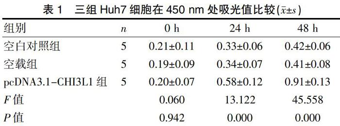
转染CHI3L1 24 h后,pcDNA3.1-CHI3L1组Huh7细胞增殖率显著高于空载组及空白对照组,差异有统计学意义(P<0.05)。见表1。
2.2 CHI3L1对Huh7细胞迁移、侵袭的影响
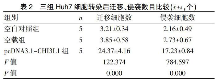
转染CHI3L1后,pcDNA3.1-CHI3L1组迁移、侵袭至下层细胞数均显著多于空载组、空白对照组,差异有统计学意义(P<0.05)。见表2。
2.3 CHI3L1促进TGF-β诱导下Smads复合物的形成
转染CHI3L1后,TGF-β刺激后形成的Smad2/3/4复合物增加,提示CHI3L1促进TGF-β诱导下Smads复合物的形成。见图1。
2.4 CHI3L1激活TGF-β信号通路的分子机制
转染CHI3L1后的Huh7细胞对TGF-β刺激上调p53非依赖的细胞周期抑制因子p21、p15,下调c-myc,抑制Rb的磷酸化,上调NOX4;TGF-β抑制剂LY364947可抑制此诱导效应。见图2。
3讨论
原发性肝细胞肝癌为恶性程度较高的肿瘤之一,患者早期临床症状不明显,临床检查发现时,多数患者已失去彻底根治的机会[9]。肝癌患者术后高复发率、高病灶转移率为影响肝癌患者预后的主要因素,使肝癌的总体疗效仍不理想,成为严重危害人类健康的重要疾病之一[10]。探讨肝癌复发、转移潜在的分子机制成为肝癌治疗的关键所在。
CHI3L1即壳多糖酶-3样蛋白1,为哺乳动物壳多糖酶家族中的成员,在血管内皮细胞的迁徙移动中发挥重要的作用[11]。由于肿瘤细胞的无限增殖为肿瘤生长、迁移、侵袭的前提条件,肿瘤转移至远处器官后,最终可导致多器官受累,加速患者死亡[12]。研究显示[13]CHI3L1在肿瘤的转移复发中起重要作用;Chen Y等[14]研究发现巨噬细胞通过分泌CHI3L1促进胃癌、乳腺癌的转移;Ngernyuang N等[5]研究发现CHI3L1可促进宫颈癌新生血管的形成,靶向CHI3L1可能是一种降低宫颈癌转移率的新型治疗手段[15]。由此猜想CHI3L1在肝癌细胞中也有类似作用,本研究通过脂质体法构建过表达CHI3L1的Huh7细胞,发现CHI3L1可促进Huh7细胞增殖、迁移、侵袭,与预期一致。
在肝细胞肝癌发生、发展中,多种信号通路的激活、异常表达具有重要的作用,这些信号通路包括:VEGFR、TGF-β、MAPK、EGFR等[16,17]。TGF-β为诱导EMT转化的生长因子,在肝细胞肝癌中可通过上调结缔组织生长因子表达水平,促进SMMC-7721细胞的迁移及侵袭[18]。在肝癌中TGF-β通路的功能较复杂,在高分化、侵袭力低的肝癌细胞中TGF-β通路激活早期应答基因,表现出抑癌效应。在低分化、侵袭力强的肝癌细胞中TGF-β激活晚期应答基因,表现出促癌效应,且由于TGF-β诱导的促癌行为主要通过TGF-β调控的非Smad通路激活引起,在TGF-β刺激下,CHI3L1可促进Smads复合物形成及TGF-β信号通路激活。本研究对过表达CHI3L1的Huh7细胞给予TGF-β刺激后,发现Smads复合物合成增加,提示CHI3L1可促进TGF-β诱导下Smads复合物的形成,从而促进Huh7细胞增殖、迁移和侵袭。p21、p15是常见的抑癌基因,其编码蛋白可与周期素依赖性蛋白激酶4结合,抑制Rb磷酸化,从而抑制细胞DNA的合成,最终下调细胞增殖活性;c-myc是永生性细胞的原癌基因,石永英等[19]研究表明TGF-β可抑制c-myc活化,并促进Smads复合物的形成,启动p21、p15的转录,抑制细胞增殖。NOX4是体内产生氧自由基的主要酶类,Ma Y等[20]研究显示TGF-β可能介导NOX4参与人脐静脉内皮细胞凋亡过程。本研究对过表达CHI3L1的Huh7细胞给予TGF-β刺激,发现p21、p15及NOX4表达显著上调,c-myc、抑制Rb磷酸化显著下调,且添加TGF-β阻断劑LY364947后可抑制上述诱导效应,提示CHI3L1可激活TGF-β信号通路,并影响下游分子表达,从而促进Huh7细胞增殖,并抑制Huh7细胞的凋亡。
综上所述,CHI3L1可促进肝细胞肝癌增殖、迁移、侵袭,具体分子途径为:①促进Smads复合物的形成,促进Huh7细胞增殖、迁移和侵袭;②激活TGF-β下游通路,促进Huh7细胞增殖、抑制其凋亡。后期还可继续应用动物模型深入研究。
[参考文献]
[1] Rathcke CN,Holmkvist J,Husmoen L LN,et al. Association of polymorphisms of the CHI3L1 gene with asthma and atopy:A populations-based study of 6514 Danish adults[J]. Plos One,2009, 4(7):e6106.
[2] Dai QH,Gong DK. Association of the polymorphisms and plasma level of CHI3L1 with Alzheimer's disease in the Chinese Han population:A case-control study[J]. Neuropsychobiology,2019,77(1):29-37.
[3] Buisson A,Vazeille E,Minetquinard R,et al. Faecal chitinase 3-like 1 is a reliable marker as accurate as faecal calprotectin in detecting endoscopic activity in adult patients with inflammatory bowel diseases[J]. Aliment Pharmacol Ther,2016,43(10):1069-1079.
[4] 李琎,曲书强. CHI3 L1基因与哮喘及其严重程度的关系[J]. 国际遗传学杂志,2016,39(5):282-285.
[5] Ngernyuang N,Shao R,Suwannarurk K,et al. Chitinase 3 like 1(CHI3L1) promotes vasculogenic mimicry formation in cervical cancer[J]. Pathology,2018,50(3):293-297.
[6] 谢而付,蒋理,凌芸,等. 血清CEA、CA19-9和CHI3L1在胰腺癌患者中的诊断价值比较[J]. 实用医学杂志, 2018,34(2):309-311.
[7] Zhou Y,He CH,Yang DS,et al. Galectin-3 interacts with the CHI3L1 axis and contributes to hermansky-pudlak syndrome lung disease[J]. J Immunol,2018,200(6):2140-2153.
[8] Libreros S,Garcia-Areas R,Iragavarapu-Charyulu V. CHI3L1 plays a role in cancer through enhanced production of pro-inflammatory/pro-tumorigenic and angiogenic factors[J]. Immunologic Research,2013,57(1-3):99-105.
[9] Dezman K,Korosec P,Rupnik H,et al. SPINK5 is associated with early-onset and CHI3L1 with late-onset atopic dermatitis[J]. Int J Immunogenet,2017,44(5):212-218.
[10] Jingjing Z,Nan Z,Wei W,et al. MicroRNA-24 Modulates Staphylococcus aureus-Induced Macrophage Polarization by Suppressing CHI3L1[J]. Inflammation,2017, 40(3):995-1005.
[11] Luo D,Chen H,Lu P,et al. CHI3L1 overexpression is associated with metastasis and is an indicator of poor prognosis in papillary thyroid carcinoma[J]. Cancer Biomark,2017,18(3):273-284.
[12] Usemann J,Frey U,Mack I,et al. CHI3L1 polymorphisms,cord blood YKL-40 levels and later asthma development[J]. BMC Pulm Med,2016,16(1):81-90.
[13] Libreros S,Garciaareas R,Shibata Y,et al. Induction of proinflammatory mediators by CHI3L1 is reduced by chitin treatment:Decreased tumor metastasis in a breast cancer model[J]. International Journal of Cancer,2012, 131(2):377-386.
[14] Chen Y,Zhang S,Wang Q,et al. Tumor-recruited M2 macrophages promote gastric and breast cancer metastasis via M2 macrophage-secreted CHI3L1 protein[J]. Journal of Hematology & Oncology,2017,10(1):36-48.
[15] 窦娴静.宫颈癌组织中CHI3L1蛋白表達及其临床意义[J].四川解剖杂志,2018,26(2):33-35.
[16] Szychlinska MA,Trovato FM,Di Rosa M,et al. Co-expression and Co-localization of cartilage glycoproteins CHI3L1 and lubricin in osteoarthritic cartilage: Morphological,immunohistochemical and gene expression profiles[J]. Int J Mol Sci,2016,17(3):359-387.
[17] Abd EA,Sadik NA,Shaker OG,et al. Are SMAD7 rs4939827 and CHI3L1 rs4950928 polymorphisms associated with colorectal cancer in Egyptian patients?[J]. Tumour Biol,2016,37(7):9387-9397.
[18] Kainulainen H. CHI3L1-a novel myokine[J]. Acta Physiol(Oxf),2016,216(3):260-261.
[19] 石永英,卢俊江,陈广原,等. p15与c-Myc在TGF-β1诱导的大鼠心肌细胞肥大中的表达及与TGF/Smad信号通路的关系[J]. 中国动脉硬化杂志,2018,26(3):245-252.
[20] Ma Y,Li W,Yin Y,et al. AST IV inhibits H2O2-induced human umbilical vein endothelial cell apoptosis by suppressing Nox4 expression through the TGF-β1/Smad2 pathway[J].International Journal of Molecular Medicine,2015,35(6):1667-1674.
(收稿日期:2019-03-18)

