miR-30c-1-3p在人结直肠癌组织中的表达及临床意义
陈澄亮 陈积贤 薛迪新 吴伟力 戴华卫 金凯 徐定银 曹高健 黄振丰 周瑞耀 吴道义 曾钰
结直肠癌的发生、发展是一个多方面、多环节综合调控的过程,与饮食、体力活动、遗传易感性等有一定的关系。结直肠黏膜细胞从正常向癌演变、从腺瘤发展为腺癌的过程中发生了一系列基因的改变,但目前关于这一系列变化的原因尚未明确。微小RNA(miRNA)是一类长度约为22个核苷酸的非编码单链RNA分子,普遍存在于动植物和病毒中。miRNA已被发现在许多肿瘤中表达异常[1-2]。因此,本实验通过探讨 miR-30c-1-3p在人结直肠癌组织中的表达及临床意义,为结直肠癌在分子水平的研究提供参考。
1 材料和方法
1.1 标本来源 实验标本来自2014年6月至2015年6月在本院行手术治疗的52例结直肠癌患者,病理诊断均为腺癌,所有患者术前均未行放化疗;其中男30例,女 22 例;年龄 38~86(64.0±14.8)岁;结直肠癌的分期依据2015年美国国立综合癌症网络(NCCN)公布的结直肠癌TNM分期法。采集每例患者经手术切除的癌组织及距离癌组织>5cm的癌旁正常黏膜组织各1份,液氮速冻后放置-80℃恒温冰箱中保存,留待提取miR-30c-1-3p。本实验经医院伦理委员会审查通过,采集标本前与所有患者签署知情同意书。
1.2 方法
1.2.1 总RNA提取 采用Trizol法,Total RNA Exteractor(生工生物工程有限公司)提取总RNA,按照说明书进行操作。提取完成的总RNA通过电泳检验,在紫外透射光下观察拍照。
1.2.2 反转录合成cDNA 利用提取的总RNA,miR-30c-1-3p加特异性引物,用焦碳酸二乙酯(DEPC)处理后的水定容至 13μl,70°C 温水浴 5min,置于冰上冰浴 10s,离心,加入4.0μl反应缓冲液、2.0μl dNTP、1.0μl RNA 酶抑制剂、2.0μl逆转录酶(浓度为 10U/μl),37°C 温水浴5min,42°C 温水浴 60min,70°C 温水浴 10min;反应完成。
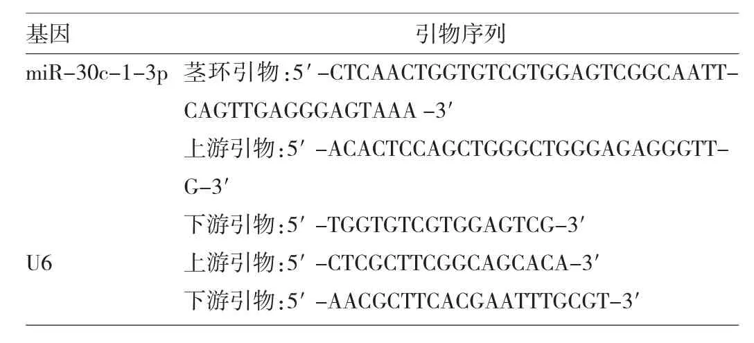
1.2.3 实时定量PCR 体系:SG Fast qPCR Master Mix(High Rox)(2X)(生工生物工程有限公司)10μl,正向引物、反向引物各 0.4μl,cDNA 模板 20μl,DEPC 处理后的水7.2μl;各基因的引物序列见表1。循环条件:初始维持 95℃ 3min;熔解 95℃ 7s,退火 57℃ 10s,延伸 72℃15s,共循环 40 次。miR-30c-1-3p 使用 U6 作为内参,计算机分析Ct值,采用2-ΔΔCt法计算相对表达量,即miR-30c-1-3p 表达水平。

表1 实时定量PCR反应引物序列
1.3 统计学处理 应用SPSS 20.0统计软件。计量资料不符合正态分布,故用 M(P25,P75)表示,组间比较采用Wilcoxon符号秩检验。P<0.05为差异有统计学意义。
2 结果
2.1 结直肠癌组织与癌旁正常黏膜组织中miR-30c-1-3p表达水平的比较 结直肠癌组织中miR-30c-1-3p表达水平为0.627(0.442,0.935),明显低于癌旁正常黏膜组织的 0.986(0.694,1.328),差异有统计学意义(P<0.05)。根据扩增曲线(图 1),估计 miR-30c-1-3p扩增效率基本一致。根据熔解曲线(图2),基本判断miR-30c-1-3p产物单一,不含引物二聚体等其他非目的性产物,熔解温度为82.08℃。

图1 miR-30c-1-3p扩增曲线

图2 miR-30c-1-3p熔解曲线
2.2 结直肠癌组织中miR-30c-1-3p表达水平与患者临床特征的关系 结直肠癌组织中miR-30c-1-3p表达水平与患者有无淋巴结转移、临床分期有关(均P<0.05),有淋巴结转移、临床分期Ⅲ~Ⅳ期的患者结直肠癌组织中miR-30c-1-3p表达水平均较低;与患者性别、年龄、肿瘤直径、肿瘤位置、分化程度、浸润深度等均无关(均 P>0.05),见表2。
3 讨论
在结直肠肿瘤中,有些基因被认为是导致肿瘤进展的癌基因如K-ras基因,有些被认为是抑癌基因如APC基因;此外,错配修复基因的突变、基因过度表达等在肿瘤进展中也起到重要作用。miRNA是近年来作为抑癌基因或原癌基因被报道较多的小分子基因,已发现其在许多肿瘤组织中表达异常,且参与肿瘤的生长[3]、增殖[4]、侵袭[5]、转移[6]、凋亡等,被认为是治疗肿瘤的新靶点[7-8]。
miR-30c是miRNA家族的成员之一。近年来研究表明miR-30c-1-3p通过3′非编码区改变靶基因细胞色素p450 3A4,从而使受体不表达[9]。相关研究报道miR-30c在乳腺癌、前列腺癌等恶性肿瘤组织中表达升高[10-11],Huang等[12]研究发现miR-30c通过靶基因ASF/SF2肿瘤蛋白剪接因子来抑制前列腺癌细胞的存活。Agostini等[13]研究认为miR-30c在卵巢肿瘤组织中表达下调。Cao等[14]研究发现miR-30c-5p通过靶点MTA1来抑制胃癌的迁移以及上皮细胞到间叶细胞的转变。Wang等[15]研究发现miR-30a-5p通过调节GRP78表达,从而抑制肾细胞癌的生长。Kawaguchi等[16]认为乳腺癌患者生存率的提高与miR-30a的高表达有关。除了肿瘤学领域,miR-30c还被发现能减少高血脂和2型糖尿病小鼠的血浆TC水平[17]。Singh等[18]研究表明恢复miR-30a的表达,能抑制神经母细胞瘤的致瘤性,且有自噬抑制作用。本研究结果发现miR-30c-1-3p在结直肠癌组织中的表达明显下调,与患者有无淋巴结转移、临床分期等有关,与患者性别、年龄、肿瘤直径、肿瘤位置、分化程度、浸润深度等无关;这提示miR-30c-1-3p可能与结直肠癌的转移侵袭相关。因此,笔者提出大胆假设:在结直肠癌的发生过程中,miR-30c-1-3p表达水平会降低;若能通过方法将miR-30c-1-3p的表达上调,有望抑制结直肠癌的进展。
综上所述,miR-30c-1-3p在人结直肠癌组织中表达下调,与有无淋巴结转移、临床分期有关;提示miR-30c-1-3p可能与结直肠癌的转移侵袭相关。

表2 结直肠癌组织中miR-30c-1-3p表达水平与患者临床特征的关系
[1]Chruscik A,Lam AK.Clinicalpathologicalimpacts of micrornas in papillary thyroid carcinoma:A crucial review[J].Experimental and molecular pathology,2015,99(3):393-398.
[2]Hollis M,Nair K,Vyas A,et al.Micrornas potential utility in colon cancer:Early detection,prognosis,and chemosensitivity[J].World journalof gastroenterology,2015,21(27):8284-8292.
[3]Xu Y,Chen J,Gao C,et al.Microrna-497 inhibits tumor growth through targeting insulin receptor substrate 1 in colorectal cancer[J].Oncology letters,2017,14(6):6379-6386.
[4]Li D,Wang H,Song H,et al.The micrornas mir-200b-3p and mir-429-5p target the limk1/cfl1 pathway to inhibit growth and motility of breast cancer cells[J].Oncotarget,2017,8(49):85276-85289.
[5]Boguslawska J,Rodzik K,Poplawski P,et al.Tgf-beta1 targets a microrna network that regulates cellular adhesion and migration in renalcancer[J].Cancer letters,2018,412:155-169.
[6]Mjelle R,Sellaeg K,Saetrom P,et al.Identification of metastasis-associated micrornas in serum from rectal cancer patients[J].Oncotarget,2017,8(52):90077-90089.
[7]Bahrami A,Aledavood A,Anvari K,et al.The prognostic and therapeutic application of micrornas in breast cancer:Tissue and circulating micrornas[J].Journal of cellular physiology,2018,233(2):774-786.
[8]Moridikia A,Mirzaei H,Sahebkar A,et al.Micrornas:Potential candidates for diagnosis and treatment of colorectal cancer[J].Journalof cellular physiology,2018,233(2):901-913.
[9]Vachirayonstien T,Yan B.Microrna-30c-1-3p is a silencer of the pregnane x receptor by targeting the 3'-untranslated region and alters the expression of its target gene cytochrome p450 3a4[J].Biochimica et biophysica acta,2016,1859(9):1238-1244.
[10]Yen MC,Shih YC,Hsu YL,et al.Isolinderalactone enhances the inhibition of socs3 on stat3 activity by decreasing mir-30c in breast cancer[J].Oncology reports,2016,35(3):1356-1364.
[11]Ling XH,Chen ZY,Luo HW,et al.Bcl9,a coactivator for wnt/beta-catenin transcription,is targeted by mir-30c and is associ-ated with prostate cancer progression[J].Oncology letters,2016,11(3):2001-2008.
[12]Huang YQ,Ling XH,Yuan RQ,et al.Mir30c suppresses prostate cancer survivalby targeting the asf/sf2 splicing factor oncoprotein[J].Molecular medicine reports,2017,16(3):2431-2438.
[13]Agostini A,Brunetti M,Davidson B,et al.Genomic imbalances are involved in mir-30c and let-7a deregulation in ovarian tumors:Implications for hmga2 expression[J].Oncotarget,2017,8(13):21554-21560.
[14]Cao JM,LiGZ,Han M,et al.Mir-30c-5p suppresses migration,invasion and epithelial to mesenchymal transition of gastric cancer via targeting mtal[J].Biomedicine&pharmacotherapy,2017,93:554-560.
[15]Wang C,Cai L,Liu J,et al.Microrna-30a-5p inhibits the growth of renal cell carcinoma by modulating grp78 expression[J].Cellular Physiology&Biochemistry International Journal of Experimental Cellular Physiology Biochemistry&Pharmacology,2017,43(6):2405.
[16]Kawaguchi T,Yan L,Qi Q,et al.Overexpression of suppressive micrornas,mir-30a and mir-200c are associated with improved survival of breast cancer patients[J].Scientific reports,2017,7(1):15945.
[17]Irani S,Iqbal J,Antoni WJ,et al.Microrna-30c reduces plasma cholesterol in homozygous familial hypercholesterolemic and type 2 diabetic mouse models[J].Journaloflipid research,2017.pii:jlr.M081299.doi:10.1194/jlr.M081299.[Epub ahead of print]
[18]Singh SV,Dakhole AN,Deogharkar A,et al.Restoration of mir-30a expression inhibits growth,tumorigenicity of medulloblastoma cells accompanied by autophagy inhibition[J].Biochemical and biophysical research communications,2017,491(4):946-952.

