
钙蛋白酶及其抑制剂对心肌肥厚模型大鼠心肌肥厚作用的研究
2016-11-29 07:18:43冯瑞赵玫
中国医科大学学报 2016年9期
冯瑞,赵玫
钙蛋白酶及其抑制剂对心肌肥厚模型大鼠心肌肥厚作用的研究
冯瑞,赵玫
(中国医科大学附属盛京医院心内科,沈阳 110004)
目的研究钙蛋白酶calpain及其抑制剂MDL28170与心肌肥厚的关系,探讨心肌肥厚可能的作用机制。方法50只280~350 g成年雄性Wistar大鼠随机分为正常对照组(NC组,n=5)、假手术组(SO组,n=5)、手术组(AO组,n=20)、手术+治疗组(AO+T组,n=20)。对AO组及AO+T组大鼠施行腹主动脉缩窄术建立心肌肥厚大鼠模型。4周后行心脏彩超检查。AO+T组大鼠MDL28170治疗2周后再行心脏彩超检查。取心脏称重,计算心重指数。Western blot检测心肌中calpain1、calpain2、CaN A的表达。结果心脏彩超证实4周后AO组及AO+T组大鼠心肌肥厚模型建立成功。药物干预后,AO+T组大鼠心肌肥厚得到控制,进展延缓,与AO组相比差距明显(P<0.05)。AO组、AO+T组大鼠心脏质量、心重指数较NC组和SO组增加,AO组增加更明显(均P<0.05)。与NC组相比,calpain1、calpain2、CaN A表达量在其余3组均有增加;而AO+T组与AO组比较calpain1、calpain2、CaN A表达量降低(P<0.05)。结论钙蛋白酶calpain与心肌肥厚关系密切,CaN途径是其作用机制之一;calpain抑制剂MDL28170对实验条件下的心肌肥厚具有明显阻遏作用。
心肌肥厚;大鼠;钙蛋白酶;calpain;抑制剂;MDL28170;信号通路;CaN
网络出版地址
细胞内游离Ca2+增加是心肌肥厚的基本信号[1⁃3]。目前大量研究[4⁃6]表明,钙蛋白酶calpain与心肌肥厚关系密切。calpain活性可影响诸多与心肌肥厚、重构有关的信号通路[钙调神经磷酸酶(calci⁃neurin,CaN)信号通路、核因子κB(nuclear factor⁃kappa B,NF⁃κB)信号通路]。这些信号通路激活后可从转录水平上介导心肌肥厚[7-8]。DOUILLARD等[9]发现过表达calpain内源性抑制剂calpastatin的大鼠与对照组相比可明显减轻药物介导的骨骼肌肥厚程度;LI等[10]发现在Ⅰ型糖尿病大鼠模型中,通过过表达calpastatin,心肌肥厚、纤维化得到明显改善。本研究通过缩窄大鼠腹主动脉的方法建立心肌肥厚大鼠模型,同时采用calpain抑制剂MDL28170对发生心肌肥厚的大鼠进行治疗,旨在探讨calpain与心肌肥厚的关系及其作用机制,为以calpain为靶点的心肌肥厚治疗提供新的思路。
1 材料与方法
1.1材料
主要包括50只280~350 g成年雄性Wistar大鼠(SPF级,中国医科大学附属盛京医院实验动物中心),MDL28170(英国Tocris Bioscience公司),兔抗大鼠calpain1单克隆抗体、兔抗大鼠calpain2单克隆抗体、兔抗大鼠CaN A单克隆抗体(英国Abcam公司)。
1.2方法
1.2.1分组、建模、给药、取材:将50只Wistar大鼠随机分为正常对照组(NC组,5只)、假手术组(SO组,5只)、手术组(AO组,20只)、手术+治疗组(AO+ T组,20只),称重标记。AO组及AO+T组大鼠用7号针头及3⁃0丝线将其腹主动脉在双侧肾动脉以上约1 cm水平处缩窄到管径为0.7 mm左右的管腔,绕线结扎。SO组大鼠只绕线不结扎。NC组大鼠不做任何处理。所有大鼠正常饲养4周后行心脏彩超检查,AO+T组大鼠腹腔注射MDL28170[20mg/(kg·d)],连续用药2周。其余3组大鼠按体质量给予同等剂量的含5%DMSO的生理盐水。6周时再次行心脏彩超检查,记录相关数据后麻醉,开胸,冰生理盐水持续心脏灌流。取心脏称重后-80℃冻存。
1.2.2心肌肥厚的判定:第4周及第6周末行心脏彩超检查。记录舒张末期室间隔厚度(interventricu⁃lar septum thickness at end⁃diastole,IVSTd)、左心室内径(left ventricular end⁃diastolic diameter,LVEDD)、左心室后壁厚度(left ventricular posterior wall thick⁃ness at end⁃diastole,LVPWTd)、左心室舒张末期容积(left ventricular end⁃diastolic volume,LVEDV)、收缩末期容积(left ventricular end⁃systolic volume,LVESV)、短轴缩短率(left ventricular fractional short⁃ening,LVFS)、射血分数(left ventricular ejection frac⁃tion,LVEF)等指标,判断大鼠心肌肥厚情况及心功能情况。
1.2.3心重指数(heart mass index,HMI)的测定:6周末取心脏称重,HMI(mg/g)=心脏质量/体质量。
1.2.4Western blot检测:取-80℃冻存的心脏标本,剪取室间隔及左心室组织块(约0.1 g),检测cal⁃pain1、calpain2、CaN A的表达,GAPDH为内参。利用图像分析软件对蛋白条带进行灰度分析。
1.3统计学分析
2 结果
2.1各组大鼠一般情况及体质量、心质量、心重指数比较
入组时(0周)各组大鼠体质量无明显差别(P>0.05),经过一段时间不同处理后,各组大鼠体质量增幅相似,不同时间(4周、6周)体质量无统计学差异(P>0.05),说明腹部手术对存活下来的动物一般生长情况不构成明显影响。在AO组和AO+T组,动物一般状态明显较NC组和SO组差。AO组及AO+T组的心脏质量、心重指数明显高于NC组及SO组(P<0.05);AO+T组心肌肥厚情况较AO组好转,心脏质量及心重指数降低(P<0.05)。见表1。
表1 不同时间大鼠体质量及心脏质量、心重指数比较Tab.1 Comparison of body weight and heart weight at different time and calculation of heart mass index
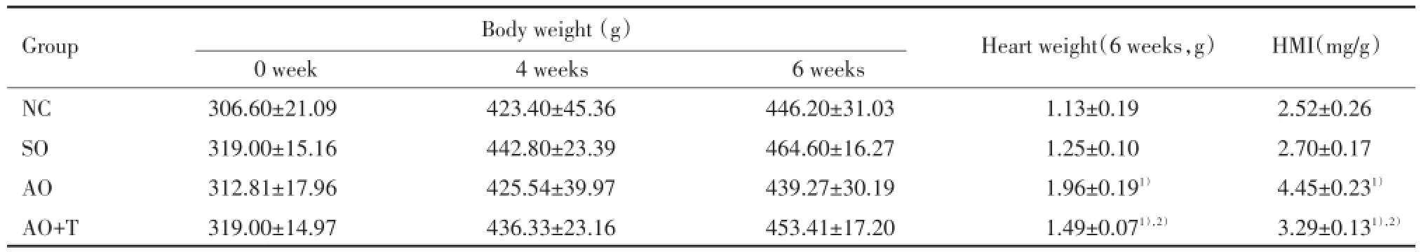
表1 不同时间大鼠体质量及心脏质量、心重指数比较Tab.1 Comparison of body weight and heart weight at different time and calculation of heart mass index
1)P<0.05 vs NC group;2)P<0.05 vs AO group.HMI,heart mass index.
Group Body weight(g) Heart weight(6 weeks,g) HMI(mg/g)0 week 4 weeks 6 weeks NC 306.60±21.09 423.40±45.36 446.20±31.03 1.13±0.19 2.52±0.26 SO 319.00±15.16 442.80±23.39 464.60±16.27 1.25±0.10 2.70±0.17 AO 312.81±17.96 425.54±39.97 439.27±30.19 1.96±0.191) 4.45±0.231)AO+T 319.00±14.97 436.33±23.16 453.41±17.20 1.49±0.071),2) 3.29±0.131),2)
2.2各组大鼠心肌肥厚及左心功能比较
心脏彩超检查显示,第4周末,与NC组、SO组比较,AO组、AO+T组大鼠室间隔、左心室壁明显增厚(P<0.05);心腔大小、左心功能等指标无统计学差异(P>0.05),证明大鼠心肌肥厚建模成功。第6周末AO组大鼠较2周前心肌肥厚进一步加重,表现为室间隔、左心室壁心肌进一步增厚,与NC组及SO组差别明显(P<0.05);而AO+T组大鼠与NC组相比仍有心肌肥厚表现(P<0.05),但肥厚程度与AO组相比有所减轻;室间隔、左心室壁厚度AO+T组与AO组有统计学差异(P<0.05)。而其他指标4组大鼠表现出不同趋势,但差异没有统计学意义(P>0.05)。见表2。
表2 术后4周、6周各组大鼠心肌肥厚及左心功能指标比较Tab.2 Comparison of indicators of myocardial hypertrophy and left ventricular function 4 weeks/6 weeks after the abdominal operation
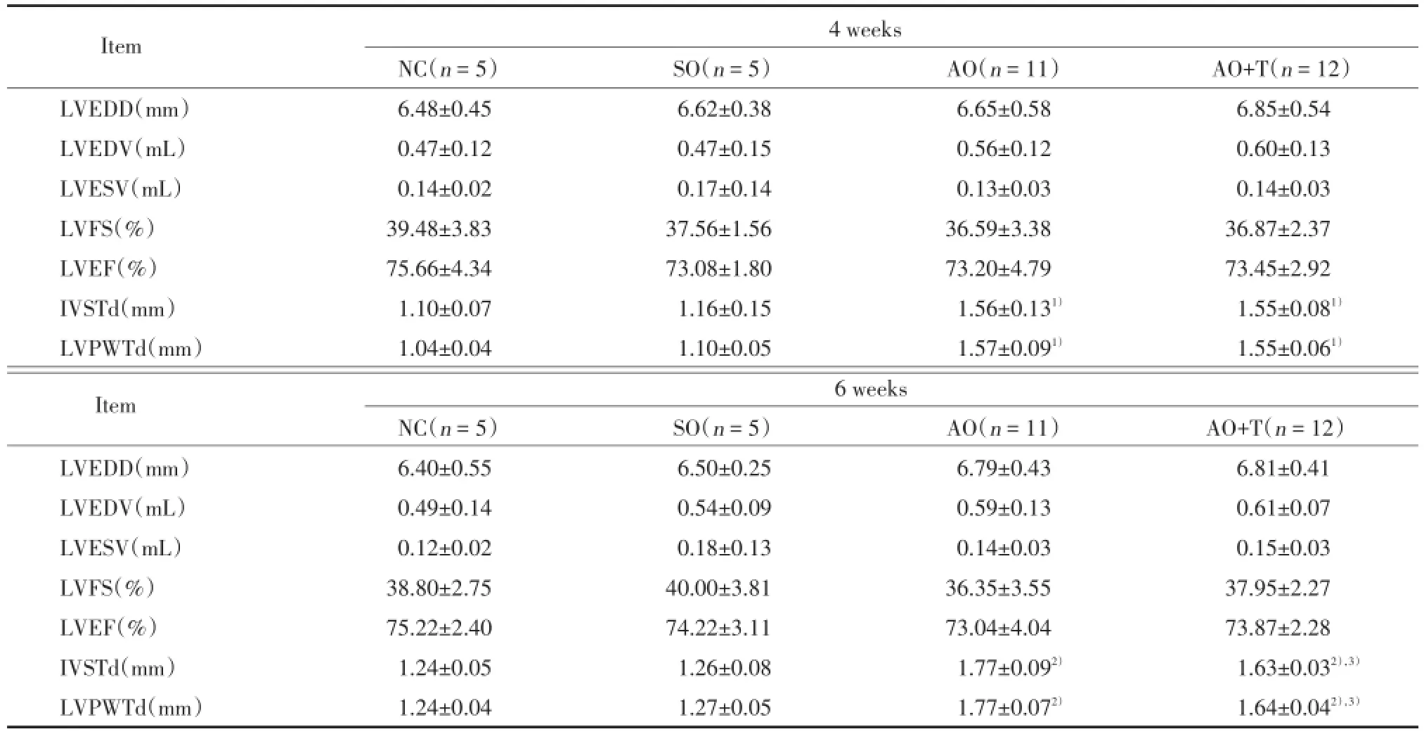
表2 术后4周、6周各组大鼠心肌肥厚及左心功能指标比较Tab.2 Comparison of indicators of myocardial hypertrophy and left ventricular function 4 weeks/6 weeks after the abdominal operation
1)P<0.05 vs NC group;2)P<0.05 vs NC group;3)P<0.05 vs AO group.LVEDD,left ventricular end⁃diastolic diameter;LVEDV,left ventricular end⁃diastolic volume;LVESV,left ventricular end⁃systolic volume;LVFS,left ventricular fractional shortening;LVEF,left ventric⁃ular ejection fraction;IVSTd,interventricular septum thickness at end⁃diastole;LVPWTd,left ventricular posterior wall thickness at end⁃dias⁃tole.
Item 4 weeks NC(n=5) SO(n=5) AO(n=11) AO+T(n=12)LVEDD(mm) 6.48±0.45 6.62±0.38 6.65±0.58 6.85±0.54 LVEDV(mL) 0.47±0.12 0.47±0.15 0.56±0.12 0.60±0.13 LVESV(mL) 0.14±0.02 0.17±0.14 0.13±0.03 0.14±0.03 LVFS(%) 39.48±3.83 37.56±1.56 36.59±3.38 36.87±2.37 LVEF(%) 75.66±4.34 73.08±1.80 73.20±4.79 73.45±2.92 IVSTd(mm) 1.10±0.07 1.16±0.15 1.56±0.131) 1.55±0.081)LVPWTd(mm) 1.04±0.04 1.10±0.05 1.57±0.091) 1.55±0.061)Item 6 weeks NC(n=5) SO(n=5) AO(n=11) AO+T(n=12)LVEDD(mm) 6.40±0.55 6.50±0.25 6.79±0.43 6.81±0.41 LVEDV(mL) 0.49±0.14 0.54±0.09 0.59±0.13 0.61±0.07 LVESV(mL) 0.12±0.02 0.18±0.13 0.14±0.03 0.15±0.03 LVFS(%) 38.80±2.75 40.00±3.81 36.35±3.55 37.95±2.27 LVEF(%) 75.22±2.40 74.22±3.11 73.04±4.04 73.87±2.28 IVSTd(mm) 1.24±0.05 1.26±0.08 1.77±0.092) 1.63±0.032),3)LVPWTd(mm) 1.24±0.04 1.27±0.05 1.77±0.072) 1.64±0.042),3)
2.3Western blot检测相关蛋白表达
检测结果表明,SO组出现了calpain1、calpain2、CaN A表达上调;AO组calpain1、calpain2、CaN A的表达较NC组及SO组明显升高(P<0.05);AO+T组3种蛋白呈现出弱表达,与AO组比较差异有统计学意义(P<0.05)。提示手术应激可能对calpain、CaN的表达产生了影响,calpain1、calpain2、CaN表达受到了特异性抑制剂的强力抑制作用。见图1、表3。
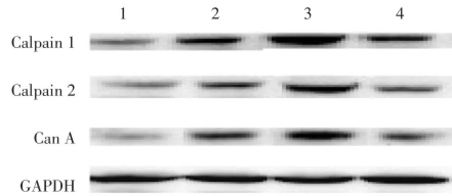
图1 各组大鼠心肌组织中相关蛋白的表达情况Fig.1 Expression of related proteins in myocardial tissue from the 4 groups
3 讨论
calpain是一类存在于细胞质中的Ca2+依赖性半胱氨酸蛋白酶,分为组织特异性和非特异性两大类。非特异性calpain包括u⁃calpain(calpain1)和m⁃calpain(calpain2),在人体内广泛表达。u⁃calpain和m⁃calpain均是由1个28×103亚基和1个80×103亚基组成的异二聚体,2种蛋白酶之间序列同源性达50%~60%,区别只在于被激活时所需要的Ca2+浓度不同:前者在微摩尔水平,后者在毫摩尔水平。由于细胞内Ca2+浓度通常波动在微摩尔水平,因此推测在生理状态下主要是calpain1在发生作用。研究[11]表明,calpain可通过介导以下2条主要的信号通路影响心肌肥厚的表达:通过水解CaN生成活性片段和水解CaN的内源性抑制剂Cain/Cabin1激活CaN信号通路;通过降解胞质中与NF⁃κB结合的抑制因子IκBα激活NF⁃kB信号通路等。
表3 各组大鼠心肌组织中相关蛋白的表达比较Tab.3 Expression of related proteins in myocardial tissue from the 4 groups
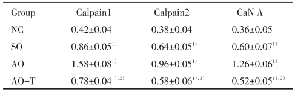
表3 各组大鼠心肌组织中相关蛋白的表达比较Tab.3 Expression of related proteins in myocardial tissue from the 4 groups
1)P<0.05 vs NC group;2)P<0.05 vs AO group.
Group Calpain1 Calpain2 CaN A NC 0.42±0.04 0.38±0.04 0.36±0.05 SO 0.86±0.051) 0.64±0.051) 0.60±0.071)AO 1.58±0.081) 0.96±0.051) 1.26±0.061)AO+T 0.78±0.041),2) 0.58±0.061),2) 0.52±0.051),2)
本研究心脏彩超证实,在发生心肌肥厚的基础上对AO+T组大鼠应用calpain特异性抑制剂—MDL28170 2周后,AO+T组大鼠心肌肥厚进程得到控制,心肌肥厚程度较AO组明显减轻。心脏标本测算后提示AO+T组心脏质量、心重指数较AO组有所减轻。证明calpain在试验条件下的心肌肥厚进程中具有促进作用,应用其特异性抑制剂可明显抑制此种条件下心肌肥厚的进展,有一定治疗效果。
Western blot检测结果表明,手术应激也可对calpain、CaN的表达产生影响,体现在SO组出现了calpain1、calpain2、CaN A表达上调。AO组3种蛋白均呈现高表达再次印证了calpain在心肌肥厚进程中的重要作用。AO+T组3种目的蛋白呈现弱表达,与AO组比较差异有统计学意义(P<0.05),提示calpain1、calpain2、CaN表达均受到了特异性抑制剂的强力抑制作用。AO组蛋白表达量从大到小依次为:calpain1、CaN A、calpain2。由此推论CaN途径是calpain致心肌肥厚进程中的一条重要信号通路,其在心肌肥厚进程中的作用不可忽视。而calpain的特异性抑制剂仍然可以通过抑制calpain的活性来抑制其下游CaN的激活。
综上所述,本实验证明了calpain与心肌肥厚进程的关系,验证了特异性抑制剂的抑制与保护作用。然而抑制剂的应用存在一个时间节点问题,本实验在心肌肥厚进展到一定程度时才给予抑制剂治疗,并未设立更早时间点来观察。而且治疗阶段仅2周,抑制剂的长期作用效果如何不得而知。另外,入组实验动物数目基数较小,结论具有一定的局限性。
[1]MIZUKAMI K,YOKOSHIKI H,MITSUYAMA H,et al.Small⁃con⁃ductance Ca2+⁃activated K+current is upregulated via the phosphory⁃lation of CaMKⅡin cardiac hypertrophy from spontaneously hyper⁃tensive rats[J].Am J Physiol Heart Circ Physiol,2015,309(6):H1066-H1074.DOI:10.1152/ajpheart.00825.2014.
[2]HORTON JS,BUCKLEY CL,ALVAREZ EM,et al.The calcium re⁃lease⁃activated calcium channel orai1 represents a crucial compo⁃nent in hypertrophic compensation and the development of dilated cardiomyopathy[J].Channels(Austin),2014,8(1):35-43.DOI:10.4161/chan.26581.
[3]LUO X,HOJAYEV B,JIANG N,et al.STIM1⁃dependent store⁃oper⁃ated Ca2+entry is required for pathological cardiac hypertrophy[J]. J Mol Cell Cardiol,2012,52(1):136-147.DOI:10.1016/j. yjmcc.2011.11.003.
[4]MEI M,TANG F,LU M,et al.AstragalosideⅣattenuates apoptosis of hypertrophic cardiomyocyte through inhibiting oxidative stress and calpain⁃1 activation[J].Environ Toxicol Pharmacol,2015,40(3):764-773.DOI:10.1016/j.etap.2015.09.007.
[5]YE T,WANG Q,ZHANG Y,et al.Over⁃expression of calpastatin in⁃hibits calpain activation and attenuates post⁃infarction myocardial remodeling[J].PLoS One,2015,10(3):e0120178.DOI:10.1371/ journal.pone.0120178.
[6]KUDO⁃SAKAMOTO Y,AKAZAWA H,ITO K,et al.Calpain⁃depen⁃dent cleavage of N⁃cadherin is involved in the progression of post⁃myocardial infarction remodeling[J].J Biol Chem,2014,289(28):19408-19419.DOI:10.1074/jbc.M114.567206.
[7]PENNANEN C,PARRA V,MORALES PE,et al.Mitochondrial fis⁃sion is required for cardiomyocyte hypertrophy mediated by a Ca2+⁃calcineurin signaling pathway[J].J Cell Sci,2014,127(12):2659-2671.DOI:10.1242/jcs.139394.
[8]GASPAR⁃PEREIRA S,FULLARD N,TOWNSEND PA,et al.The NF⁃κB subunit c⁃Rel stimulates cardiac hypertrophy and fibrosis[J].Am J Pathol,2012,180(3):929-939.DOI:10.1016/j.aj⁃path.2011.11.007.
[9]DOUILLARD A,GALBES O,BEGUE G,et al.Calpastatin overex⁃pression in the skeletal muscle of mice prevents clenbuterol⁃in⁃duced muscle hypertrophy and phenotypic shift[J].Clin Exp Phar⁃macol Physiol,2012,39(4):364-372.DOI:10.1111/j.1440⁃1681.2012.05677.x.
[10]LI Y,MA J,ZHU H,et al.Targeted inhibition of calpain reduces myocardial hypertrophy and fibrosis in mouse models of type 1 dia⁃betes[J].Diabetes,2011,60(11):2985-2994.DOI:10.2337/ db10⁃1333.
[11]LI X,LUO R,CHEN R,et al.Cleavage of IκBα by calpain induces myocardial NF⁃κB activation,TNF⁃α expression,and cardiac dys⁃function in septic mice[J].Am J Physiol Heart Circ Physiol,2014,306(6):H833-H843.DOI:10.1152/ajpheart.00893.2012.
(编辑武玉欣)
Myocardial Hypertrophy Process in Rat Model of Myocardial Hypertrophy Mediated by Calpain and Its Inhibitor
FENG Rui,ZHAO Mei
(Department of Cardiology,Shengjing Hospital,China Medical University,Shenyang 110004,China)
ObjectiveTo investigate the relationship between calpain and myocardial hypertrophy,and further explore the underlying mecha⁃nism.MethodsA total of 50 adult male Wistar rats(280⁃350 g)were randomly divided into 4 groups:normal control group(NC,n=5),sham operation group(SO,n=5),abdominal aortic operation group(AO,n=20),abdominal aortic operation plus treatment group(AO+T,n=20). AO group and AO+T group
the surgery of abdominal aortic coarctation.After 4 weeks,rats of AO+T group were given MDL28170 for ther⁃apy for 2 weeks.Then echocardiography examination was performed.The heart mass and calculate the ratio of heart mass and body weight in each group were recorded.The expression of calpain1,calpain2 and CaN A of myocardial tissue were determined by Western blot.ResultsThe myo⁃cardial hypertrophy model was successfully established in both AO and AO+T group 4 weeks after operation,as echocardiography indicated.After drug intervention,rats of AO+T group developed a much slower progress of myocardial hypertrophy and the progressing was under control,which showed a significant gap compared with AO group(P<0.05).The heart mass and the heart mass index(HMI)were increased significantly in AO and AO+T group than the other two groups,especially in AO group which was more serious(P<0.05).Compared with NC group,the expression level of calpain1,calpain2,and CaN A were increased in the other 3 groups,while AO+T group showed a reduced expression by contrast of AO group.The differences between these 2 groups were statistically significant(P<0.05).ConclusionCalpain is closely related with myocardial hypertrophy,and CaN signaling pathway is one of the most important mechanisms;calpain inhibitor MDL28170 has an obvious inhibition effect on myocardial hypertrophy under the experimental conditions.
myocardial hypertrophy;rat;calpain;inhibitor;MDL28170;signaling pathway;CaN
R542.2
A
0258-4646(2016)09-0769-04
10.12007/j.issn.0258⁃4646.2016.09.001
国家自然科学基金(81100161)
冯瑞(1988-),女,医师,硕士.
赵玫,E-mail:zhaom1@sj⁃hospital.org
2015-12-23
网络出版时间:
猜你喜欢
昆明医科大学学报(2021年4期)2021-07-23 01:22:00
文苑(2018年22期)2018-11-19 02:54:30
中国医疗保险(2017年5期)2017-05-17 08:26:39
新农业(2016年18期)2016-08-16 03:28:31
三峡大学学报(自然科学版)(2016年6期)2016-04-16 05:02:56
西南军医(2016年6期)2016-01-23 02:21:21
中国康复理论与实践(2015年10期)2015-12-24 05:42:46
现代电生理学杂志(2015年1期)2015-07-18 11:02:16
医学研究杂志(2015年4期)2015-06-10 06:42:43
食品工业科技(2014年7期)2014-03-11 18:14:47
