
硫氧还蛋白-1高表达的幽门螺杆菌慢性感染蒙古沙土鼠模型的建立与评价
2016-11-23 02:19:22张贺军刘琳娜石岩岩丁士刚
北京大学学报(医学版) 2016年5期
张贺军,刘琳娜,张 超,石岩岩,丁士刚△
(1. 北京大学第三医院消化科,幽门螺杆菌感染及上胃肠疾病防治研究北京市重点实验室,北京 100191; 2. 华北理工大学附属医院消化内科,河北唐山 063000)
·论著·
硫氧还蛋白-1高表达的幽门螺杆菌慢性感染蒙古沙土鼠模型的建立与评价
张贺军1,刘琳娜1,张 超2,石岩岩1,丁士刚1△
(1. 北京大学第三医院消化科,幽门螺杆菌感染及上胃肠疾病防治研究北京市重点实验室,北京 100191; 2. 华北理工大学附属医院消化内科,河北唐山 063000)
目的:建立硫氧还蛋白-1(thioredoxin-1,Trx1)基因高表达的幽门螺杆菌(Helicobacterpylori,Hp)慢性感染蒙古沙土鼠模型,评价Trx1在Hp长期定植导致胃黏膜的病变及其致癌性中的作用。方法:健康雄性蒙古沙土鼠75只,随机分为3组:Trx1高表达Hp组(30只),Trx1低表达Hp组(30只)和对照组(15只)。分别采用Trx1高表达和低表达Hp菌株灌胃,建立Hp长期感染蒙古沙土鼠腺胃模型。在接种Hp后第4、20、34、48、70和90周分批处死动物,行快速尿素酶实验和Warthin-Starry银染判定Hp定植情况,HE染色观察胃黏膜病理改变。结果:(1)成功建立了Trx1高表达和低表达Hp长期感染蒙古沙土鼠腺胃的动物模型。(2)Trx1高表达和低表达Hp组胃黏膜病变随时间延长而逐渐加重,Trx1高表达Hp组出现黏膜不平及结节性病变的时间分别为第20周和第34周,早于Trx1低表达Hp组(分别为第48周和第70周);Trx1高表达Hp组从第48周起观察到黏膜糜烂(3只),而Trx1低表达Hp组和对照组在整个实验过程中均未见明显的黏膜糜烂改变。Trx1高表达Hp组出现病变的时间和病变范围明显早于/重于Trx1低表达Hp组。(3)Trx1高表达Hp组在接种后第34周可观察到上皮细胞的轻度异型增生,明显早于Trx1低表达Hp组(第48周)。第90周,Trx1高表达Hp组有71.4%(5/7)、Trx1低表达Hp组有42.8%(3/7)检出管状腺癌(P=0.592),均为早期癌,且Trx1高表达Hp组有1例为黏膜下癌,此外,Trx1高表达Hp组还观察到2例上皮细胞重度异型增生(28.6%,2/7),而在Trx1低表达Hp组仅观察到3例上皮细胞中度异型增生(42.8%,3/7)。结论:Trx1高表达和低表达的Hp可稳定定植于蒙古沙土鼠腺胃,出现类似于人感染Hp后出现的各种病理变化,并诱发腺癌的发生;Trx1高表达Hp组胃黏膜病变明显重于Trx1低表达Hp组,Trx1可能是我国人群Hp致病及致癌的毒力因子。
硫氧还蛋白质类;螺杆菌,幽门;胃肿瘤;模型,动物;沙鼠亚科
胃癌是危害我国人民健康的重大疾病之一,幽门螺杆菌(Helicobacterpylori,Hp)感染与胃癌密切相关[1]。Hp已被世界卫生组织国际癌症研究机构列为Ⅰ类致癌原,但其与胃癌关系的确立主要依据人群流行病学资料。多项研究表明,Hp通过其毒力因子[如细胞毒素相关蛋白A(cytotoxin-associated protein A, cagA)和空泡毒素A(vacuolating cytotoxin A, vacA)等]的作用参与胃癌的发生与发展[2],但这种关系在我国人群中并不明确[3-4]。我们前期的研究工作发现,硫氧还蛋白-1(thioredoxin-1,Trx1)可能是我国人群感染Hp致病及致癌的一种新的毒力因子[5-7]。本研究通过建立Trx1高表达Hp长期感染蒙古沙土鼠(Mongolian gerbil, MG)模型,观察Trx1高表达Hp长期感染引起的胃黏膜病理改变及肿瘤的发生情况,为后续的相关研究提供方法。
1 材料与方法
1.1 材料
Hp菌株:选用本研究实验室保种的从胃癌患者胃内分离的Trx1高表达Hp株和从胃炎患者胃内分离的Trx1低表达Hp株[8]。
实验动物:3周龄MGs(清洁级,远交封闭群)75只,雄性,体重25~35 g,购自上海生物制品研究所有限责任公司[实验动物生产许可证号:SCXK(沪)2009-0020]。置于北京大学医学部实验动物科学部[实验动物使用许可证号:SYXK(京)2011-0039]饲养,室温为21~25 ℃,相对湿度为50%~60%,每日光照、黑暗交替各12 h。自由进食、饮水,饲料及饮水均经高压灭菌处理。
1.2Hp菌液制备
将-80 ℃冻存的菌种在含5%(体积分数)的Columbia琼脂平板上复苏,转种于添加15%(体积分数)胎牛血清和抗生素的布氏肉汤(Brucella broth)中。微氧[5% O2、8% CO2、80% N2(均为体积分数)]条件下孵育72 h进行增菌,37 ℃下振荡(120 r/min)培养60~72 h。取少量培养液涂片,革兰染色后观察细菌形态,检测尿素酶阳性后,将培养液离心,沉淀菌体混悬于灭菌磷酸缓冲盐溶液(phosphate buffer saline,PBS)(pH=7.2)中,细菌浓度应用比浊分光光度计调整到1×10⁹菌落形成单位(colony-forming unit, CFU)/mL,立即接种动物。
1.3 动物模型的制备
给予MGs含氟哌酸0.5 g/L、制霉菌素0.5万 U/mL和万古霉素0.1 g/L的饮水2周,给予致癌剂N-甲基-N’-硝基-N-亚硝基胍(MNNG,浓度200×10-6)1周。随机将MGs分为3组:Trx1高表达Hp组30只,Trx1低表达Hp组30只,对照组15只。所有MGs接种前禁食、禁水24 h,实验组经口灌入浓度为1×10⁹CFU/mL的Hp菌液0.5 mL,对照组以灭菌PBS代替,每周灌胃1次,连续灌胃5周。在接种Hp后分别于第4、20、34、48、70、90周分批处死MGs,观察Hp定植情况及病理学改变。
1.4 模型的检测和评价
MGs处死前禁食、禁水24 h,1%(质量分数)戊巴比妥钠腹腔注射麻醉(30 mg/kg),打开腹腔游离全胃,沿大弯侧剪开,肉眼观察胃黏膜外观并记录病变部位、数目、大小、形态。从幽门区取一小块胃黏膜用于快速尿素酶试验(rapid urease test, RUT)鉴定,其余胃组织用10%(体积分数)甲醛溶液固定,标本沿胃的纵轴连续切成3 mm大小的组织条块,石蜡包埋,制成4 μm的连续切片,分别行苏木素-伊红染色(hematoxylin and eosin, HE)及Warthin-Starry银染。
以RUT和Warthin-Starry银染同时阳性为判定Hp感染的标准。HE染色显微镜下观察胃黏膜病理变化,包括炎症、萎缩、肠上皮化生、异型增生及胃癌等。
1.5 统计学分析
利用SPSS 13.0统计分析软件,采用χ2检验进行统计学分析,以α=0.05为检验水准。
2 结果
2.1 有效动物及Hp定植情况
在实验过程中,共有13只MGs死亡,余62只动物均完成实验,其中Trx1高表达Hp组25只,Trx1低表达Hp组24只,对照组13只(表1)。
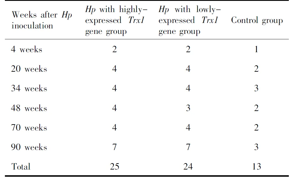
表1 有效实验动物分批处死情况Table 1 The numbers of valid experimental animals
所有实验组MGs从接种Hp后第4周起胃黏膜经RUT及Warthin-Starry银染均为阳性,Warthin-Starry银染可见细菌分布于在胃黏膜黏液层及小凹中,随时间的延长,在部分腺腔内可见细菌定植。对照组MGs无Hp检出。
2.2 胃黏膜大体变化
整个实验过程中,对照组MGs胃黏膜均未见红斑、出血、糜烂、溃疡及息肉等明显肉眼病变。
Trx1高表达Hp组MGs在接种Hp后第20周胃黏膜可见花斑样改变,第34周可见结节样病变(2/4),第48周出现黏膜糜烂出血(2/4)及结节样病变(1/4),第70周可见多发结节(3/4,图1A)和胃体多发糜烂(1/4),第90周均可见明显的多发性结节样隆起(7/7),最大直径达1 cm(图2A)。
Trx1低表达Hp组MGs在接种Hp后第48周可见胃体黏膜粗糙不平(1/3),第70周可见结节性病变(3/4),第90周胃黏膜可见散在的结节隆起(7/7),最大直径达0.5 cm(图3A)。
余时间点两个实验组均未见红斑、出血、糜烂、溃疡及息肉等明显肉眼病变。
2.3 胃黏膜组织学变化
整个实验过程中对照组MGs胃黏膜组织学观察显示胃黏膜各层结构完整、排列整齐,黏膜固有层及黏膜下层偶见个别炎细胞浸润。
Trx1高表达Hp组MGs在接种Hp后第20周胃黏膜固有层内可见少量急、慢性炎细胞浸润,小凹上皮略增生。第34周胃黏膜炎症加重,固有层以及黏膜下层均可见淋巴细胞及中性粒细胞聚集,小凹上皮增生明显,可见肠上皮化生,部分上皮细胞具有轻度异型性。少量胃体腺增生,偶见个别腺管扩张。第48周可见黏膜糜烂坏死,固有层内淋巴细胞及中性粒细胞浸润明显增加,个别腺管囊性扩张,部分区域腺体减少,可见肠上皮化生,呈萎缩性胃炎表现,可见部分上皮呈轻至中度异型增生。第70周胃黏膜可见重度急、慢性炎细胞浸润,可见黏膜糜烂、坏死,黏膜下层可见灶状淋巴细胞浸润,小凹上皮增生明显,小凹弯曲变长,增生性息肉形成(图1B),部分肠上皮化生细胞呈中度异型增生(图1B)。第90周可见5只(71.4%)发生高分化管状腺癌(其中1例癌组织侵及黏膜下层,另4例为黏膜内癌,图2B),2例增生性息肉伴有重度异型增生。
Trx1低表达Hp组MGs在接种Hp后第48周黏膜固有层内可见急、慢性炎细胞浸润,小凹上皮增生,可见肠上皮化生,部分上皮细胞具有轻度异型性,胃体腺增生。第70周可见萎缩性胃炎表现伴部分腺管扩张,可见肠上皮化生,部分上皮呈轻至中度异型增生。第90周可见3只(42.8%)高分化管状腺癌(均为黏膜内癌,图3B),3只发生增生性息肉伴中度异型增生,余1只仅见轻度慢性炎细胞浸润。两实验组MGs检出胃癌的差异无统计学意义(P=0.592)。
3 讨论
Trx是一类广泛分布于真核生物和原核生物的小分子多功能蛋白,主要分为Trx1和Trx2两型。Trx1通过其氧化还原活性区域可逆性催化氧化还原反应,具有清除自由基、抗氧化[9]及抗细胞凋亡的作用[10]。在胃癌[11]、前列腺癌[12]、胆管癌[13]、甲状腺癌[14]等多种肿瘤中的研究表明,肿瘤组织中Trx1表达明显上调,其可能通过抑制细胞凋亡、促进细胞增殖从而促进肿瘤的发生[15],此外,Trx1还与卵巢癌[16]、多发性骨髓瘤[17]等多种肿瘤细胞的耐药性发生有关。
Hp的Trx基因根据其长度可分为Trx1和Trx2,分别编码蛋白Trx1和Trx2[18]。Trx1作为Hp的抗氧化系统,与其胃内存活、抑制宿主胃内氧化应激和氮化应激密切相关[19]。我们的前期工作发现,胃癌患者胃内Hp的Trx1在核酸和蛋白质水平的表达均明显高于胃良性疾病[5-6],Hp分泌的Trx1可能通过上调cyclin D1的表达从而促进胃癌细胞的生长,是我国人群Hp致病、致癌的候选毒力因子[7]。
图1 Trx1高表达Hp组MGs在接种Hp后第70周,胃窦黏膜多发结节病变(A),病理学检查显示为增生性息肉(B, 左侧箭头),可见肠上皮化生细胞呈中度异型增生(B, 右侧箭头),黏膜固有层内可见急、慢性炎细胞浸润,黏膜下层可见淋巴组织增生(HE ×40) 图2 Trx1高表达Hp组MGs在接种Hp后第90周,胃窦黏膜可见多发结节样隆起(A),病理学检查显示为高分化腺癌,部分癌组织突破黏膜肌层至黏膜下层,黏膜下层可见淋巴组织增生(B, HE ×40) 图3 Trx1低表达Hp组MGs在接种Hp后第90周,胃窦黏膜可见多发结节样隆起(A),病理学检查显示为高分化腺癌,癌组织局限于黏膜固有层内,黏膜下层可见淋巴组织增生(B, HE ×40)
Figure 1 The macroscopic and microscopic appearance of the gastric mucosa of Mongolian gerbil at 70 weeks after Hp inoculation in Hp with highly-expressed Trx1 gene group. The multiple nodules could be seen in the gastric antrum (A), the histology revealed hyperplasic polyp (B, left arrow). Mixed inflammatory cells infiltration was present within the lamina propria, and the epithelia with intestinal metaplasia showed moderate dysplasia (B, right arrow). The hyperplasia of lymphoid tissue could be observed in the submucosa (HE ×40) Figure 2 The macroscopic and microscopic appearance of the gastric mucosa of Mongolian gerbil at 90 weeks after Hp inoculation in Hp with highly-expressed Trx1 gene group. The multiple nodules could be seen in the gastric antrum (A), the histology revealed well-differentiated adenocarcinoma, some adenocarcinoma invaded into submucosa, the hyperplasia of lymphoid tissue could be observed in the submucosa (B, HE ×40) Figure 3 The macroscopic and microscopic appearance of the gastric mucosa of Mongolian gerbil at 90 weeks after Hp inoculation in Hp with lowly-expressed Trx1 gene group. The multiple nodules could be seen in the gastric antrum (A), the histology revealed well-differentiated adenocarcinoma, the tissue of adenocarcinoma was limited within the lamina propria, the hyperplasia of lymphoid tissue could be observed in the submucosa (B, HE ×40)
MGs具有自然患胃炎的概率低、对Hp易感且感染后胃黏膜的病理变化与人相似、动物寿命较长适合长期观察等优点,而且国内外多项研究已用Hp感染MGs成功诱发出胃腺癌[20-25],因此本研究利用Trx1高表达和低表达Hp分别感染MGs,整个实验过程中各实验组均可见Hp的稳定定植,成功建立了Trx1高表达和低表达Hp稳定感染的动物模型。
Correa[26]提出Hp感染后胃癌的发生遵循“Hp感染-慢性胃炎-萎缩、肠化-异型增生-腺癌”途径。本研究建立的Hp稳定感染的MGs模型观察到类似于Hp感染人胃黏膜后出现的各种肉眼及病理变化,着重观察了异型增生与腺癌的发生。胃黏膜肉眼形态上,实验组胃黏膜病变随时间延长而逐渐加重,出现花斑、增生性结节、糜烂等病变,Trx1高表达Hp组出现黏膜不平及结节性病变的时间分别为第20周和第34周,而Trx1低表达Hp组则分别出现在第48周和第70周;Trx1高表达Hp组从第48周起观察到黏膜糜烂(3只),而Trx1低表达Hp组和对照组在整个实验过程中均未见明显的黏膜糜烂改变。Trx1高表达Hp组出现肉眼所见病变的时间和病变的范围明显早于/重于Trx1低表达Hp组。组织病理学上,Trx1高表达Hp组在接种后第34周可观察到上皮细胞的轻度异型增生,明显早于Trx1低表达Hp组(第48周)和其他类似的研究(第40周或第45周)[23-24,27]。随着观察时间的延长,第90周Trx1高表达和低表达Hp组分别有71.4%和42.8%检出胃腺癌,Trx1高表达Hp组腺癌的发生率明显高于国内外的文献报道(18%~40%)[20-22,27],分析其原因一是与所用的菌株有很大关系,本研究采用的是从胃癌患者胃内分离的Trx1高表达Hp株,而其他文献中采用的是国际标准菌株[20,22,25,27]或从胃溃疡患者胃内分离的临床菌株[21],这也证实了Trx1可能是Hp致癌的一种毒力因子;其次可能与观察时间有关[23-25],本研究观察周期达到了90周,而其他文献报道的观察时间为62~84周不等[20-22,27],亦有文献报道观察周期达到24个月[25];此外,目前沙土鼠尚无品系,对其遗传学背景缺乏深入研究,故即使应用相同菌株在相同条件下也可能出现不同的反应。本研究中Trx1高表达和低表达Hp组检出胃腺癌的差异虽然无统计学意义(71.4%vs. 42.8%,P=0.592),但是在Trx1高表达Hp组还是观察到2只动物发生上皮细胞的重度异型增生(28.6%),而在Trx1低表达Hp组仅观察到3只发生上皮细胞的中度异型增生,在Trx1高表达Hp组中有1例为黏膜下癌,故有必要进一步延长观察时间并扩大样本量进行观察。
总之,本研究首次建立了Trx1高表达Hp感染MGs诱发胃癌模型,证实了Trx1可能是Hp致病、致癌过程中的毒力因子,此模型的建立为后续进行Trx1致癌机制的在体研究提供了重要的动物模型及相关的理论和实验依据。
[1]Prinz C, Schwendy S, Voland P. H pylori and gastric cancer: shifting the global burden [J]. World J Gastroenterol, 2006, 12(34): 5458-5464.
[2]Wen S, Moss SF. Helicobacter pylori virulence factors in gastric carcinogenesis [J]. Cancer Lett, 2009, 282(1): 1-8.
[3]Fock KM, Talley N, Moayyedi P, et al. Asia-Pacific consensus guidelines on gastric cancer prevention [J]. J Gastroenterol Hepatol, 2008, 23(3): 351-365.
[4]Zhang Y, Liu H, Zhou K. Lack of correlation of vacA genotype, cagA gene ofHelicobacterpyloriand their expression products with various gastroduodenal diseases [J].Chin Med J (Engl), 2001, 114(7): 703-706.
[5]张静, 丁士刚, 钟丽君, 等. 消化性溃疡、胃炎与胃癌患者幽门螺杆菌蛋白质组的差异分析[J]. 中华医学杂志, 2006, 86(38): 2690-2694.
[6]石岩岩, 丁士刚, 鲁凤民, 等. 硫氧还蛋白1对胃癌细胞系BCG823生长的影响[J]. 中国微创外科杂志, 2011, 11(11): 1030-1033.
[7]Shi Y, Liu L, Zhang T, et al. The involvement ofHelicobacterpylorithioredoxin-1 in gastric carcinogenesis [J]. J Med Microbiol, 2013, 62(Pt8): 1226-1234.
[8]石岩岩, 丁士刚, 鲁凤民, 等. 胃癌和消化性溃疡患者幽门螺杆菌临床菌株硫氧还蛋白-1表达量分析[J]. 胃肠病学, 2011, 16(10): 601-604.
[9]Becana M, Matamoros MA, Udvardi M, et al. Recent insights into antioxidant defenses of legume root nodules [J]. New Phytol, 2010, 188(4): 960-976.
[10]Lillig CH, Holmgren A. Thioredoxin and related molecules—from biology to health and disease [J]. Antioxid Redox Signal, 2007, 9(1): 25-47.
[11]Noda N, Ochiai A, Miyazaki K, et al. Detection of thioredoxin in gastric cancer: association with histological type [J]. Antioxid Redox Signal, 2000, 2(3): 519-528.
[12]Shan W, Zhong W, Zhao R, et al. Thioredoxin 1 as a subcellular biomarker of redox imbalance in human prostate cancer progression [J]. Free Radic Biol Med, 2010, 49(12): 2078-2087.
[13]Yoon BI, Kim YH, Yi JY, et al. Expression of thioredoxin during progression of hamster and human cholangiocarcinoma [J]. Cancer Sci, 2010, 101(1): 281-288.
[14]Lincoln DT, Al-Yatama F, Mohammed FM, et al. Thioredoxin and thioredoxin reductase expression in thyroid cancer depends on tumour aggressiveness [J]. Anticancer Res, 2010, 30(3): 767-775.
[15]Tonissen KF, Di Trapani G. Thioredoxin system inhibitors as mediators of apoptosis for cancer therapy [J]. Mol Nutr Food Res, 2009, 53(1): 87-103.
[16]Wang J, Yang H, Li W, et al. Thioredoxin 1 upregulates FOXO1 transcriptional activity in drug resistance in ovarian cancer cells [J]. Biochim Biophys Acta, 2015, 1852(3): 395-405.
[17]Raninga PV, Di Trapani G, Vuckovic S, et al. Inhibition of thioredoxin 1 leads to apoptosis in drug-resistant multiple myeloma [J]. Oncotarget, 2015, 6(17): 15410-15424.
[18]Comtois SL, Gidley MD, Kelly DJ. Role of the thioredoxin system and the thiol-peroxidases Tpx and Bcp in mediating resistance to oxidative and nitrosative stress inHelicobacterpylori[J]. Micro-biology, 2003, 149(Pt1): 121-129.
[19]McGee DJ, Kumar S, Viator RJ, et al.Helicobacterpylorithioredoxin is an arginase chaperone and guardian against oxidative and nitrosative stresses [J]. J Biol Chem, 2006, 281(6): 3290-3296.
[20]Honda S, Fujioka T, Tokieda M, et al. Development ofHelicobacterpylori-induced gastric carcinoma in Mongolian gerbils [J]. Cancer Res, 1998, 58(19): 4255-4259.
[21]Watanabe T, Tada M, Nagai H, et al.Helicobacterpyloriinfection induces gastric cancer in Mongolian gerbils [J]. Gastroente-rology, 1998, 115(3): 642-648.
[22]郑青, 陈晓宇, 施尧, 等. 幽门螺杆菌长期感染蒙古沙土鼠建立胃癌模型的研究[J]. 中华消化杂志, 2003, 23(2): 92-96.
[23]姚永莉, 张万岱, 徐波, 等. 幽门螺杆菌长期感染蒙古沙土鼠腺胃模型的建立及评价[J]. 中华消化杂志, 2001, 21(11): 654-657.
[24]金哲, 胡伏莲, 魏红, 等. 幽门螺杆菌长期感染蒙古沙土鼠腺胃模型的建立及评价[J]. 中华医学杂志, 2008, 88(22): 1518-1522.
[25]赵艳, 谢渊, 陈娴, 等. 幽门螺杆菌感染蒙古沙鼠胃癌模型的建立及蛋白质组学研究[J]. 中华病理学杂志, 2014, 43(12): 820-826.
[26]Correa P.Helicobacterpyloriand gastric carcinogenesis [J]. Am J Surg Pathol, 1995, 19(Suppl 1): S37-S43.
[27]李琦, 刘宁宁, 赵成根, 等. 幽门螺杆菌长期感染诱发C57BL/6小鼠胃癌模型的建立及对血管新生的影响[J]. 世界华人消化杂志, 2010, 18(16): 1637-1642.
(2015-07-16收稿)
(本文编辑:任英慧)
Evaluation and establishment of Mongolian gerbil model of long-term infection of Helicobacter pylori with highly-expressed thioredoxin-1 gene
ZHANG He-jun1, LIU Lin-na1, ZHANG Chao2, SHI Yan-yan1, DING Shi-gang1△
(1. Department of Gastroenterology, Peking University Third Hospital, Beijing Key Laboratory forHelicobacterpyloriInfection and Upper Gastrointestinal Diseases, Beijing 100191, China; 2. Department of Gastroenterology, North China University of Science and Technology Affiliated Hospital, Tangshan 063000, Hebei, China)
Objective: To establish a Mongolian gerbils model by long-term infection ofHelicobacterpylori(Hp) with highly-expressedthioredoxin-1 (Trx1) gene and to investigate the histopathological findings of gastric mucosa in Mongolian gerbils. Methods: In this study, 75 healthy male Mongolian gerbils were randomly divided into 3 groups:Hpwith highly-expressedTrx1 gene group (n=30),Hpwith lowly-expressedTrx1 gene group (n=30), and control group (n=15). The animals underwent gastric perfusion ofHpsuspension once a week for 5 weeks. The animals were sacrificed at the end of 4, 20, 34, 48, 70, and 90 weeks after inoculation for detectingHpcolonization by rapid urease test and Warthin-Starry silver staining and histological examination, respectively. Results: (1) The Mongolian gerbil model of long-term infection ofHpwith highly-expressedTrx1 gene and lowly-expressedTrx1 gene were successfully established. (2) The macroscopic mucosal lesions, including erythema, uneven, erosion, nodules,etc. could be observed in experimental groups. The severity of lesions and the time when lesions occurred inHpwith highly-expressedTrx1 gene group were heavier/earlier than that inHpwith lowly-expressedTrx1 gene group. (3) Histopathologically, the gastric mucosa ofHpwith highly-expressedTrx1 gene group showed the mild dysplastic hyperplasia of epithelial cells 34 weeks after theHpinoculation, and the time was in the 48th week inHpwith lowly-expressedTrx1 gene group. At the end of the 90th week afterHpinoculation, the gastric adenocarcinoma could be detected in the two experimental groups (71.4%vs. 42.8%). The difference between the two experimental groups did not reach statistical significance (P=0.592), which might be due to the small sample capacity and/or short observation time. In addition, there were 2 cases with severe epithelial dysplastic hyperplasia inHpwith highly-expressedTrx1 gene group, and only 3 cases with moderate epithelial dysplastic hyperplasia inHpwith lowly-expressedTrx1 gene group. The uninfected control animals showed no abnormal findings throughout the entire observation period. Conclusion:Hpwith highly-expressed/lowly-expressedTrx1 gene colonizes stably in the glandular gastric mucosa of Mongolian gerbils. The histological changes after infection are similar to those of theHpinfected human being, andHpwith highly-expressedTrx1 gene cause the injury of gastric mucosa and the occurrence of gastric adenocarcinoma. Trx1 maybe the virulence factor that participates in the pathogenesis of gastric cancer andHpexpressing high levels ofTrx1 should be highly toxic for gastric diseases in China.
Thioredoxins;Helicobacterpylori; Stomach neoplasms; Models, animal; Gerbillinae
国家自然科学基金(30770980、81270475)资助 Supported by the National Natural Science Foundation of China (30770980,81270475)
时间:2016-9-5 9:33:56
http://www.cnki.net/kcms/detail/11.4691.R.20160905.0933.018.html
R735.2
A
1671-167X(2016)05-0766-05
10.3969/j.issn.1671-167X.2016.05.003
△ Corresponding author’s e-mail, dingshigang222@163.com
猜你喜欢
西藏医药(2021年4期)2021-08-31 13:23:12
读者·校园版(2020年20期)2020-10-13 05:32:58
家庭医学(下半月)(2019年10期)2019-11-16 08:59:52
文萃报·周五版(2019年12期)2019-09-10 07:22:44
中国现代药物应用(2018年14期)2018-08-27 07:05:20
散文选刊·下半月(2018年2期)2018-02-23 21:11:50
家庭用药(2018年11期)2018-01-23 12:29:52
现代检验医学杂志(2016年2期)2016-11-14 02:38:20
水科学与工程技术(2016年3期)2016-07-10 15:12:44
中国继续医学教育(2015年16期)2015-12-26 04:49:18
