扩散峰度成像与宫颈癌病理学特征相关性的初步探索
闫坤,胡莎莎,杨品,黎金葵,翟亚楠,雷军强
扩散峰度成像与宫颈癌病理学特征相关性的初步探索
闫坤,胡莎莎,杨品,黎金葵,翟亚楠,雷军强*
目的 评价对比磁共振扩散加权成像(DWI)与扩散峰度成像(DKI)在鉴别宫颈癌病理类型及分化程度的价值。材料与方法 搜集39例经病理确诊的宫颈癌患者,包括鳞癌组(31例)与腺癌组(8例),鳞癌组分为高、中、低分化组(分别为7、19、5例)。所有患者行常规、DWI和DKI序列扫描,对比ADC、MK及MD值在鳞癌、腺癌与高、中、低分化宫颈鳞癌内的差别,经ROC曲线评价对比ADC、MK及MD值鉴别宫颈鳞癌、腺癌与高、中、低分化鳞癌的能力。结果 (1) ADC值与MD值鳞癌均低于腺癌, MK值鳞癌高于腺癌,差异均具有统计学意义(P<0.001)。MK鉴别宫颈鳞癌与腺癌的ROC曲线下面积AUC (0.968)最大,其次为MD (0.940)、ADC (0.915)。(2) ADC、MK、MD值在低中高分化鳞癌中差异均具有统计学意义(P<0.05 )。在低、中与中、高分化鳞癌的鉴别中,MK具有最佳鉴别诊断效能,ROC曲线下面积AUC (0.905,P=0.003;0.940,P<0.001),其次是MD (AUC=0.884,P=0.009;AUC=0.887,P=0.002)、ADC (AUC=0.853,P=0.012;AUC=0.842,P=0.003)。结论 在宫颈鳞癌与腺癌、高中低分化鳞癌的鉴别上,DKI优于DWI。
子宫颈肿瘤;弥散磁共振成像;病理学
在女性恶性肿瘤中,宫颈癌具有高发病率与死亡率(分别居第二、三位)[1],近十几年来,其发病率与死亡率在我国呈逐年增高的趋势,对我国女性健康形成极大威胁[2]。宫颈癌的病理类型与分级、分期等情况是临床医师决定治疗方案、判断预后的关键因素,因此准确判断宫颈癌的病理特征非常重要[3-4]。对于不进行手术的患者,穿刺活检可能存在误差,因此,需要影像手段对癌组织定性加以补充。
扩散加权成像(diffusion-weighted imaging,DWI)能通过测量表观扩散系数(apparent diffusion coefficient, ADC)值对异常病变组织内水分子扩散变化进行定量分析进而反映病变组织特征。现今关于ADC值与宫颈癌病理特点的研究不多且存在较多差异,因此ADC值在宫颈癌病理类型及病理分级中的鉴别价值有待商榷。DWI技术的理论基础是假定水分子扩散符合高斯分布模型[5]。然而在人体大多数复杂的组织结构中,由于细胞自身及细胞内外复杂微环境等多种因素的影响,水分子的扩散呈现非高斯分布特征[6-7],以高斯模型为基础的DWI并不能真实的观察复杂微环境下人体组织内水分子的扩散。DKI (diffusional kurtosis imaging,DKI)以非高斯分布模型为基础,创新拓展于DWI技术基础之上[6],相比DWI,能更加真实、准确的把握人体组织微观结构特点,为临床提供更丰富的信息[8-10],目前,已有相关研究显示DKI在肿瘤病理类型鉴别与分级上优于DWI。
因此本研究采用以非高斯模型为基础的DKI技术,初步探索其对不同病理学类型与分级宫颈癌的鉴别能力,并与以高斯模型为理论基础的DWI加以对照。
1 材料与方法
1.1研究对象
连续搜集2015年10月至2016年3月我院妇产科、放疗科收治的宫颈癌患者,均为手术病理或宫颈活检初次证实;纳入标准:(1)原发性宫颈癌;(2) MR检查前未经任何治疗;排除标准:(1)磁共振检查禁忌者:如幽闭恐惧症以及体内有金属支架、心脏起搏器、宫内节育器等金属物;(2)病灶最大径小于1.0 cm;(3)宫颈腺鳞癌、小细胞肿瘤等较少见肿瘤。
1.2盆腔MRI扫描设备及检查方法
采用西门子 Magnetom Skyra 3.0 T磁共振扫描仪,线圈为标准体部相控阵8通道。接受扫描前8 h禁食,保持膀胱适度充盈,进床体位为仰卧位、头先进,呼吸保持自然放松状态,使用腹带减小运动呼吸伪影。常规序列:轴面 TSE-T1WI:TR 536 ms,TE 20 ms, FOV 420 mm×420 mm,矩阵 384×269,层厚4.5 mm,层数30,NEX为1;轴面FS-TSE-T2WI:TR 6450 ms,TE 66 ms,FOV 420 mm×420 mm,矩阵320×224,层厚4.5 mm,层数 30,NEX为2;矢状面TSE-T2WI:TR 6630 ms,TE 77 ms,FOV 350 mm×350 mm,矩阵320×224,层厚4.0 mm,层数30,NEX为2;矢状面FS-TSE-T2WI:TR 5172 ms,TE 106 ms,FOV 350 mm×350 mm,矩阵320×224,层厚4.0 mm,层数25,NEX为2;冠状面TSE-T2WI:TR 4000 ms,TE 50 ms,FOV 340 mm×341 mm,矩阵320×182,层厚4.0 mm,层数30,NEX为1。常规DWI序列采用单次激发平面回波(SS-EPI)序列轴面成像,扫描参数:TR 4600 ms,TE 57 ms,FOV 258 mm×420 mm,矩阵160×95,层厚4.5 mm,层数121,NEX为1、6,扩散敏感梯度b值取0、800 s/mm2。DKI序列:采用SS-EPI序列轴面成像,扫描参数:TR 5630 ms,TE 89 ms,FOV 1966 mm ×1885 mm,矩阵160×95,层厚4.5 mm,层数121,NEX为1,扩散敏感梯度b值取0、500、1000、1500、2000 s/mm2,30个均匀分布的弥散方向。
1.3DKI及DWI数据的测量与分析
所有MRI常规、DWI、DKI序列图像均由2名放射科主治医师在不了解患者病理与临床资料的情况下单独分析,当出现分歧时,协商寻求一致意见,分歧较大时,由高年资决定。
DWI、DKI图像处理及数据测量:通过Siemens Workstation后处理DWI图像得到ADC图;通过DKE、mricron后处理DKI图像得到MK、MD图。对比观察常规T1WI、T2WI图像,在ADC、MD和MK图上沿肿瘤边缘画出感兴趣区,避开病灶内的坏死、出血及囊变区等,且每个感兴趣区面积不小于50 mm2。全部测量层面肿瘤的ADC、MD、MK均值作为该病灶最终值。
1.4数据统计学分析
通过SPSS 19.0和Graph Pad Prism 5进行统计学分析。通过Bland-Altman分析检测2名测量者DKI与DWI各参数测量的一致性;通过Kolmogorov-Smirnov检验,分析测得各参数值,均满足正态分布,采用独立样本t检验,对不同病理类型宫颈癌的DWI与DKI参数进行比较;采用单因素方差分析(one-way ANOVA),对不同分化程度宫颈鳞癌的DWI与DKI参数进行比较,采用LSD检验对组与组两两之间进行比较。对差异有统计学意义的参数,通过ROC曲线比较评价其在鉴别宫颈癌不同病理学特征的能力。差异有统计学意义的标准为P<0.05。

表1 不同病理学类型宫颈癌DWI与DKI各参数对比Tab. 1 DWI and DKImetric of different cervical cancer pathological type

表2 高、中、低分化宫颈鳞癌DWI、DKI各参数对比Tab. 2 DWI and DKI metric of high, medium and low differentiation cervical squamous cell cancer

表3 DWI、DKI参数鉴别宫颈鳞癌与腺癌的ROC曲线对比Tab. 3 ROC curve of DWI, DKI metric indifferentiating cervical cancer pathological type
2 结果
2.1纳入与排除患者资料
本研究最终入组39例宫颈癌患者,年龄33~79岁[(51.10±11.13)岁],13例在磁共振扫描后10 d内进行手术,术后癌组织送检,病理确诊为宫颈癌,26例通过穿刺活检确诊。
2.2不同病理学特征宫颈癌DWI与DKI各参数对比
通过Bland-Altman检测发现两位测量者所测ADC、MD、MK值均具有较好的一致性,在95%一致性界限之内的点分别为38/39(97.4%),37/39(94.8%), 38/39(97.4%),将高年资主治医师所测数据作为最终分析评价指标。
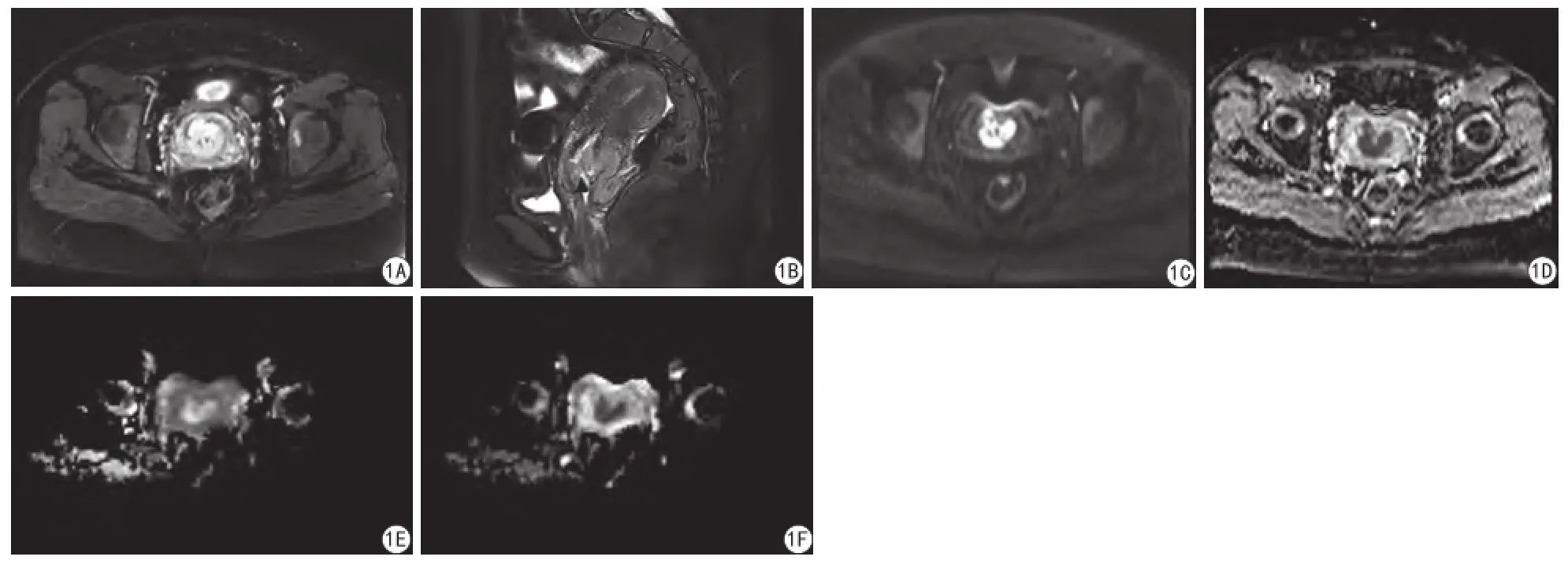
宫颈鳞癌的ADC、MD值均低于宫颈腺癌,MK值宫颈鳞癌高于宫颈腺癌,差异均具有统计学意义(P<0.001)(表1)。高分化、中分化、低分化宫颈鳞癌的ADC、MK、MD值差异有统计学差异(P<0.001)(表2),LSD两两比较,ADC、MK、MD值在高与中、中与低分化宫颈鳞癌中均具有统计学差异(P<0.001)(图1)。
2.3DWI与DKI鉴别宫颈鳞癌、腺癌与高、中、低分化宫颈鳞癌效能对比
3个参数对比,在鉴别宫颈鳞癌与腺癌方面,MK具有最优鉴别能力, ROC曲线下面积AUC最大(0.968),高于ADC与MD,差异具有明显统计学意义(P<0.001),各参数敏感度、特异度见表3(图2)。
鉴别低、中分化与中、高分化宫颈鳞癌时,MK值均具有最有诊断效能,曲线下面积分别为0.905(P=0.003)(表3)、0.940(P<0.001),其次依次为MD、ADC。鉴别低、中分化宫颈鳞癌时,各参数敏感度、特异度见表4;鉴别中、高分化宫颈鳞癌时,各参数敏感度、特异度见表5(图3,4)。
3 讨论
本研究首次对DKI在宫颈癌病理类型(腺癌、鳞癌)中的鉴别能力进行了探讨,结果显示宫颈鳞癌的MK值明显高于宫颈腺癌,宫颈鳞癌的MD、ADC值明显低于宫颈腺癌值,ROC曲线下面积MK最大,均高于以高斯模型为基础的传统DWI参数ADC值和MD值(P<0.001);高、中、低分化的宫颈鳞癌的MK、MD、ADC值,两两对比,差异均具有统计学意义(P<0.05),通过分析ROC曲线发现,MK在鉴别低中、中高分化鳞癌上均具有最大曲线下面积,其次为MD、ADC。虽然目前尚未见DKI关于宫颈癌的相关研究,但国内外有关鉴别脑、鼻咽、乳腺、肝脏、胆管、膀胱、前列腺病变良恶性和肿瘤病理类型及分级的研究[11-17]初步表明,非高斯模型中的扩散峰度参数诊断价值高于高斯模型中的扩散加权参数,与本研究结果相似。

图1 宫颈中分化鳞癌。轴面(A)及矢状面(B) T2WI抑脂像癌组织为稍高信号;常规DWI (C)为高信号,ADC图(D)为低信号;MK图(E)为高信号;MD图(F)为低信号Fig. 1 Cervical medium differentiation squamous cell cancer. axial view (A) and sagittal view (B) T2WI-FScancer tissue presents lightly high signal. DWI (C) presents high signal, ADC (D) presents lightly high signal. MK (E) presents high signal. MD (F) presents low signal.

图2 DWI、DKI各参数鉴别宫颈鳞癌、腺癌的ROC曲线 图3 DWI、DKI各参数鉴别宫颈低、中分化鳞癌的ROC曲线 图4 DWI、DKI各参数鉴别宫颈中、高分化鳞癌的ROC曲线Fig. 2 ROC curve of DWI, DKI metric in differentiating cervical squamous cell carcinoma, cervical adenocarcinoma. Fig. 3 ROC curve of DWI, DKI metric in differentiating low, medium differentiation cervical squamous cell carcinoma. Fig. 4 ROC curve of DWI, DKI metric in differentiating medium, high differentiation cervical squamous cell carcinoma.

表4 DWI、DKI各参数鉴别宫颈低、中分化鳞癌的ROC曲线对比Tab. 4 ROC curve of DWI, DKI metric in differentiating low, medium differentiation cervical squamous cell carcinoma

表5 DWI、DKI各参数鉴别宫颈中、高分化鳞癌的ROC曲线对比Tab. 5 ROC curve of DWI, DKI metric in differentiating medium,high differentiation cervical squamous cell carcinoma
MK、MD及ADC值三种参数从根本上来说均体现出癌组织细胞内外水分子的扩散运动状况。而癌组织细胞内外水分子的扩散运动状况受包括细胞核、细胞器的改变、核浆比、细胞密度、细胞内外水分子的比例等多种因素影响[18-19]。
在诸多因素中,细胞密度最为关键,为肿瘤病理类型与分级的重要评价参数。许多研究表明细胞密度是决定组织内水分子扩散情况的最关键因素,与正常组织相比,恶性肿瘤细胞增殖速度快、密度大、分布紧凑、外周空间缩小,造成组织中水分子扩散显著受限[20-21]。
从病理类型来看,鳞癌内细胞分布紧凑,细胞密度比腺癌大而均匀,又因无腺管样结构,故肿瘤组织内水分子扩散受限较为明显,而腺癌内细胞分布相对分散,细胞密度较低,又具有较多腺管样结构,间质成分较多,因此腺癌内水分子弥散相对自由,受限较弱。Matoba等[22]发现肺鳞癌细胞密度高于腺癌,间接支持本研究结果。
肿瘤细胞密度不仅与肿瘤病理类型密切相关,同时与肿瘤分化程度关系紧密。分化程度越低的鳞癌组织内,细胞异型性越大,伴随胞内核浆比变高,细胞器数量变大,造成细胞内水分子扩散受限;另一方面,分化程度越低,癌组织细胞密度越高,细胞外空间越狭窄,细胞外水分子扩散运动受限越明显。所以推测可知,高、中、低分化宫颈鳞癌MK、MD以及ADC值差别的病理学基础主要源于细胞密度的差异。
MK为DKI最关键的参数,代表空间各梯度方向的扩散峰度平均值[23],其与组织复杂程度呈正比,结构复杂度越高(如癌细胞病理分级越高、分布越紧凑) ,水分子运动阻碍则越显著,MK值越高[24]。
部分DKI在胶质瘤的研究结果显示,MK值同肿瘤恶性程度表现为正相关,ROC曲线分析发现,相比于DWI参数以及其他DKI参数,MK 在区分高、低级别胶质瘤中具有最佳诊断效能。这些研究[11,24-25]间接支持了本研究结果。在本研究结果中,与ADC与MD值相比,MK值在宫颈鳞、腺癌、宫颈鳞癌高中低分化鉴别中均具有最优诊断效能,也更加印证了宫颈癌病灶内水分子扩散特点符合非高斯模型。
MD值代表平均扩散率,与ADC值相比,反映多个扩散敏感梯度方向上的水分子的扩散特征,因此,从理论上讲,MD值在鉴别不同病理类型及分化程度的宫颈癌也较ADC值更具有优势,而本研究结果也支持这一理论假设。
目前尚未见DKI与宫颈癌病理特征相关性的报道,本研究结果显示在宫颈癌病理特征鉴别上,DKI优于DWI,与其他部位DKI研究结果相似,也更加印证了DKI在细微结构的观察能力优于DWI。
本研究也存在较多局限性:总体样本量较小,尤其鳞癌高、低分化组以及宫颈腺癌的病例数较少,目前关于DKI在宫颈癌病理类型及病理分级的研究尚无文献报道,需要更大样本量的研究进一步证实DKI在宫颈癌中的价值;由于目前DKI在宫颈癌方面的研究尚处于空白阶段,因此很难确定在宫颈扫描的最佳DKI序列参数值,如b值的多少、b值大小及扩散梯度多少方向的选择等等,故仍需更多学者参与其中进行研究。
[References]
[1] Torre LA, Bray F, Siegel RL, et al. Global cancer statistics, 2012. CA Cancer J Clin, 2015, 65(2): 87-108.
[2] Hu SY, Zheng RS, Zhao FH, et al. Trend analysis of cervical cancer incidence and mortality rates in Chinese women during 1989-2008. Acta Academiae Med Sinicae, 2014, 36(2): 129-135.胡尚英, 郑荣寿, 赵方辉,等. 1989至2008年中国女性子宫颈癌发病和死亡趋势分析. 中国医学科学院学报, 2014, 36(2): 129-135.
[3] Katanyoo K, Sanguanrungsirikul S, Manusirivithaya S. Comparison of treatment outcomes between squamous cell carcinoma and adenocarcinoma in locally cervical cancer. Gynecol Oncol, 2012, 125(2): 292-296.
[4] Chepovetsky J, Kalir T, Weiderpass E. Clinical applicability of microarray technology in the diagnosis, prognostic stratification, treatment and clinical surveillance of cervical adenocarcinoma. Curr Pharm Des, 2013, 19(8): 1425-1429.
[5] Hui ES, Cheung MM, Qi L, et al. Towards better MR characterization of neural tissues using directional diffusion kurtosis analysis. Neuro image, 2008, 42(1): 122-134.
[6] Jensen JH, Helpern JA, Ramani A, et al. Diffusional kurtosis imaging: the quantifcation of non-gaussian water diffusion by means of magnetic resonance imaging. Magn Reson Med, 2005, 53(6): 1432-1440.
[7] Jensen JH, Helpern JA. MRI quantification of non-Gaussian water diffusion by kurtosis analysis. NMR Biomed, 2010, 23(7): 698-710.
[8] Nilsson M, Wirestam R, Stahlberg F, et al. Regional values of diffusional kurtosis imaging estimates in the healthy brain. J MagnReson Imaging, 2013, 37(3): 610-618.
[9] Yoshida M, Hori M, Yokoyama K, et al. Diffusional kurtosis imaging of normal-appearing white matter in multiple sclerosis: Preliminary clinical experience. Jpn J Radiol, 2013, 31(1): 50-55.
[10] Dang YX, Wang XM. Present research situation of diffusion kurtosis imaging and intravoxel incoherent motion of the brain. Chin J Magn Reson Imaging, 2015, 6(2): 145-150.党玉雪, 王晓明. 磁共振新技术DKI和IVIM在中枢神经系统的研究现状. 磁共振成像, 2015, 6(2): 145-150.
[11] Jiang R, Jiang J, Zhao L, et al. Diffusion kurtosis imaging can efficiently assess the glioma grade and cellular proliferation. Oncotarget, 2015, 6(39): 42380-42393.
[12] Roethke MC, Kuder TA, Kuru TH, et al. Evaluation of diffusion kurtosis imaging versus standard diffusion imaging for detection and grading of peripheral zone prostate cancer. Invest Radiol, 2015, 50(8): 483-489.
[13] Nogueira L, Brandao S, Matos E, et al. Application of the diffusion kurtosis model for the study of breast lesions. Eur Radiol, 2014, 24(6): 1197-1203.
[14] Suo S, Chen X, Ji X. Investigation of the non-Gaussian water diffusion properties in bladder cancer using diffusion kurtosis imaging: a preliminary study. J Comput Assist Tomogr, 2015, 39(2): 281-285.
[15] Goshima S, Kanematsu M, Noda Y. Diffusion kurtosis imaging to assess response to treatment in hypervascular hepatocellular carcinoma. AJR Am J Roentgenol, 2015, 20(5): W543-549.
[16] Chen Y, Ren W, Zheng D. Diffusion kurtosis imaging predicts neoadjuvant chemotherapy responses within 4 days in advanced nasopharyngeal carcinoma patients. J Magn Reson Imaging, 2015, 42(5): 1354-1361.
[17] Xu ML, Xing CH, Chen HW, et al. Application value of DKI in grading of extrahepatic cholangiocarcinoma. Chin J Magn Reson Imaging, 2016, 7(1): 34-39.徐蒙莱, 邢春华, 陈宏伟, 等. DKI技术在肝外胆管癌分级中的应用价值. 磁共振成像, 2016, 7( 1): 34-39.
[18] Motoshima S, Irie H, Nakazono T, et al. Diffusion-weighted MR imaging in gynecologic cancers. J Gynecol Oncol, 2011, 22(4): 275-287.
[19] Woodhams R, Kakita S, Hata H, et al. Diffusion-weighted imaging of mucinous carcinoma of the breast: evaluation of apparent diffusion coeffcient and signal intensity in correlation with histologic fndings. AJR Am J Roentgenol, 2009, 193(1): 260-266.
[20] Matsumoto Y, Kuroda M, Matsuya R, et al. In vitro experimental study of the relationship between the apparent diffusion coeffcient and changes in cellularity and cell morphology. Oncol Rep, 2009, 22(3): 641-648.
[21] Manenti G, Roma D, Mancino S, et al. Malignant renal neoplasms: correlation between ADC values and cellularity in diffusion weighted magnetic resonance imaging at 3 T. Radial Med, 2008, 113(2): 199-213.
[22] Matoba M, Tonami H, Kondou T, et al. Lung carcinoma: diffusion weighted MR imaging preliminary evaluation with apparent diffusion coeffcient. Radiology, 2007, 243(2): 570-577.
[23] Hui ES, Cheung MM, Qi L, et al. Advanced MR diffusion characterization of neural tissue using directional diffusion kurtosis analysis. Conf Proc IEEE Eng Med Biol Soc, 2008: 3941-3944.
[24] Raab P, Hattingen E, Franz K, et al. Cerebral gliomas: diffusional kurtosis imaging analysis of microstructural differences. Radiology, 2010, 254(3):876-881.
[25] Van Cauter S, De Keyzer F, Sima DM, et al. Integrating diffusion kurtosis imaging, dynamic susceptibility-weighted contrast-enhanced MRI, and short echo time chemical shift imaging for grading gliomas. Neuro Oncol, 2014, 16(7): 1010-1021.
The relationship between DKI and pathological features of cervical cancer: a primary study
YAN kun, HU Sha-sha, YANG Pin, LI Jin-kui, ZHAI Ya-jun, LEI Jun-qiang*
Department of Radiology, the First Hospital of Lanzhou University, Lanzhou 730000, China
*Correspondence to: Lei JQ, E-mail: leijq1990@163.com
Objective: To evaluate and contrast diffusion-weighted imaging (DWI) and diffusion kurtosis imaging (DKI) in differentiating cervical cancer pathological type and degree of differentiation. Materials and Methods: Collecting 39 patients with cervical cancer diagnosed by pathological examination, 31 were cervical squamous cell cancer (7 high, 19 medium and 5 low differentiation cervical squamous cell cancer), and the remaining 8 patients were cervical adenocarcinoma. Everyone underwent cervical magnetic resonance imaging (MRI) examination, the sequences included conventional, DWI and DKI. Compared mean value of ADC (apparent diffusion coefficient), MK (mean kurtosis) and MD (mean diffusion) of cervical squamous cell cancer, cervical adenocarcinoma and high, medium and low differentiation cervical squamous cell cancer. Evaluate the discriminability of ADC, MK and MD in cervical squamous cell cancer, cervical adenocarcinoma and high, medium and low differentiation cervical squamous cell cancer by receiver operating characteristics (ROC) curves. Results: (1) The mean values of ADC, MD were lower in cervical squamous cell carcinoma contrast to adenocarcinoma (P<0.001), but the mean values of MK in cervical squamous cell carcinoma were higher contrast to adenocarcinoma (P<0.001). When differentiating cervical squamous cell carcinoma from cervical adenocarcinoma, mean value of MK possessed a biggest AUC (0.968), followed MD (0.940) and ADC (0.915). (2) When differentiating poorly from medium , medium from high differentiated squamous cell carcinoma, the mean value of MK had best ability with largest AUC (0.905, P=0.003. 0.940, P<0.001), followed MD (AUC=0.884, P=0.009. AUC=0.887, P=0.002) and ADC (AUC=0.853, P=0.012. AUC=0.842, P=0.003). Conclusions: DKI is better than DWI ondiscriminating cervical squamous cell carcinoma, cervical adenocarcinoma and high, medium and low differentiated subtypes of cervical squamous carcinoma.
Uterine cervical neoplasms; Diffusion magnetic resonance imaging; Pathology
9 Apr 2016, Accepted 26 May 2016
甘肃省兰州市兰州大学第一医院,730000
雷军强,E-mail:leijq1990@163.com
2016-04-09
接受日期:2016-05-26
R445.2;R737.33
A
10.12015/issn.1674-8034.2016.09.009
闫坤, 胡莎莎, 杨品, 等. 扩散峰度成像与宫颈癌病理学特征相关性的初步探索. 磁共振成像, 2016, 7(9): 683-688.

