磷酸二氢钠体系中316L不锈钢表面微孔结构的制备
张艳梅*,朱用洋,许增才,张晓华,吴一凡,陈玲,揭晓华
(广东工业大学材料与能源学院,广东 广州 510006)
磷酸二氢钠体系中316L不锈钢表面微孔结构的制备
张艳梅*,朱用洋,许增才,张晓华,吴一凡,陈玲,揭晓华
(广东工业大学材料与能源学院,广东 广州 510006)
在0 °C下采用磷酸二氢钠的水溶液对316L不锈钢进行阳极氧化,以制备高密度、有序的微孔结构。研究了NaH2PO4浓度、氧化电压、氧化时间、丙三醇添加量等工艺参数对氧化膜多孔结构的影响。结果表明,氧化液中NaH2PO4浓度低于0.10 mol/L时,氧化膜表面仅能形成极浅的凹坑。氧化电压低于30 V时,氧化膜的微孔结构分布稀疏。NaH2PO4浓度或氧化电压过高都会使基体溶解速率过快,氧化膜的多孔结构消失。最佳NaH2PO4浓度和氧化电压分别为0.30 mol/L和30 ~ 40 V。在该条件下对316L不锈钢阳极氧化20 min,可获得有序排列的蜂窝状微孔结构,孔径为180 ~ 260 nm,孔密度约为1.5 × 10⁹个/cm2。氧化液中添加10%(体积分数)丙三醇后,所得微孔结构的均匀性提高,但是孔变浅,因此不建议在氧化液中加丙三醇。
不锈钢;阳极氧化;多孔结构;磷酸二氢钠;丙三醇
First-author's address: Faculty of Materials and Energy, Guangdong University of Technology, Guangzhou 510006,China
近年来,多孔结构在磁学、电学、光学、传感装置、生物材料等领域获得广泛应用。医用316L不锈钢常用来制作骨板、骨钉、血管支架等医疗器件[1-3]。在 316L不锈钢表面构建纳米多孔结构,利用多孔结构作为药物载体(吸附药物)来实现局部的、长效的、可控的药物释放,具有药物用量少而全身毒副作用小的优点[4]。此外,表面多孔组织还能加速内皮化[5-6]。因此,不锈钢表面多孔结构在医用生物材料中具有广泛的应用前景。
表面多孔结构的制备方法主要有喷砂[7]、激光雕刻[8]、化学腐蚀[9]和阳极氧化[10-14]。其中,阳极氧化法具有操作简单、成本低等特点,主要用于铝[10]、钛[11]、钽[12]、钨[13]、锆[14]等阀金属表面多孔结构的制备,但近年来已逐渐拓展到纯铁[15-17]和不锈钢[18-21]基体的表面处理。Kure等[18]报道了采用氟化铵-乙二醇体系可在 304不锈钢表面制得多孔结构,但孔结构形貌未进行详细介绍。Lu[19]、Martin[20]、Zhan[22]等采用高氯酸-乙二醇体系,分别在 AISI 304、904L、304L不锈钢表面获得了多孔结构,但孔都较浅,深度只有 20 nm左右。目前采用阳极氧化法在不锈钢表面制备多孔结构的文献相对较少,且均以乙二醇作为溶剂,制备的多孔结构形貌尚不理想。为了进一步优化多孔结构形貌,电解液配方、氧化工艺、多孔结构的形成机理等研究尚待深入。
本文拟采用NaH2PO4水溶液在316L不锈钢表面制备多孔结构,研究多孔结构的形貌特点及工艺参数对孔结构的影响,并进行工艺优化。
1 实验
1. 1 预处理
基体材料为40 mm × 20 mm × 0.2 mm的316L不锈钢。先用800#至2000#碳化硅砂纸逐级打磨,再用去离子水冲洗干净,并干燥。然后依次用丙酮和863金属清洗液超声清洗15 min除油。最后以不锈钢为阳极,高纯石墨为阴极,进行电化学抛光,具体参数为:硫酸400 mL/L,磷酸600 mL/L,阳极电流密度50 A/dm2,温度85 °C,极间距50 mm,时间3 ~ 4 min。
1. 2 阳极氧化
采用0.03 ~ 0.45 mol/L NaH2PO4水溶液体系,经预处理的316L不锈钢为阳极,高纯石墨为阴极,工艺条件为:电压15 ~ 60 V,温度0 °C(采用上海勒顿DFY-5/10 °C型低温恒温水浴仪恒温),时间5 ~ 60 min。
1. 3 阳极氧化膜的微观结构分析
采用日本日立公司的SU8010型场发射扫描电镜(SEM)分析不锈钢阳极氧化后的表面形貌。采用Image-pro plus 6.0软件分析孔径和孔密度。
2 结果与讨论
2. 1 磷酸二氢钠浓度对微孔结构的影响
图1为电压30 V下采用不同浓度NaH2PO4溶液对316L不锈钢阳极氧化处理20 min后的表面形貌。从图1a可知,在0.03 mol/L NaH2PO4电解液中阳极氧化后,不锈钢表面形成一些极浅的凹坑(见白色箭头所指)。这是因为此时NaH2PO4浓度较低,对孔底氧化膜的侵蚀较慢,底部基体的溶解较慢,孔深生长缓慢。从图1b可知,NaH2PO4浓度为0.10 mol/L时,凹坑加深,但孔分布仍较分散且不均匀(图1b中A、B区)。从图1c可知,NaH2PO4浓度为0.30 mol/L时,由于NaH2PO4浓度较高,腐蚀较快,基体表面生成了高密度蜂窝状排列的多孔结构阵列,孔径为180 ~ 260 nm,孔密度约为1.5 × 10¹⁰个/cm2。从图1d可知,NaH2PO4浓度为0.45 mol/L时,由于孔壁溶解加快,孔变浅,孔壁减薄,孔径增大到220 ~ 450 nm,孔密度减小,甚至有连孔现象(图1d中白线区域)。因此适宜的NaH2PO4浓度为0.30 mol/L。仔细观察图1c可发现,316L不锈钢上的多孔结构与纯铝表面的六方形纳米多孔结构[10]不同,微孔呈不规则的圆形,并且孔径大小不一。可能是因为不锈钢基体为合金,在阳极氧化过程中一些元素会优先溶解,而这种优先溶解与微观结构关系密切[23]。

图1 316L不锈钢在不同浓度NaH2PO4溶液中阳极氧化后的表面形貌Figure 1 Surface morphologies of 316L stainless steel anodized in aqueous solution containing different contents of NaH2PO4
2. 2 氧化电压对微孔结构的影响
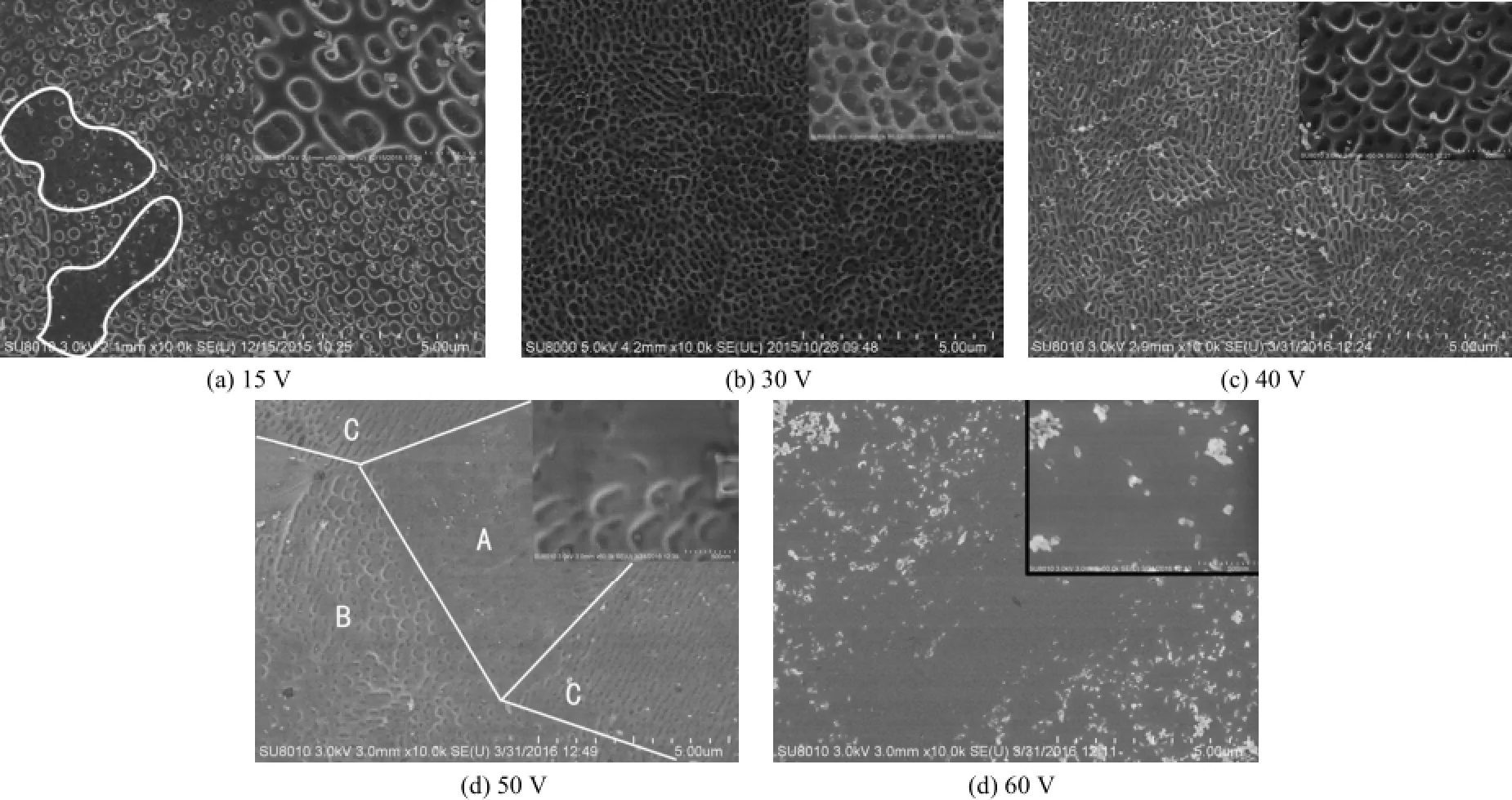
图2是在不同电压下采用0.30 mol/L NaH2PO4溶液对316L不锈钢阳极氧化20 min后的表面形貌。从图2可知,电压为15 V时,不锈钢表面的多孔结构分布不均(如图2a圈线处的孔小而浅)且较稀疏,呈圆形或椭圆形,孔径为130 ~ 260 nm。电压为30 V时,不锈钢表面生成了排列紧密的蜂窝状多孔结构,孔径为180 ~ 260 nm,孔边缘清晰。电压为40 V时的表面形貌与30 V时相近,但孔径略大,在220 ~ 300 nm范围内,孔略浅。当电压增至50 V时,多孔结构的溶解速率加快,图2d中A区域的溶解较为严重,多孔结构几乎消失,只剩下极少的凹坑;B区域很明显是溶解后剩下的极浅孔洞;C区域的长条沟壑状是溶解后孔洞相连形成的。当电压增大到60 V时,多孔结构完全溶解,表面变得光滑平整。从宏观上看,当氧化电压<50 V时,316L不锈钢表面呈黄色,氧化电压≥50 V时则呈白色。可见,电压对316L不锈钢表面多孔结构形貌的影响较大,电压在30 ~ 40 V之间时能形成高密度有序的多孔结构。

图2 316L不锈钢在不同电压下阳极氧化后的表面形貌Figure 2 Surface morphologies of 316L stainless steel anodized at different voltages
2. 3 氧化时间对微孔结构的影响
其他参数同2.2,在电压30 V下,阳极氧化不同时间后不锈钢表面多孔结构的形貌如图3所示。从图3可知,氧化5 min时,氧化层表面在微观上不平整。氧化10 min时,不锈钢表面呈现出向微孔过渡的形貌,但孔结构还不清晰,深度小,边缘形状不规则。氧化15 min时,不锈钢表面有较浅的多孔结构形成,但孔边缘形状不规则。氧化20 min时,不锈钢表面多孔结构排列规整,形状较为规则,孔边缘清晰,深度较大。当氧化时间延长至60 min后,多孔结构变化不大。所以,氧化20 min时,基体表面已形成了稳定的多孔形貌,延长时间对多孔形貌的影响不大。

(d) 20 min (e) 60 min图3 316L不锈钢阳极氧化不同时间后的表面形貌Figure 3 Surface morphologies of 316L stainless steel anodized for different time
2. 4 添加丙三醇对微孔结构的影响
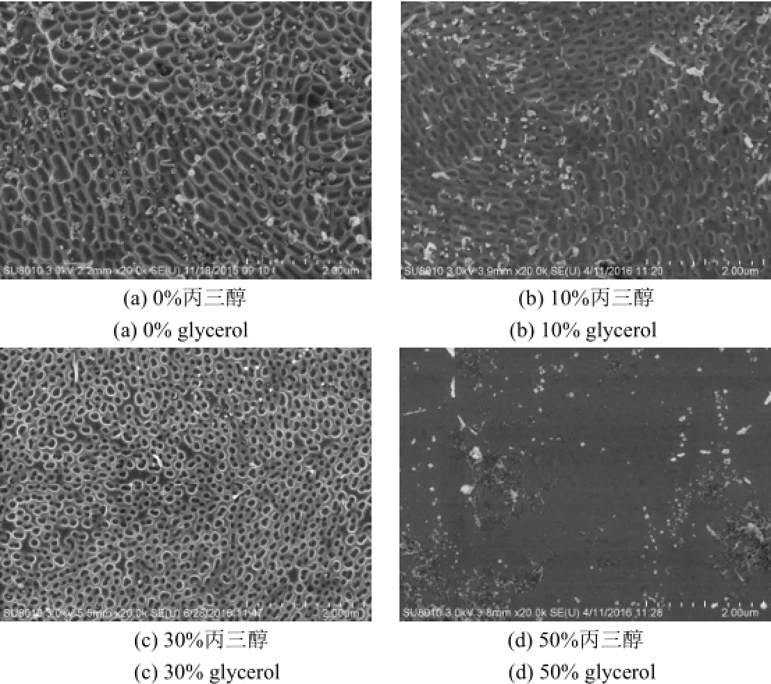
将丙三醇添加到阳极氧化电解液中可以提高电解液黏度,降低电导率,减少氧化物在电解液中的溶解,有利于阀金属表面形成有序的多孔结构[26-27]和促进不锈钢在INCO电解液中形成多孔结构[28]。因此在电解液中添加不同含量的丙三醇,在上文确定的较优条件(NaH2PO4浓度0.30 mol/L,电压30 V,温度0 °C,时间20 min)下对316L不锈钢进行阳极氧化,以研究丙三醇对316L不锈钢表面纳米多孔结构形成的影响,结果见图4。

图4 316L不锈钢在不同含量丙三醇溶液中阳极氧化后的表面形貌Figure 4 Surface morphologies of 316L stainless steel anodized in aqueous solution with different contents of glycerol
从图4可知,与未添加丙三醇时相比,添加10%(体积分数,下同)丙三醇后,孔密度变化不大,孔结构均匀性改善,但孔变浅、变厚,孔径减小。丙三醇用量为30%时,孔的形状接近规则的圆形,孔径减小至100 nm左右,孔密度增大至3.3 × 10⁹个/cm2。添加50%丙三醇后,表面平整,无纳米多孔结构存在。
综上可知,虽然添加适量丙三醇可改善多孔结构的均匀性,但也使孔变浅,这不利于实际应用,因此不建议在阳极氧化液中添加丙三醇。
3 结论
(1) 电解液中添加适量丙三醇有助于改善不锈钢阳极氧化膜多孔结构的均匀性,但添加过多不利于表面多孔结构的形成。
(2) 316L不锈钢表面制备多孔结构的最优工艺为:NaH2PO4浓度0.30 mol/L,电压30 V,温度0 °C,时间20 min。该条件下,可在316L不锈钢上得到有序的蜂窝状多孔结构,孔较深,孔径为180 ~ 260 nm,孔密度为109~ 1010个/cm2。
[1] CARDENAS L, MACLEOD J, LIPTON-DUFFIN J, et al. Reduced graphene oxide growth on 316L stainless steel for medical applications [J]. Nanoscale, 2014,6 (15): 8664-8670.
[2] ELTER P, SICKEL F, EWALD A. Nanoscaled periodic surface structures of medical stainless steel and their effect on osteoblast cells [J]. Acta Biomaterialia,2009, 5 (5): 1468-1473.
[3] LATIFI A, IMANI M, KHORASANI M T, et al. Electrochemical and chemical methods for improving surface characteristics of 316L stainless steel for biomedical applications [J]. Surface and Coatings Technology, 2013, 221: 1-12.
[4] GULTEPE E, NAGESHA D, CASSE B D F, et al. Sustained drug release from non-eroding nanoporous templates [J]. Small, 2010, 6 (2): 213-216.
[5] MAROIS Y, SIGOT-LUIZARD M-F, GUIDOIN R. Endothelial cell behavior on vascular prosthetic grafts: effect of polymer chemistry, surface structure, and surface treatment [J]. ASAIO Journal, 1999, 45: 272-280.
[6] GOLDEN M A, HANSON S R, KIRKMAN T R, et al. Healing of polytetrafluoroethylene arterial grafts is influenced by graft porosity [J]. Journal of Vascular Surgery, 1990, 11 (6): 838-845.
[7] 崔晓明, 陈良建, 郑遥. 不同方法制备钛种植体对成骨细胞黏附影响的体外研究[J]. 山西医科大学学报, 2013, 44 (1): 75-79.
[8] 顾兴中, 倪中华. 微孔结构血管支架的激光切割工艺[J]. 华中科技大学学报(自然科学版), 2007, 35 (增刊1): 143-146.
[9] 董何彦, 马宗民, 程增兵. 新型无聚合物微盲孔载药支架的研究[J]. 辽宁师范大学学报(自然科学版), 2012, 35 (1): 93-97.
[10] VARGHESE O K, GONG D W, PAULOSE M, et al. Highly ordered nanoporous alumina films: effect of pore size and uniformity on sensing performance [J]. Journal of Materials Research, 2002, 17 (5): 1162-1171.
[11] MACAK J M, TSUCHIYA H, TAVEIRA L, et al. Smooth anodic TiO2nanotubes [J]. Angewandte Chemie International Edition, 2005, 44 (45): 7463-7465.
[12] WEI W, MACAK J M, SCHMUKI P. High aspect ratio ordered nanoporous Ta2O5films by anodization of Ta [J]. Electrochemistry Communications, 2008,10 (3): 428-432.
[13] TSUCHIYA H, MACAK J M, SIEBER I, et al. Self-organized porous WO3formed in NaF electrolytes [J]. Electrochemistry Communications, 2005, 7 (3): 295-298.
[14] LEE W J, SMYRL W H. Zirconium oxide nanotubes synthesized via direct electrochemical anodization [J]. Electrochemical and Solid-State Letters, 2005, 8 (3): B7-B9.
[15] HABAZAKI H, KONNO Y, AOKI Y, et al. Galvanostatic growth of nanoporous anodic films on iron in ammonium fluoride-ethylene glycol electrolytes with different water contents [J]. The Journal of Physical Chemistry C, 2010, 114 (44): 18853-18859.
[16] HABAZAKI H, OIKAWA Y, FUSHIMI K, et al. Importance of water content in formation of porous anodic niobium oxide films in hot phosphate-glycerol electrolyte [J]. Electrochimica Acta, 2009, 54 (3): 946-951.
[17] LATEMPA T J, FENG X J, PAULOSE M, et al. Temperature-dependent growth of self-assembled hematite (α-Fe2O3) nanotube arrays: rapid electrochemical synthesis and photoelectrochemical properties [J]. The Journal of Physical Chemistry C, 2009, 113 (36): 16293-16298.
[18] KURE K, KONNO Y, TSUJI E, et al. Formation of self-organized nanoporous anodic films on Type 304 stainless steel [J]. Electrochemistry Communications,2012, 21: 1-4.
[19] LU W, ZOU D, HAN Y, et al. Self-organised nanoporous anodic films on superaustenitic stainless steel [J]. Materials Research Innovations, 2014, 18 (sup4): 747-750.
[20] MARTIN F, FRARI D D, COUSTY J, et al. Self-organisation of nanoscaled pores in anodic oxide overlayer on stainless steels [J]. Electrochimica Acta, 2009, 54 (11): 3086-3091.
[21] 张艳梅, 黄家强, 许增才, 等. 430不锈钢表面微纳多孔结构阳极氧化膜的制备及疏水性研究[J]. 电镀与涂饰, 2015, 34 (12): 662-667.
[22] ZHAN W T, NI H W, CHEN R S, et al. Formation of nanopore arrays on stainless steel surface by anodization for visible-light photocatalytic degradation of organic pollutants [J]. Journal of Materials Research, 2012, 27 (18): 2417-2424.
[23] TSUCHIYA H, MACAK J M, GHICOV A, et al. Nanotube oxide coating on Ti-29Nb-13Ta-4.6Zr alloy prepared by self-organizing anodization [J]. Electrochimica Acta, 2006, 52 (1): 94-101.
[24] ZHANG B W, NI H W, CHEN R S, et al. A two-step anodic method to fabricate self-organised nanopore arrays on stainless steel [J]. Applied Surface Science,2015, 351: 1161-1168.
[25] FATTAH-ALHOSSEINI A, SAATCHI A, GOLOZAR M A, et al. The transpassive dissolution mechanism of 316L stainless steel [J]. Electrochimica Acta, 2009,54 (13): 3645-3650.
[26] LEE B G, CHOI J W, LEE S E, et al. Formation behavior of anodic TiO2nanotubes in fluoride containing electrolytes [J]. Transactions of Nonferrous Metals Society of China, 2009, 19 (4): 842-845.
[27] MURATORE F, BARON-WIECHEĆ A, GHOLINIA A, et al. Comparison of nanotube formation on zirconium in fluoride/glycerol electrolytes at different anodizing potentials [J]. Electrochimica Acta, 2011, 58: 389-398.
[28] BERVIAN A, LUDWIG G A, KUNST S R, et al. The influence of the glycerin concentration on the porous structure of ferritic stainless steel obtained by anodization [J]. DYNA, 2015, 82 (190): 46-52.
[ 编辑:周新莉 ]
Preparation of microporous structure on 316L stainless steel in aqueous sodium dihydrogen phosphate solution
ZHANG Yan-mei*, ZHU Yong-yang, XU Zeng-cai, ZHANG Xiao-hua, WU Yi-fan, CHEN Ling, JIE Xiao-hua
316L stainless steel was anodized in sodium dihydrogen phosphate aqueous solution at 0 °C to form an orderly and highly-dense microporous structure on its surface. The effects of process parameters including NaH2PO4concentration,oxidation voltage and time, and dosage of glycerol on the microporous structure of oxidation film were studied. It was found that only an oxidation film with extremely shallow pits is formed in anodizing solution with a NaH2PO4concentration below 0.10 mol/L. The micropores are sparsely distributed on the oxidation film obtained at anodizing voltages below 30 V. Too high NaH2PO4concentration and anodizing voltage result in too fast dissolution of substrate and the disappearance of microporous structure of oxidation film. The optimal NaH2PO4concentration and anodizing voltage is 0.30 mol/L and 30-40 V, respectively. An orderly and highly-dense honeycomb-like microporous structure with a pore diameter of 180-260 nm and a density of 1.5 × 10⁹pores/cm2can be obtained by anodizing under the given conditions for 20 min. The uniformity of microporous structure is improved by adding 10vol% glycerol to anodizing solution, but the pores become shallow. It is not recommended to add glycerol to anodizing solution.
stainless steel; anodization; porous structure; sodium dihydrogen phosphate; glycerol
TQ153.6
A
1004 - 227X (2016) 16 - 0853 - 05
2016-05-17
2016-06-28
广东省科技计划项目(2015A010105027);揭阳市产学研结合项目(201416)。
张艳梅(1972-),女,山西高平人,博士,副教授,主要从事材料表面改性、新材料制备及材料性能方面的研究工作。
作者联系方式:(E-mail) zhyanmei2006@126.com。

