内皮祖细胞在实体瘤治疗中的作用研究进展
黄 睿,李 静,王 丽,杨举伦
内皮祖细胞在实体瘤治疗中的作用研究进展
黄 睿,李 静,王 丽,杨举伦
内皮祖细胞;实体瘤;靶点;基因载体
1997年,Asahara等[1]运用免疫磁珠分选法首次从成人外周血中分离出了一种CD34+/VEGFR-2+、可分化为成熟内皮细胞(endothelial cells,ECs)的单个核细胞,将之命名为内皮祖细胞(endothelial progenitor cells,EPCs)。内皮祖细胞参与胚胎时期血管发生(vasculogenesis),在成年个体可通过血管发生和血管生成(angiogenesis)两种方式参与血管新生[2-3]。内皮祖细胞主要存在于骨髓中,此外,脐带血、外周血、脂肪、脾脏、胎肝等组织中也有储存。这为体外分离、诱导培养及研究内皮祖细胞提供了可能。目前,内皮祖细胞已被广泛应用于缺血性疾病防治的研究,有关内皮祖细胞在实体瘤生长中的作用也引起了人们的重视。本文将就内皮祖细胞在实体瘤治疗中的作用研究进展进行阐述。
1 内皮祖细胞的生物学特点
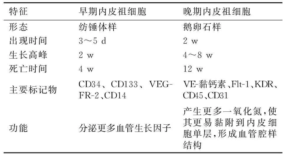
内皮祖细胞是胚胎时期由造血母细胞(hemangioblast)在卵黄囊中分化而来。在体外可采用免疫磁珠分选法、流式细胞分选法及密度梯度离心法等从不同组织中分离得到内皮祖细胞,并采用直接贴壁法或间接贴壁法进行诱导培养[4]。Hur等[5]在内皮祖细胞体外诱导培养过程中发现了早期内皮祖细胞(early EPC)和晚期内皮祖细胞(late EPC)(表1)。目前尚缺乏特异性标记物对内皮祖细胞进行鉴定,需联合采用形态学、表型鉴定、功能检测等方法。一般将同时表达CD34、CD133、VEGFR-2的细胞认定为内皮祖细胞[6]。其中,CD133随内皮祖细胞分化为成熟内皮细胞转为阴性。此外,还有学者认为CD14也可作为内皮祖细胞的鉴别标志之一[7]。功能检测:内皮祖细胞可吞噬乙酰化低密度脂蛋白(AcLDL),并与荆豆凝集素-1(UEA-1)结合[8];在Matrigel凝胶上能够形成血管腔样结构[9];可表达一氧化氮合酶及产生一氧化氮。

表1 早期内皮祖细胞与晚期内皮祖细胞的比较
2 内皮祖细胞与实体瘤血管新生
2.1 实体瘤血管新生 实体瘤血管新生将导致其侵袭力、转移力增强。病理组织学研究显示,在肿瘤组织中,血管周围的细胞能获得充足的养分,生长良好;而远离血管的细胞因缺乏营养而死亡。
2.2 内皮祖细胞参与实体瘤血管新生 受实体瘤生长刺激,内皮祖细胞从储存组织被动员至外周血中发挥作用。这是一个复杂的过程,涉及多种生长因子、酶、配体和细胞表面受体的参与,其具体机制尚不清楚。有研究表明,基质金属蛋白酶-9(MMP-9)在此过程中发挥关键作用[10]。MMP-9的激活可使膜结合Kit配体(mKitL)转化为可溶性Kit配体(sKitL),促进cKit+的干/祖细胞向骨髓微环境的血管区移动。受此作用,血管内皮生长因子(VEGF)、基质细胞衍生因子(SDF-1)的表达水平上调,促进内皮祖细胞从组织中动员至外周血。此外,促红细胞生成素(EPO)、粒细胞集落刺激因子(G-CSF)、PPARγ激动剂等也有促进内皮祖细胞动员的作用。在一些细胞因子及趋化因子(如:VEGF[11]、SDF-1等[12])的介导下,内皮祖细胞可迁移至肿瘤周边部位,经过黏附、侵袭、分化等一系列过程参与实体瘤新生血管的形成。此外,VEGF/VEGFR-2信号通路、SDF-1/CXCR-4信号通路[13]、Notch信号通路[14]及PI3K/Akt信号通路等[15]被认为在此过程中起着十分重要的作用。
3 内皮祖细胞在实体瘤治疗中的作用
3.1 内皮祖细胞作为实体瘤治疗的潜在靶点 通过对肺癌[16]、乳腺癌[17]、非小细胞肺癌[18]、胰腺癌[19]及恶性胶质细胞瘤等[20]实体瘤患者外周血中内皮祖细胞的数量进行检测后发现,其含量增加与疾病分期及进展相关,提示内皮祖细胞是这些肿瘤治疗的潜在靶点。目前,尚无直接作用于内皮祖细胞的药物。但可通过抑制与其功能相关的因子及信号通路,间接达到治疗肿瘤的目的。这样虽不能直接杀死肿瘤细胞,但可使肿瘤长期处于无进展状态,对于延长患者的生存期大有裨益。
Gao等[21]发现,抑制分化抑制因子-1(inhibitor of differentiation-1,Id1)的表达,可以抑制内皮祖细胞促血管生成的功能,延长肺癌荷瘤鼠的生存周期。Willett等[22]在对接受抗血管生成治疗的直肠癌患者的研究中发现,应用基于VEGF的抗血管生成药物贝伐单抗后,患者体内肿瘤血管密度和内皮祖细胞数量明显减少。
3.2 内皮祖细胞作为实体瘤靶向治疗的基因载体 Wang等[23]将标记后的内皮祖细胞注入人乳腺癌荷瘤鼠模型体内,发现内皮祖细胞能被募集到瘤床部位,参与肿瘤血管新生。王鈜等[24]用携带增强型绿色荧光蛋白的腺病毒(Ad5-EGFP)感染内皮祖细胞,将其注入结肠癌荷瘤鼠模型体内,结果仅在肿瘤周边组织发现荧光细胞的分布,证实了内皮祖细胞的肿瘤靶向性;而后又进用携带人伽马干扰素(hγIFN)基因的腺病毒(Ad5-hγIFN)感染内皮祖细胞,结果发现这些内皮祖细胞(EPCs- hγIFN)在动物体内可抑制肿瘤生长。Arbab等[25]及Varma等[26]在研究神经胶质瘤,袁献温等[27]在研究肝癌的过程中均得出了类似的结论。以上研究结果提示,内皮祖细胞具备作为实体瘤靶向治疗基因载体的潜能。选用内皮祖细胞作为基因载体,能使药物快速准确达到肿瘤部位,减少因血液循环带来的药物损失,维持局部药物浓度,提高疗效。
3.3 内皮祖细胞作为化疗疗效的预测指标 Roodhart等[28]及Fürstenberger等[29]在研究中运用流式细胞术对接受化疗后患者的外周血进行检测,发现内皮祖细胞数量增加与抗肿瘤治疗的效果及预后有关。这可能与在接受化疗时,机体自身的抵抗机制有关。在化疗中联合采用抗血管生成疗法将有助于提高肿瘤治疗的效果。栾文强等[30]检测了非小细胞肺癌(NSCLC)患者化疗前后外周血中内皮祖细胞的数量,发现联合应用化疗及抗血管生成治疗后,患者外周血中内皮祖细胞的数量下降幅度大于单用化疗者;且化疗联合抗血管生成治疗对非小细胞肺癌的临床有效率及收益率均优于单用化疗。
3.4 内皮祖细胞改善放疗后局部组织血液供应 在恶性肿瘤的治疗中,有很大一部分患者需要联合采用放疗以提高疗效。射线虽能促使肿瘤细胞发生凋亡,但也会造局部组织缺血坏死。已有相关研究表明,通过移植内皮祖细胞可改善放疗后组织的血液供应。这为治疗放疗后组织缺血提供了一种新的途径。
4 总结与展望
内皮祖细胞是一类干/组细胞,在肿瘤发生时能从不同组织中被动员至外周血,靶向分布到肿瘤周边组织,以血管发生和血管生成两种方式参与实体瘤血管新生,具备作为实体瘤治疗靶点及基因载体的潜能。然而,内皮祖细胞应用于临床的有效性及安全性尚待评估。首要问题就是找到一种能在体外大量分离、诱导培养内皮祖细胞的方法。此外,如何对内皮祖细胞进行鉴定;经基因改造的内皮祖细胞是否会改变其祖细胞特性;基于内皮祖细胞的疗法与其他一些抗肿瘤治疗方法联合应用等一系列问题仍需进一步研究。本实验室已成功构建了一种能够在肿瘤细胞中特异性表达抗p21ras单链抗体的腺病毒载体。如果以内皮祖细胞搭载此种腺病毒载体治疗肿瘤,可能在很大程度上提高靶向治疗的精准性和安全性。虽然目前关于内皮祖细胞在实体瘤治疗中的作用尚处于实验研究阶段,有很多问题有待解决。但相信随着关于内皮祖细胞研究的深入,内皮祖细胞在实体瘤治疗领域将具有广阔的应用前景。
[1] Asahara T,Murohara T,Sullivan A,et al.Isolation of putative progenitor endothelial cells for angiogenesis[J].Science,1997,275:964-967.
[2] Simons M.Angiogenesis:where do we stand now[J]?Circulation,2005,111:1556-1566.
[3] Skalak TC.Angiogenesis and microvascular remodeling:a brief history and future roadmap[J].Microcirculation,2005,12:47-58.
[4] Eggermann J,Kliche S,Jarmy G,et al.Endothelial progenitor cell culture and differentiation in vitro:a methodological comparison using human umbilical cord blood[J].Cardiovasc Res,2003,58:478-486.
[5] Hur J,Yoon CH,Kim HS,et al.Characterization of two types of endothelial progenitor cells and their different contributions to neovasculogenesis[J].Arterioscler Thromb Vasc Biol,2004,24:288-293.
[6] Gehling UM,Ergun S,Schumacher U,et al.In vitro differentiation of endothelial cells from AC133-positive progenitor cells[J].Blood,2000,95:3106-3112.
[7] Fujiyama S,Amano K,Uehira K,et al.Bone marrow monocyte lineage cells adhere on injured endothelium in a monocyte chemoattractant protein-1-dependent manner and accelerate reendothelialization as endothelial progenitor cells[J].Circ Res,2003,93:980-989.
[8] Loomans CJ,Wan H,de Crom R,et al.Angiogenic murine endothelial progenitor cells are derived from a myeloid bone marrow fraction and can be identified by endothelial NO synthase expression[J].Arterioscler Thromb Vasc Biol,2006,26:1760-1767.
[9] Harris LJ,Zhang P,Abdollahi H,et al.Availability of adipose-derived stem cells in patients undergoing vascular surgical procedures[J].J Surg Res,2010,163:e105-112.
[10] Heissig B,Hattori K,Dias S,et al.Recruitment of stem and progenitor cells from the bone marrow niche requires MMP-9 mediated release of kit-ligand[J].Cell,2002,109:625-637.
[11] Murayama T,Tepper OM,Silver M,et al.Determination of bone marrow-derived endothelial progenitor cell significance in angiogenic growth factor-induced neovascularization in vivo[J].Exp Hematol,2002,30:967-972.
[12] Hiasa K,Ishibashi M,Ohtani K,et al.Gene transfer of stromal cell-derived factor-1alpha enhances ischemic vasculogenesis and angiogenesis via vascular endothelial growth factor/endothelial nitric oxide synthase-related pathway:next-generation chemokine therapy for therapeutic neovascularization[J].Circulation,2004,109:2454-2461.
[13] Petit I,Jin D,Rafii S.The SDF-1-CXCR4 signaling pathway:a molecular hub modulating neo-angiogenesis[J].Trends Immunol,2007,28:299-307.
[14] Chen JY,Feng L,Zhang HL,et al.Differential regulation of bone marrow-derived endothelial progenitor cells and endothelial outgrowth cells by the Notch signaling pathway[J].PLoS One,2012,7:e43643.
[15] Su Y,Gao L,Teng L,et al.Id1 enhances human ovarian cancer endothelial progenitor cell angiogenesis via PI3K/Akt and NF-kappaB/MMP-2 signaling pathways[J].J Transl Med,2013,11:132.
[16] Nowak K,Rafat N,Belle S,et al.Circulating endothelial progenitor cells are increased in human lung cancer and correlate with stage of disease[J].Eur J Cardiothorac Surg,2010,37:758-763.
[17] Nolan DJ,Ciarrocchi A,Mellick AS,et al.Bone marrow-derived endothelial progenitor cells are a major determinant of nascent tumor neovascularization[J].Genes Dev,2007,21:1546-1558.
[18] Hilbe W,Dirnhofer S,Oberwasserlechner F,et al.CD133 positive endothelial progenitor cells contribute to the tumour vasculature in non-small cell lung cancer[J].J Clin Pathol,2004,57:965-969.
[19] Vizio B,Novarino A,Giacobino A,et al.Pilot study to relate clinical outcome in pancreatic carcinoma and angiogenic plasma factors/circulating mature/progenitor endothelial cells:preliminary results[J].Cancer Sci,2010,101:2448-2454.
[20] Rafat N,Beck G,Schulte J,et al.Circulating endothelial progenitor cells in malignant gliomas[J].J Neurosurg,2010,112:43-49.
[21] Gao D,Nolan DJ,Mellick AS,et al.Endothelial progenitor cells control the angiogenic switch in mouse lung metastasis[J].Science,2008,319:195-198.
[22] Willett CG,Boucher Y,di Tomaso E,et al.Direct evidence that the VEGF-specific antibody bevacizumab has antivascular effects in human rectal cancer[J].Nat Med,2004,10:145-147.
[23] Wang XY,Ju S,Li C,et al.Non-invasive imaging of endothelial progenitor cells in tumor neovascularization using a novel dual-modality paramagnetic/near-infrared fluorescence probe[J].PLoS One,2012,7:e50575.
[24] 王鋐,刘广贤,徐建明,等.内皮祖细胞作为干扰素基因载体靶向肿瘤的实验研究[J].中国肿瘤临床,2008,35:761-765.
[25] Arbab AS,Pandit SD,Anderson SA,et al.Magnetic resonance imaging and confocal microscopy studies of magnetically labeled endothelial progenitor cells trafficking to sites of tumor angiogenesis[J].Stem Cells,2006,24:671-678.
[26] Varma NR,Janic B,Iskander AS,et al.Endothelial progenitor cells(EPCs)as gene carrier system for rat model of human glioma[J].PLoS One,2012,7:e30310.
[27] 袁献温,葛夏青,孙喜太,等.内皮祖细胞介导TRAIL基因对小鼠肝细胞癌的治疗作用[J].世界华人消化杂志,2012,20:2986-2991.
[28] Roodhart JM,Langenberg MH,Vermaat JS,et al.Late release of circulating endothelial cells and endothelial progenitor cells after chemotherapy predicts response and survival in cancer patients[J].Neoplasia,2010,12:87-94.
[29] Furstenberger G,von Moos R,Lucas R,et al.Circulating endothelial cells and angiogenic serum factors during neoadjuvant chemotherapy of primary breast cancer[J].Br J Cancer,2006,94:524-531.
[30] 栾文强,毕文静,岳保红,等.非小细胞肺癌重组人血管内皮抑素治疗与循环内皮祖细胞水平研究[J].中国实用医刊,2012,39:60-62.
650032 昆明,昆明医科大学成都军区昆明总医院临床学院(黄 睿);昆明理工大学医学院(李 静);成都军区昆明总医院病理科(王 丽,杨举伦)
R 730.261
A
1004-0188(2014)02-0229-03
10.3969/j.issn.1004-0188.2014.02.050
2013-09-22)

