Protective effects of Ginseng mixture on myocardial fibrosis in rats
*
1Tangshan Workers Hospital affiliated to Hebei Medical University, Tangshan 063000, China
2Anatomy Teaching and Research Section of Hebei Union University, Tangshan 063009, China
Protective effects of Ginseng mixture on myocardial fibrosis in rats
Chun-Lai Zhang1, Yue-Hong Li1*, Hong-Xia Zhou2, Yu-Xin Zhang2, Yong-Sheng Wang2, Zhi-Yong Zhang2, Ling-Li Meng2, Xiao-Ming Shang1
1Tangshan Workers Hospital affiliated to Hebei Medical University, Tangshan 063000, China
2Anatomy Teaching and Research Section of Hebei Union University, Tangshan 063009, China
Objective:To explore the protective effects of ginseng mixture on myocardial fibrosis (MF) in rats.Methods:A total of 60 Wistar rats were randomly divided into control group without modeling operation, and another 4 groups using subcutaneous injections of isopropyl adrenaline for 10 d to set up the MF model: model group with saline lavage treatment after modeling, captopril group with captopril lavage, ginseng mixture group A and group B with low and high dose mixture treatment respectively. After treatment for 14 d, abdominal aorta and myocardial tissue were extracted to observe the pathological morphological changes and heart weight index in each group.Results:The left ventricular weight and heart heavy index of captopril group and group B were significantly lower than that of model group and group A (P<0.05); Model group and group A showed a higher hydroxyproline (Hyp) content in myocardial tissue than the control group and lower catalase (CAT) activity than control group (P<0.05); captopril group and group B showed a lower Hyp content and higher CAT activity compared with group A and model group (P<0.05), a significantly lower level of serum glutathione peroxidase (GSH-PX) and CAT and a higher level of serum creatine kinase, lactate dehydrogenase and H2O2in model group and group A were observed compared with the control group (P<0.05). A higher level of GSH-PX and CAT and a lower level of creatine kinase, lactate dehydrogenase and H2O2in captopril group and group B were observed compared with group A and model group (P<0.05); and histopathological examination showed that in captopril group and group B, secretion of collagen fiber was significantly inhibited and myocardial injury was significantly lighter than that of model group.
Conclusions:Ginseng mixture plays a protective effect on myocardium by inhibiting antioxidant process of MF.
ARTICLE INFO
Article history:
Received 10 March 2014
Received in revised form 15 May 2014
Accepted 15 July 2014
Available online 20 September 2014
Ginseng mixture
1. Introduction
Myocardial fibrosis (MF) often occur in the process of myocardial remodeling hypertensive heart disease, rheumatic heart disease and other diseases after myocardial infarction with a rising trend of fatality rate[1-3]. MF is a heart disease caused by collagen component change under increasing cardiac tissue collagen fibers due to all kinds of pathogenic factors[4]. Its occurrence and development accompanied by myocardial interstitial network reconfiguration and decreased cardiac function, can cause function decline of myocardial contraction and relaxation, seriously affecting the patients health[5]. The pathogenesis of MF is not entirely clear, its regulation also involves the renin-angiotensin aldosterone system, a variety of cytokines, cell apoptosis, and other systems. Studies have shown that[6], the oxidative stress plays an important role in the process of MF pathogenesis, therefore, reversal of MF by regulating oxidative stress is of great significance. Clinical treatment of anti myocardial fibrosis rely mainly on angiotensin-converting enzyme inhibitors (ACEI), AT1 receptor antagonist, endothelin receptor antagonist and β -blockers, of which the receptor antagonists of ACEI, AT1 are most commonly used yet with long-term side effects, and these antagonists can’t completely reverse myocardial fibrosis process[7]. With the deepening of the motherlandmedicine research on MF pathogenesis, great progress has been made in the treatment of MF. On the theory basis of “cut the nut, conducting qi and blood in order to harmonize them” (“Plain question. To really theory”), ginseng mixture can promote blood circulation to remove the blood stasis, and activate the blood circulation. To observe the improvement of MF by ginseng mixture, the author selected Wistar rat to set up MF model treated with ginseng mixture lavage. The heart weight parameters, myocardial biochemical indexes and tissue morphology were observed to analyze the protection mechanism of ginseng mixture against MF in rats.
2. Materias and methods
2.1. Experimental animals
A total of 60 clean level Wistar rats aged 2 months, male and female unlimited, (216.1±12.3) g, were provided by the Animal Experiment Center, and bred with free food and water at room temperature (22±1) ℃, the experimental process handling of animals was strictly followed by the regulations of experimental animals administration.
2.2. Instrument and reagent
Automatic biochemical analyzer (Shanghai Schindler MedicalInstrument Company); Olympus BH- type 2 microcope (Japan); BI-2000 immunohistochemical analysis system; Isopropyl adrenaline hydrochloride injection (1 mg, batch number: H31021344), produced by Shanghai Hefeng Pharmaceutical Co., LTD.; Ginseng mixture provided by the traditional Chinese medicine center; Captopril (12.5 mg/ tablet, batch number: H31022986) manufactured by Shanghai Squibb Co., LTD.; reagents including catalase (CAT), glutathione peroxidase (GSH-PX), hydroxyproline (Hyp), creatine kinase (CK), hydrogen peroxide (H2O2) and lactate dehydrogenase (LDH) were provided by Nanjing Institute of Biological Engineering.
2.3. Modeling
Isopropyl epinephrine injection was injected to set up the MF model as follows: subcutaneous injection 20.0 mg/kg for the first time, 10.0 mg/kg on the 2nd day, 5.0 mg/kg on the 3rd day, 3.0 mg/kg from 4th to 10th day. During the modeling period, rats were provided with free access to food and water. Modeling criteria: each 2 rats were randomly killed after modeling for 10 d, myocardial tissue was extracted for histological observation, once the microscopic result shows hyperplasia of a large number of collagen fibers between the endocardial myocardial fibers, the MF model was regarded as set up.
2.4. Grouping
A total of 60 Wistar rats were randomly divided into control group without modeling operation, and another 4 groups had subcutaneous injections of isopropyl adrenaline for 10 d to set up the MF model: model group with 2 mL saline lavage treatment after modeling for 2 d, captopril group with captopril lavage (0.45 mg/2 mL) after modeling for 2 d, ginseng mixture group A and group B with low (20 g/kg) and high dose (80 g/kg) mixture treatment respectively. After treatment for 14 d, abdominal aorta and myocardial tissue were extracted for observing the pathological morphological changes and heart weight index in each group. The lavage treatment were conducted for 14 consecutive d (1 time/d).
2.5. Observation indexes
At the end of the treatment, the rats were kept fasting for 24 h, anesthesed using intraperitoneal injection of 3% sodium pentobarbital, 5 mL blood was extracted from abdominal aorta for observing the changes of CAT, GSH-PX, Hyp, CK, H2O2, LDH and other indexes, operation process were strictly followed by the kit manual. After the extraction of blood, atrium, atrial, adipose tissue and valves were eliminated followed by PBS washing, ventricular weight/body weight index was calculated according to the heart weight and body weight. Then the left ventricular myocardium was fixed using 10% formalin followed by gradient ethanol dehydration, and embedding and sectioning using paraffin, dyeing, Masson collagen fiber was HE dyed for histopathological observation.
2.6. Statistical analysis
SPSS19.0 statistical software was used, measurement data were expressed with (mean ± sd), and analyzed byttest.P< 0.05 was regarded as significant difference.
3. Results
3.1. Comparison between groups in the rat heart weight index
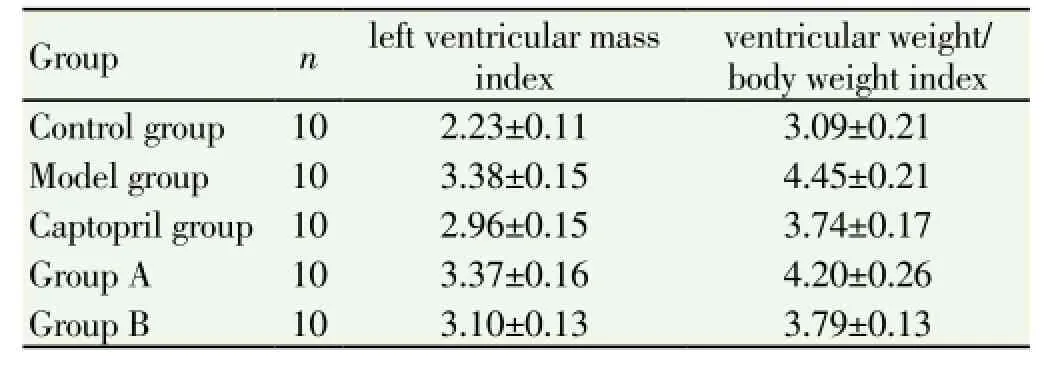
The left ventricular mass index and ventricular weight/ body weight index of model group, captopril group and groups A and B were significantly higher than that of control group (P<0.05); left ventricular mass index and ventricularweight/body weight index of captopril group and group B were significantly lower than that of model group and group A (P<0.05), left ventricular mass index of captopril group was the minimum among all the group (Table 1).

Table 1 Comparison between groups in the rat heart weight index (mg/g).
3.2. Hyp level and CAT activity in myocardial tissue of each group
Model group and ginseng group A showed higher Hyp content and lower CAT activity in myocardial tissue compared with control group (P<0.05). The captopril group and group B showed lower Hyp content and higher CAT activity compared with model group and group A (P<0.05). There was no significant difference in Hyp content and CAT activity between captopril group, group B and the control group (P>0.05) (Table 2).

Table 2 Comparision of Hyp level and CAT activity in myocardial tissue between groups.
3.3. Serum GSH-PX, CAT, CK and LDH activity and H2O2content between groups
Model group and group A showed a significantly lower serum GSH-PX, CAT activity and a higher CK, LDH and H2O2content than the control group (P<0.05). There was no significant difference in f GSH-PX, CAT, LDH activity and H2O2content between captopril group and group B (P>0.05) (Table 3).

Table 3 Serum GSH-PX, CAT, CK and LDH activity and H2O2content between groups.
3.4. Myocardial tissue morphology observation
Microscopic observation showed control group with normal myocardial fibers, round or oval myocardial nuclei, and a small number of collagen fibers between myocardial tissue could be observed with occasional inflammatory cells. In model group, collagen fiber hyperplasia, vasodilation congestion with inflammatory cells infiltration were visible between myocardial fibers; the group A showed more collagen fiber hyperplasia, but without angiectasis hyperemia; the captopril group and group B showed a small number of collagen fibers in heart periosteal tissue microscopically (Figure 1, 2).

Figure 1. Morphology observation of myocardial tissue (HE, ×200).

Figure 2. Morphology observation of myocardial tissue (Massom×400).
4. Discussion
Myocardial tissue, which mainly consists of myocardial cells and collagen secreted by normal myocardial fibroblasts form a fiber network connection, which plays an important role in maintaining cardiac structure and function[8-12]. As pathogenic factors induce MF occurs, excessive activation and proliferation of myocardial fibroblasts can lead to extracellular matrix deposition, myocardial stiffness, and electrophysiological disorders[13]. In addition, increased concentration of myocardial interstitial collagen reduces capillary density, limits the oxygen diffusion between myocardial cells, increases myocardial ischemia, forming a vicious cycle, which eventually leads to refractory congestive heart failure, so the MF is a necessary procedure to endstage heart disease[14]. Therefore, to prevent or reverse MF is of great significance to patients’ health. Establishment methods commonly used for MF model in rats are: subcutaneously injection of catecholamine in abdomen aorta and coronary ligation method and subcutaneous injection of high-dose isopropyl adrenaline method[15-17]. The author established Wistar rats MF model by using subcutaneous injection of high-dose isopropyl adrenaline, and two rats of each group were randomly selected and executed after 10 d of modeling, microscopic observation of myocardial tissue showed a large number of collagen fibers, indicating that this method to establish the rat MF model is effective.
There is no explicitly specify of MF in Chinese trandition medicine. MF is generally belong to the “bi”, “palpitation”,“edema” because of its main symptoms of chest pain, chest condition, palpitations, shortness of breath, breathing difficulties, and so on[18]. MF pathogenesis is associated with lung, liver, spleen and kidney, mainly on the heart arteries and veins. Therefore, appropriate treatment should replenish blood and qi. Based on the “plain question. To really theory” main ingredients of ginseng mixture include: salvia miltiorrhiza, radix astragali, radix paeoniae rubra, angelica, peach kernel, safflower, earthworm, leeches, pinellia, etc. By promoting blood circulation to remove blood stasis as the therapeutic principles.
Salvia miltiorrhiza activates the circulation of bloodstream. Astragalus membranaceus can greatly tonify the spleen and stomach, quickly removes the blood stasis. Radix paeoniae rubra can promote blood circulation; Angelica invigorates the circulation of blood and blood stasis without harming body; Rhizoma ligustici wallichii invigorates good blood and qi; Peach kernel, safflower, have strong effect of huoxue quyu, the auxiliary medicine salvia miltiorrhiza can enhance the main medicine. Lumbricus can only strengthen body fluid and remove the adverse effect of blood stasis.
Studies have confirmed that[19], Hyp content in myocardial tissue can be used as an important index of judging the degree of myocardial fibrosis. In addition, HW/BW, and left ventricular weight (LVW)/BW ratio were positively associated with the degree of myocardial fibrosis, also serve as important indexes of judging the degree of myocardial fibrosis[20]. In this experiment, the model group showed significantly higher level of HW/BW, LVW/BW and Hyp in the myocardial tissue compared with control group; the reduced HW/BW, LVW/BW and Hyp of captopril group and group B suggest that the mixture can inhibit myocardial interstitial collagen formation and relieve the myocardial fibrosis. In MF occurrence and development process, a large number of oxygen free radicals can be produced during generating H2O2in a short period of time, leading to excessive consumption of GSH-PX and CAT, so the abnormal changes of H2O2content can also reflect the myocardial cell damage degree in process of MF, and the CAT and GSH-PX content changes, also can reflect the body’s ability to remove reactive oxygen species[21-23].
In this study, the serum H2O2content of model group rats increased significantly, showed that myocardial cell was greatly damaged; Activity of GSH-PX and CAT at the same time, suggest ability to remove reactive oxygen species H2O2by GSH-PX and CAT decreased severely; The captopril group and group B can improve the activity of GSH-PX and CAT, increase H2O2clearance. The experiment results showed that ginseng mixture in high dose with significant effect, no statistical difference was observed compared with captopril group (P>0.05). It indicates that ginseng mixture can raise the activity of GSH-PX and CAT, reduce lipid peroxidation damage by H2O2, improve cell normal function, so as to achieve resisting myocardial fibrosis[24,25]. According to histopathology study, endocardial collagen fiber hyperplasia of model group was serious, combined with small amount of inflamatory cells infiltration; only a small amount of collagen fibers were observed in group B, with occasional inflammatory cells infiltration. It indicates that a certain dose of ginseng mixture can inhibit myocardial collagen from secretion for MF rat, thus inhibiting the occurrence and development of MF.
This experiment shows that ginseng mixture is a effective drug for treatment of MF by inhibiting antioxidant effect during myocardial fibrosis process, which reflects not only the traditional Chinese medicine yiqi huoxue therapy principles of MF, but also the guidelines of modern medical treatment.
Conflict of interest statement
We declare that we have no conflict of interest.
[1] Wang YC, Ding YS, Liu ML. Molecular mechanism of cause myocardial fibrosis by different factors. J Med Rev 2012; 18(17): 2736-2739..
[2] Xie YQ, Li XH. Relationship between TGF-β1 and CTGF in myocardial fibrosis during chronic heart failure process. J Chengde Med Coll 2011; (2): 181-183.
[3] Aupperle H, Baldauf K, Marz I. An immunohistochemical study of feline myocardial fibrosis. J Comp Pathol 2011; 145(2-3): 158-173.
[4] Chen R, Xie ML. application prospect of TGF -β/Smads signaling pathways in treatment of myocardial fibrosis. Chin Pharm Bull 2012; 28(9): 1189-1192.
[5] Lan HY. Diverse roles of TGF-β/Smads in renal fibrosis and inflammation. Int J Biol Sci 2011; 7(7): 1056-1067.
[6] Chang WJ, Cai H. The role of connective tissue growth factor in myocardial fibrosis. Circulation Mag 2012; 22(4): 62-66.
[7] Luo TY, Liu XH. Role of connective tissue growth factor in myocardial fibrosis. Progr Cardiovascular Epidemiol 2009; (S1): 8-10.
[8] Zhang Y, Niu XL, Shang L. dynamic observation of the expression of connective tissue growth factor in the process of spontaneously hypertensive in rats. J Heart 2011; (4): 446-449.
[9] Li G, Yang SQ. Role of PI3K/Akt pathway in myocardial cell hypoxia reoxygenation injury by hydrogen sulfide post-processing. J Third Military Med Univ 2011; 33(17): 1803-1807.
[10] Zhang W, Liu GH, Zhang LH. Akt and myocardial protection. Progr Cardiovascular Epidemiol 2011; 32(4): 537-540.
[11] Zhang Y, Zhang J. PI3K/AKT pathway in HKCs mesenchymal transdifferentiation process induced by TGF -β1. J Fuzhou General Hospital 2011; (1): 20-21.
[12] Sun B, Yin Li, Xia WC, Miao CS, Li XN, Ren LQ, et al. The influence of apocynum extracton Ang Ⅱ induced myocardial fibroblasts. Chin Trop Med 2011; (9): 1113-1115.
[13] Massare J, Berry JM, Luo X, Rob F, Johnstone JL, Shelton JM, et al. Diminished cardiac fibrosis in heart failure is associated with altered ventricular arrhythmia phenotype. J Cardiovasc Electrophys 2010; 21(9): 1031-1037.
[14] Chu JX, Li GM, Han SY, Shi RF, Zhu LS. In vitro inhibitory effect Of rutin in buckwheat leaves on myocyte hypertrophy induced by angiotensin Ⅱ in the newborn rat. J West Chin Pharm 2010, (4) : 428-430.
选取了粗糙表面的金属铝和铜样品,在1 064 nm波长下开展了材料反射特性的测量实验,并利用几何光学近似的方法对测量结果进行了验证。
[15] Wang YP, Xu M, Gao W. Research progress on myocardial fibrosis related biomarkers. Progr Physiol Sci 2010; 41(6): 461-463.
[16] McManus DD, Piacentine SM, Lessard D, Gore JM, Yarzebski J, Spencer FA, et al. Thirty-year (1975 to 2005) trends in the incidence rates, clinical features, treatment practices, and shortterm outcomes of patients <55 years of age hospitalized with an initial acute myocardial infarction. Am J Cardiol 2011; 108(4): 477-482.
[17] Su Y, Wang L, Zhang MZ. Epidemiological research of acute myocardial infarction. J Trad Chin Combined Western Med Cardio-cerebrovascular Dis 2012; 10 (4): 467-469.
[18] Liu SY, Xing WP, Wu ZY, Zuo HH, Qiu YZ. Effect of Puerarin on expression of transforming growth factor -β1 and connective tissue growth factor in left ventricular myocardial tissue for myocardial fibrosis model in rats. J Clin Basic Res 2010; 26(12): 942-926.
[19] Nguyen DT, Ding C, Wilson E, Marcus GM, Olgin JE. Pirfenidone mitigates left ventricular fibrosis and dysfunction after myocardial infarction and reduces arrhythmias. Heart Rhythm 2010; 7(10): 1438-1445.
[21] Zhou D, Li Z, Zhang L, Zhan C. Inhibitory effect of tanshinone ⅡA on TGF Ⅱ-beta1-induced cardiac fibrosis. J Huazhong Univ Sci Tech-nolog Med Sci 2012; 32(6): 829-833.
[22] Wu AM, Zhang DM, Lou LX, Zhai JY, Lu XY, Chai LM, et al. Effects of Chinese herbal medicines Shengmai injection and Xuesaitong injection on ventricular fibrillation threshold and connexin 43 expression in rats with myocardial infarction. J Chin Integrative Med 2011; 9(7): 775-782.
[23] Jellis C, Martin J, Narula J, Marwick TH. Assessment of nonischemic myocardial fibrosis. J Am Coll Cardiol 2010; 56(2): 89-97.
[24] Small EM, Thatcher JE, Sutherland LB, Kinoshita H, Gerard RD, Richardson JA, et al. Myocardin-related transcription factor-a controls myofibroblast activation and fibrosis in response to myocardial infarction. Circ Res 2010; 107(2): 294-304.
[25] Kapur NK, Wilson S, Yunis AA, Qiao X, MacKey E, Parurchuri A, et al. Reduced endoglin activity limits cardiac fibrosis and improves survival in heart failure. Circulation 2012; 125(22): 2728-2738.
ment heading
10.1016/S1995-7645(14)60125-5
*Corresponding author: Yue-Hong Li, M.D., Associate Chief Physician, Tangshan Workers Hospital affiliated to Hebei Medical University, Tangshan 063000, China.
Tel: 15831509792
E-mail: lyht1970@163.com
Foundation project: It is supported by Tangshan Science and Technology Research Key Project (grant No. 12150222B-15)
Captopril
Myocardial fibrosis
Protection
 Asian Pacific Journal of Tropical Medicine2014年9期
- Asian Pacific Journal of Tropical Medicine的其它文章
- Sonic Hedgehog signaling pathway in primary liver cancer cells
- Runx3 might participate in regulating dendriti cell function in patients with irritable bowel syndrome
- Protection effect of Emodin pretreatment on intestinal I - RI damage of intestinal mucosa in ratsa
- Protective effect and mechanism of lithium chloride pretreatment on myocardial ischemia-reperfusion injury in rats
- Effect of siRNA interference on nerve growth factor in intervertebral disc inflammation rats
- Nerve protective effect of rhTPO and G-CSF on hypoxic ischemic brain damage in rats
