
Effect of anesthesia on cognitive status and MMP-2 expression in rats
2014-03-23 01:27:30HongTuLiQuanJunCaoXiangJieQiWeiLingLu
Hong-Tu Li, Quan-Jun Cao, Xiang-Jie Qi, Wei-Ling Lu,3*
1Department of Anesthesiology, People's Hospital of Linzi District, Zibo 255400, Shandong Province, China
2Department of Urology, People's Hospital of Linzi District, Zibo 255400, Shandong Province, China
3Operating Room, People's Hospital of Linzi District, Zibo 255400, Shandong Province, China
Effect of anesthesia on cognitive status and MMP-2 expression in rats
Hong-Tu Li1, Quan-Jun Cao1, Xiang-Jie Qi2, Wei-Ling Lu1,3*
1Department of Anesthesiology, People's Hospital of Linzi District, Zibo 255400, Shandong Province, China
2Department of Urology, People's Hospital of Linzi District, Zibo 255400, Shandong Province, China
3Operating Room, People's Hospital of Linzi District, Zibo 255400, Shandong Province, China
Objective: To investigate the effect of anesthesia on the cognitive status damage and MMP-2 expression in rats. Methods: A total of 120 healthy rats were selected and randomly divided into the control group, CF3-CH(OCH2F)-CF3 (Sevoflurane) group and CF3-CH2-O-CHF-CF3 group (Sevoflurane) (n=40). After training for 3 d by the Morris water maze, the control group were injected with fentanyl for analgesia, the CF3-CH(OCH2F)-CF3 group and the CF3-CH2-O-CHFCF3 group were anesthesia with CF3-CH (OCH2F)-CF3 and CF3-CH2-O-CHF-CF3 on the basis of fentanyl, then rats in three groups underwent open surgery and suture conventional incision. Morris water maze was used to measure the rats' cognitive ability in three groups on the 1st d, 3rd d, 5th d and 7th d, and the brain tissue MMP-2 expression was detected. Results: After 1 d/7 d of the surgery, Morris water maze performance and MMP-2 expression were not significantly different among three groups (P>0.05); After 3 d/5 d of the surgery, compared with the control group, the Morris water maze test result was significantly worsened, MMP-2 expression levels were significantly increased (P<0.05); After 3 d/5 d of the surgery, compared with the CF3-CH2-O-CHF-CF3 group, Morris water maze test result of CF3-CH(OCH2F)-CF3 group was significantly worsened,MMP-2 expression levels were significantly increased (P<0.05). Conclusions: Anesthesia can cause some injury on cognitive status, different anesthetic drugs may cause different injury, and the cognitive status injury is related to the MMP-2 expression.
ARTICLE INFO
Article history:
Received 10 December 2013
Received in revised form 15 January 2014
Accepted 15 February 2014
Available online 20 April 2014
Anesthesia
1. Introduction
Postoperative cognitive dysfunction (POCD) refers to the neurocognitive dysfunction caused by anesthesia, which is a common postoperative neurological complication. The clinical symptoms were that patients have personality changes in a few days to several weeks or months postoperative, with decreased memory, attention, social adaptability, cognitive ability and comprehension abilit, even long-term cognition disorders, which severely affects patients’ life quality[1]. Recently the pathogenesis of POCD is not very clear, but numerous studies showed that anesthesia can suppress brain function temporality, and this effect is reversible[2]. This study selected adult rats as the research object to explore the impact of anesthesia on the cognitive status and matrix metalloproteinase-2 (MMP-2) expression.
2. Materials and methods
2.1. General information
A total of 120 healthy 14-month-old male rats weighing 346-382 g were selected. They were randomly divided into the control group, the CF3-CH(OCH2F)-CF3 (Sevoflurane) group and the CF3-CH2-O-CHF-CF3 group(Sevoflurane) (n=40).
2.2. Anesthesia methods
The control group were injected with fentanyl foranalgesia, then underwent open operation 20 min later. On the basis of fentanyl, self-made closed circulating organic glass box was used in the CF3-CH(OCH2F)-CF3 (Sevoflurane) group and the CF3-CH2-O-CHF-CF3 group (Sevoflurane), each with one hole on both sides in the box. One hole was connected to the Drager fabius anesthesia machine (Draeger produced), and the other hole was connected to F-MCl type gas monitors (USA DateOhmeda produced). Concentration of 3% CF3-CH(OCH2F)-CF3 (Sevoflurane) and the CF3-CH2-O-CHF-CF3 (Sevoflurane) was continuously imported respectively, at the speed of 1.5 L/min[3]. After 10 min of sustained anesthesia, the depth of anesthesia with pain stimulation was assessed. The rats then underwent open surgery with 1 cm incision, suture conventional incision after 3 min. During surgery, the monitor was open to ensure the concentration of anesthetic gas[4].
2.3. Morris water maze test
Before 1d of the experiment, all rats swam in the water maze in batches for three times, 2 min times to adapt to the water maze. Morris water maze was produced by Beijing Pusheng Science and Technology Ltd. The water maze was constituted of circular pool with 130 cm diameter and 50 cm height and cylindrical platform with l0 cm diameter and 29 cm height. The wall was marked with four cardinal points and the pool was divided into four quadrants. In exercise and experimental tests, the water depth was 30 cm and the temperature was (26±1.3) ℃, the outer wall was pasted with reference object. Morris water maze test was performed after 1 d, 3 d, 5 d and 7 d of the operation. Tests included three parts: place navigation test, spatial probe test and visible platform task training, 10 rats were randomly selected in each group every day to participate in the experiment.
Pace navigation test was as follows[1]: A total of 30 rats in three groups were put into the water from four quadrants in sequence, the swim tracks of the rats were recorded as the latency (escape latency). If the rats did not climb the platform Ttill 120 s, it was still counted as 120s[5]. This test was carried out every morning and afternoon, average value was calculated on that day.
Spatial probe test was as follows[2]: after the place navigation test, the platform was removed, the rats were put into the pool at the point and swam for 120 s. The times of crossing the former platform was measured. The average value of the total number from the 4 points was counted as rats’ individual score, the mean value of 10 individual scores was counted as the spatial probe test result on that day[6,7]. Exercise test experiments[8] was as follows: platform was removed, foreleg of the rats was used as the observed target, the times of two front leg drive in 1 min with a video camera was recorded.
2.4. MM P-2 expression of rat brain tissue
After the Morris water maze test, anesthesia method was performed the same as above. Then the neck was brokento obtain the brain tissue. Tissue was made into 10% homogenate with saline in batches, MMP-2 expression levels were detected by ELISA.
2.5. Data analysis
Excel 2007was used, data were expressed as mean±SD values and analyzed with Spss18.0 software then t test. Each rate was expressed by percentage and was compared byx2test. Measurement data were compared with nonparametric test (Z test). P<0.05 was considered as statistical significant difference.
3. Results
3.1. Morris water maze test results
3.1.1 Rat escape latency
After 1 d/7 d of the surgery, three groups escape latency had no statistically significant difference(P> 0.05); After 3 d/5 d of the surgery, compared with the control group, the escape latency of the CF3-CH2-O-CHF-CF3 group and the CF3-CH(OCH2F)-CF3 group were significantly increased (P <0.05); After 3 d/5 d of the surgery, compared with the CF3-CH2-O-CHF-CF3 group, the escape latency of the CF3-CH(OCH2F)-CF3 group was significantly decreased (P<0.05) (Table 1).
3.1.2. Times of rats crossed the location of platform
After 1 d/7 d of the surgery, the times of rats crossed thelocation of platform in three groups had no statistically significant difference(P>0.05); After 3 d/5 d of the surgery, compared with the control group, the times of rats crossed the location of platform of the CF3-CH2-O-CHF-CF3 group and the CF3-CH(OCH2F)-CF3 group were significantly decreased (P<0.05); After 3 d/5 d of the surgery, compared with the CF3-CH2-O-CHF-CF3 group, the escape latency of the CF3-CH(OCH2F)-CF3 group was significantly increased (P<0.05) (Table 2).

Table 1 Comparison of the escape latency of rats in three groups (mean±SD, s).
3.1.3. Exercise test experiments of the rats
The times of two front leg drive of rats in three groups were (158.69±25.41) times, (161.36±25.82) times and (159.80± 25.35) times respectively, the difference was not statistically significant (P>0.05).
3.2. MM P-2 expression of rat brain tissues:
After 1 d/7 d of the surgery, the difference in MMP-2 expressions of three groups were not significant (P>0.05); After 3 d/5 d of the surgery, compared with the control group, the MMP-2 expression levels of the CF3-CH2-OCHF-CF3 group and the CF3-CH(OCH2F)-CF3 group were significantly increased (P<0.05); After 3 d/5 d of the surgery, compared with the CF3-CH2-O-CHF-CF3 group, the MMP-2 expression levels of the CF3-CH(OCH2F)-CF3 group was significantly increased (P<0.05) ( Table 3).
4. Discussion
POCD is the central nervous system complication postanesthetic, which occurs in adult and elderly patients. This disorder may persist for a long time and have negative influence on the rehabilitation and quality of life in patients. The currently foreign researches showed that in elderly patients, the probability of POCD after 7 d of general anesthesia non-cardiac surgery is higher than 25%[9], the probability of occurrence of POCD after three months is higher than 9%; in adult patients, the probability of POCD after 7 d of general anesthesia abdominal operation or plastic surgery is higher than 19%, the probability of occurrence of POCD after 3 months more than 6%[10]. The pathogenesis of POCD is not yet clear, but a lot of studies have shown that it related to perioperative anesthesia. Results of correlation study in China such as Wuet al[11] that surgery anesthetic (particularly inhaled anesthetics) will inhibit the cholinergic system and damage the cognitive status. The results of Egiet al[12], Jinget al[13] and Niet al[14] showed that the damage of cognitive status is related to the amount of anesthetic and the duration, and the damage of cognitive status is positively correlated to the amount of anesthetic, while Ni et al believe that different anesthetics can cause different degrees of cognitive status damage[14].
The brain stem reticular activating system now considered to be maintain the excitement of the cerebral cortex, so that the body can in a waking state and with clear conscience. Its dysfunction and tissue damage is the important mechanism of cognition disorders. Rats’ brain tissue structure is similar with human brain tissue, and the hippocampus is an important part for general anesthesia[15]. Hippocampus can receive visual, auditory, tactile, pain and other information by brain stem reticular formation and subcutaneous fibrous. MMPs are a class of proteolytic enzyme with similar composition to amino acid and depend on zinc ion. Recently it is found that MMPs involved in tissue damages of aortic aneurysm, rheumatoid arthritis, gastric ulcer, myocardial disease and fibrotic lung disease and the nervous system infections and other diseases. MMPs entry into cellular and finished endocytosis, then participate in the proteolytic cleavage at cytoplasm and organelle.MMP-2 is particularly active in the brain tissue of the rat with transient focal cerebral ischemia, and directly lead to neuronal apoptosis and brain damage.MMP-2 has high expression in cerebralischemia, brain injury, hemodynamic changes, inflammation and so on, while MMP-2 has a low expression in normal rat brain. Kimet al[16] and others considered that MMP-2 may be related to the recovery after ischemic injury, which is closed to the results of Istaphanouset al[17].
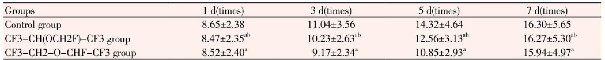
Table 2 Comparison of times of rats crossed the location of platform in three groups (mean±SD).

Table 3 Comparison of MMP-2 expression of rat brain tissues in three groups(mean±SD, mg/L)
In this study, after 1 d/7 d of the surgery , three groups Morris water maze performance and MMP-2 expression were not significantly different, the difference had no statistically significant (P>0.05), which is consistent with the results of Liet al[8]. After 3 d/5 d of the surgery, compared with the control group, the Morris water maze test of the sevoflurane groups were significantly decreased ,these differences have statistically significant (P<0.05). This is consistent with the results of Tanget al[18], Luet al[9]. MMP-2 expression levels of the sevoflurane groups were significantly increased, which is consistent with the results of Kimet al[16] and Istaphanouset al[17]. After 3 d/5 d of the surgery, compared with the CF3-CH2-O-CHF-CF3 group, the CF3-CH(OCH2F)-CF3 Morris water maze test was significantly decreased, MMP-2 expression levels were significantly increased, these differences were statistically significant (P<0.05). This is consistent with results of Liet al[4]. The exercise test experiments of the rats in three groups had no significant difference (P>0.05), which indicated that the escape latency is not related to the exercise ability.
In summary, anesthesia can cause some damage on the cognitive status, different anesthetic drugs may cause different damages, and the cognitive status injury is related to the MMP-2 expression. But there are lots of different anesthetic drugs and usually combined application in clinical, we will do further research on the cognitive status injury of the combined application of different anesthetics.
Conflict of interest statement
We declare that we have no conflict of interest.
[1] Hu ZH, Ou YW. Postoperative cognitive dysfunction and alzheimer’s disease from pathogenesis. Med phil 2011; 32(2): 69-71.
[2] Krenk L, Rasmussen LS, Kehlet H. New insights into the pathophysiology of postoperative cognitive dysfunckion. Acta Anaesthesiol Stand 2010; 54(8):951-956.
[3] Lombard FW, Mathew JP. Neurocognitive dysfunction following cardiac surgery. Semin Cardiothorac Vasc Anesth 2010; 14(2):102.
[4] Li CY. Effects on spatial learning and memory in newborn rats following propofol and sevoflurane anesthesia. Anatomy Res 2011; 33(6): 438-440.
[5] Wu H, Xu PC, Yang JX. The concentration of sevoflurane anesthesia on cognitive function in adult rats and the hippocampal dentate gyrus neurogenesis. Shandong Med 2011; 51(9): 28-29.
[7] Wen HM, Lin SY, Gao J, Yin ZL, He Q, Dai DM,et al. The influence and mechanism of Xingnaojing Injection to elderly rats ketamine anesthesia learning and memory. Guangdong Med 2012; 33(11): 1546-1548.
[8] Li Q, Yu HL, Wang GL. The study of affecting factors for postoperative cognitive dysfunction after cardiac surgery. Med Rev 2010; 16(1): 79-82.
[9] Lu GL, Chen XL, Song YY, Mao XD, Liao L. The influence of Sevoflurane inhalation anesthesia of newborn rat hippocampal neurogenesis and cognitive development. Chin J New Drugs Clin 2011; 30(8): 615-618.
[10] Wang DY, Zhang X, Yang TD, Ye JN, Jia XB. Effect of orexin-A on learning-memory ability and ChAT expression in basal forebrain of aged rats after ketamine anesthesia. Chongqing Med 2009; 38(15): 1885-1889.
[11] Wu SH. The influence of different anesthesia methods on cognitive function after surgery for elderly patients. Jilin Med 2012; 33(1): 93-94.
[12] Egi M, Bellomo R, Stachowski E, French CJ, Hart G, Stow P. Hypoglycemia and outcome in critically ill patients. Mayo Clinproc 2010; 85(4): 217-221.
[13] Jing YM, Zhang XX, Ji SX, Liu YM, Lan JS. The research progress of fluorine marcie effects on postoperative cognitive function. J Clin Anesthesiol 2012; 28(11): 1136-1138.
[14] Ni X, Martin-Caraballo M. Dillerential ellectol glutamate receptor blockade on dendriti coutgrowth in chicken lumba rmotoneurons. Neuro Pharmacol 2010; 8(3): 593-604.
[15] Mochizuki N, Takagi N, Kurokawa K, Kawai T, Besshoh S, Tanonaka K, et al. EIIect of NMDA receptor antagonist on proliferation of neurospheres from embryonic brain. Neurosci Lett 2010; 417(2): 143-148.
[16] Kim YJ, Lee H, Kim CH, Lee GY, Baik HJ, Han JI. Effect of Ilumazenil on recovery from anesthesia and the bispectrol index after sevoflurane/fentanyl general anesthesia in unpreme-dicated pations. Korean J Anesthesiol 2012; 62(1): 19-23.
[17] Istaphanous Gk, Howard J, Nan X, Hughes EA, McCann JC, McAuliffe JJ, et al. Comparison of the neuroapoptotic properties of equipotent anesthetic concentrations of desflurane,isoflurane,or sevoflurane in neonatal mice. Ancsthcsiol 2011; 114(3): 578-587.
[18] Tang DM, Zhang YJ, Chen G. Effect of sevoflurane anesthesia on spatial learning and memory function and hippocampus cholinergic neurone activity in infant rats. J Chin Pract Diagn Ther 2012; 26(4): 326-328.
ment heading
10.1016/S1995-7645(14)60051-1
*Corresponding author: Wei-Ling Lu, People's Hospital of Linzi District, Zibo 255400, Shandong Province, China.
Tel: 15666703780
E-mail: Lht030506@sina.com
Foundation project: It is supported by Shandong Research Programs Projects (2006GG2202028) and Key Technologies R&D Programme Projects of Shandong (2006GG2202028).
Cognition
Matrix metalloproteinase-2
Influence study
 Asian Pacific Journal of Tropical Medicine2014年4期
- Asian Pacific Journal of Tropical Medicine的其它文章
- Effect of bone marrow mesenchymal stem cells on the Smad expression of hepatic fibrosis rats
- Correlation of expression of STAT3, VEGF and differentiation of Th17 cells in psoriasis vulgaris of guinea pig
- Ultrasonic diagnosis and vasoactive substances examination in patients with cirrhosis
- Effect of low intensity pulsed ultrasound on repairing the periodontal bone of Beagle canines
- Effect of RSCs combined with COP-1 on optic nerve damage in glaucoma rat model
- Expression of PI3-K, PKB and GSK-3β in the skeletal muscle tissue of gestational diabetes mellitus