结直肠癌细胞外泌体的制备及其促单核细胞增殖作用
董 超,刘 迪,冯业童,刘朋飞,吴 璇,吴昊昱,周余来
(吉林大学药学院生物工程实验中心,吉林 长春 130021)
结直肠癌细胞外泌体的制备及其促单核细胞增殖作用
董 超,刘 迪,冯业童,刘朋飞,吴 璇,吴昊昱,周余来
(吉林大学药学院生物工程实验中心,吉林 长春 130021)
目的:从结直肠癌细胞系中分离鉴定外泌体,初步研究结直肠癌来源的外泌体潜在的免疫学特性,为外泌体在结直肠癌免疫学治疗中的应用提供依据。方法:超速离心法从HCT116和SW480 2种结直肠癌细胞系培养上清中分离提取正常细胞外泌体和热休克细胞外泌体,进一步用220nm的滤膜进行纯化获得纯度较高的外泌体。采用透射电镜观察所提取外泌体表征形态,聚丙烯酰胺凝胶电泳法分析细胞和外泌体的蛋白组成,检测外泌体促进外周血单个核细胞(PBMCs)的增殖功能。结果:透射电镜下正常的外泌体和细胞热休克获得的外泌体形态相近,均呈现典型的圆形或椭圆形外泌体结构,直径为50~100nm。聚丙烯酰胺凝胶电泳,结直肠癌细胞裂解物与结直肠癌细胞系来源外泌体的蛋白组成不同,与未处理和经热休克处理的结直肠癌细胞比较,外泌体对PBMCs的增殖有更强的促进作用(P<0.05),热休克作用可使外泌体诱导的PBMCs的增殖程度显著升高(P<0.05)。结论 :超速离心法和滤膜过滤方法可有效地从结直肠癌细胞系中提取外泌体,细胞裂解物和外泌体的蛋白组成差异可能是造成免疫学特性差异的主要原因,将细胞经热休克处理可增强外泌体的免疫原性。
结直肠肿瘤;外泌体;热休克;免疫治疗
胃肠道恶性肿瘤中结直肠癌发病率仅次于胃癌和食道癌,发病率较高,且近年来发病率仍呈现上升的趋势[1]。尽管手术治疗和相应的辅助治疗方法发展较快,但患者的术后存活率并没有得到显著改善[1-2]。与传统治疗方法比较,肿瘤免疫治疗通过提高自身免疫力杀死肿瘤细胞,不仅副作用小而且更加安全,已成为最具潜力的结直肠癌治疗方法[3]。外泌体是一种可由多种细胞分泌的膜泡结构,来自于细胞的多泡内含体。不同来源的外泌体具有与来源相关的特殊功能[4]。普遍存在于不同来源外泌体的蛋白有黏附蛋白和热休克蛋白等,这些蛋白在外泌体介导信号传导中起着重要作用。外泌体表面的蛋白可通过抗原提呈细胞被T细胞识别并激活免疫系统[5-6]。因此肿瘤细胞分泌外泌体被认为是提呈肿瘤抗原的载体,在肿瘤免疫治疗中具有巨大的应用前景[7-8]。本研究分离鉴定了结直肠癌细胞系来源的外泌体并对其免疫学特性进行了初步探讨,特别是外泌体与细胞在高相对分子质量蛋白方面的差异,可能是二者促单核细胞增殖能力不同的原因。类似研究在国内外尚未见报道。
1 材料与方法
1.1 细胞、主要试剂及仪器 人结直肠癌细胞系HCT116和SW480细胞株由吉林大学药学院生物工程实验中心提供;DMEM高糖培养、RPMI 1640培养基、胎牛血清购自美国Gibco公司,胰蛋白酶、对钠十二烷基硫酸盐(SDS)购自美国Sigma公司,丙烯酰胺、甲叉双丙烯酰胺和预染蛋白Marker购自北京鼎国生物技术有限责任公司,二喹啉甲酸(BCA)蛋白浓度测定试剂盒、CCK-8试剂盒购自中国碧云天生物技术研究所;JEM-2000EX透射电子显微镜购自日本电子公司,OptimaTM LE-80K低温超速离心机购自美国Beckman Coulter公司。
1.2 细胞培养 人结直肠癌细胞系HCT116和SW480均培养于含10%胎牛血清的DMEM高糖培养基中,置于5%CO2、37℃培养箱常规培养。细胞于43℃处理1h后恢复常规培养,进行热休克处理。人外周血采自健康献血志愿者,通过密度梯度离心法分离单个核细胞,培养于含10%胎牛血清、1%抗生素的RPMI 1640培养基中。
1.3 结直肠癌细胞系来源的外泌体的分离 在培养细胞汇合度达80%后,PBS洗2遍,在含10%胎牛血清的DMEM高糖培养基中培养48h,通过3次离心去除细胞培养上清中的细胞和细胞碎片,条件分别是300g离心10min,1 200g离心20min,10 000g离心30min。然后在100 000g离心60min的超速离心条件下外泌体聚集至管底,用PBS重悬洗涤1次,最终的沉淀通过220nm滤膜过滤获得纯度较高的外泌体,保存温度为-80℃。
1.4 外泌体超微形态的观察 将从细胞培养液上清中超离获得的外泌体加在铜网上,被吸附在铜网上的外泌体被2%的磷酸钨酸染色并在透射电镜下观察其形态。
1.5 蛋白浓度的测定 结直肠癌细胞系和分离的外泌体在蛋白裂解液中裂解。加入裂解液后充分混匀,置于冰上30min,100℃、10min,然后15 000r·min-1离心20min去除细胞碎片。用二喹啉甲酸(BCA)方法测定蛋白浓度。相同质量的细胞蛋白和外泌体通过聚丙烯酰胺凝胶电泳后用考马斯亮蓝染色。分离胶浓度为12%。
1.6 共培养外周血单个核细胞(PBMCs)增殖率测定 将外周血单个核细胞与结直肠癌细胞蛋白裂解液和结直肠癌细胞系来源的外泌体共培养。外周血单个核细胞的浓度为2×10⁶mL-1,细胞蛋白和外泌体浓度为200mg·L-1。PBMCs和蛋白样品均在培养基中重悬,各组体积一致,均为200μL(表1)。共培养 60h 后,采用 CCK-8 法测定PBMCs增殖率。增殖率与绝对吸光度(A)值成正比。细胞增殖率=(各实验组A值-空白组A值)×100%。
1.7 统计学分析 采用SPSS 17.0统计软件进行统计分析。各组单核细胞A值以±s表示,组间比较采用单因素方差分析。

表1 PBMCs共培养实验分组方案Tab.1 Grouping plan of PBMCs in co-culture experiment
2 结 果
2.1 透射电镜下结肠癌细胞系来源外泌体的形态学特征 在透射电镜下可观察到超速离心获得的沉淀由直径约100nm的膜泡结构组成。热休克处理后细胞分泌的外泌体与未处理细胞分泌的外泌体均为圆形或椭圆形,形态较为相近。见图1。

图1 透射电镜下未处理细胞和热休克结直肠癌细胞来源外泌体的形态学(×80 000)Fig.1 The morphology of exosomes from untreated and heat shock treated colorectal carcinoma cells under transmission electron microscope(×80 000)
2.2 结直肠癌细胞全蛋白与外泌体全蛋白一维变性凝胶电泳 正常外泌体和经热休克处理的外泌体在聚丙烯酰胺凝胶电泳中有着相近的蛋白条带,将相同含量的外泌体(10μg)和细胞全蛋白 (10μg)分别加入加样孔进行电泳。正常细胞和热休克处理细胞的电泳结果中蛋白条带同样相近 (图2A和B)。然而细胞全蛋白与外泌体比较在电泳条带上差别较大,尤其是相对分子质量较大的蛋白条带。聚丙烯酰胺凝胶电泳结果通过Quantity One 4.62 furthermore分析,外泌体中相对分子质量高的蛋白含量较细胞系中相对分子质量高的蛋白含量更多,而相对低分子质量蛋白条带相近 (图2C和D)。
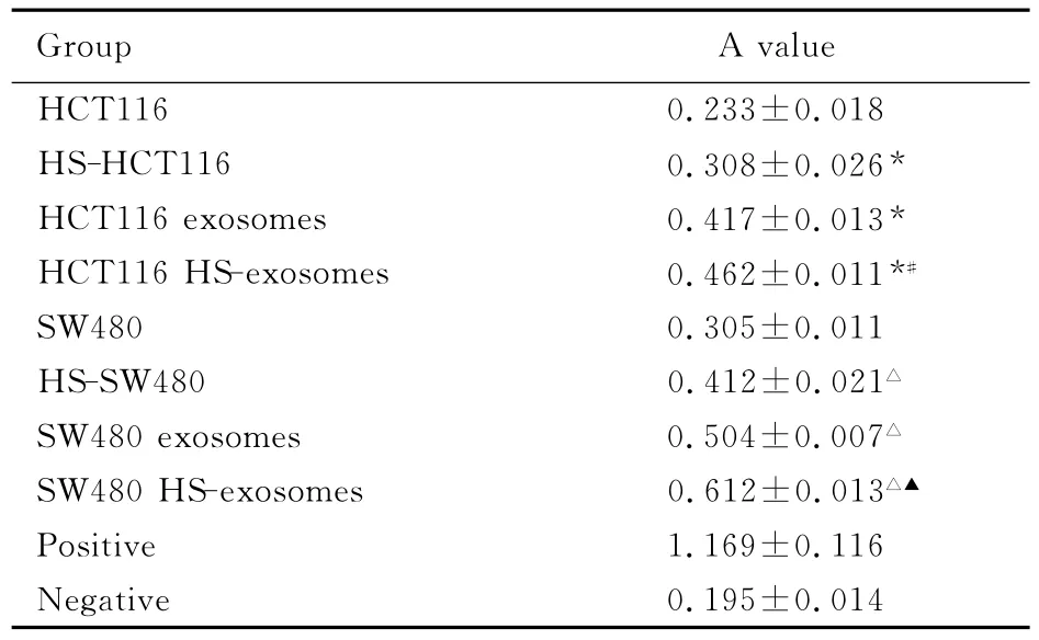
2.3 共培养外周血单个核细胞增殖实验 与未处理和经热休克处理的结直肠癌细胞比较,外泌体对PBMCs的增殖有更强的促进作用 (P<0.05)。结直肠癌细胞系和外泌体的PBMCs增殖能力均可通过热休克加强。见表2。
3 讨 论
外泌体是由包括肿瘤细胞在内的多种细胞分泌的膜泡结构,包含多种膜蛋白和胞质蛋白,这种外泌体在多种体液中都有存在[9],在肿瘤细胞迁移和转移中起到重要作用。很多与肿瘤迁移信号通路相关的蛋白均存在于肿瘤细胞分泌的外泌体中,从而传递信号导致肿瘤的转移等恶性表型[10],有报道[11-12]表 明:外 泌 体 同 时 可 以 传 递 mRNA 和 miRNA。

图2 结直肠癌细胞全蛋白和对应来源外泌体全蛋白聚丙烯酰胺凝胶电泳图Fig.2 Electrophoregram of SDS-PAGE of full proteins of colorectal carcinoma cells and matching exosomes
表2 不同肿瘤抗原刺激物作用下PBMCs增殖结果Tab.2 Proliferation results of PBMCs stimulated with different tumor antigens (±s)

表2 不同肿瘤抗原刺激物作用下PBMCs增殖结果Tab.2 Proliferation results of PBMCs stimulated with different tumor antigens (±s)
*P<0.05 vs HCT116group;△P<0.05 vs SW480group;#P<0.05 vs HCT116exsomes group;▲P<0.05 vs SW480 exosomes group.
Group A value HCT116 0.233±0.018 HS-HCT116 0.308±0.026*HCT116exosomes 0.417±0.013*HCT116HS-exosomes 0.462±0.011*#SW480 0.305±0.011 HS-SW480 0.412±0.021△SW480exosomes 0.504±0.007△SW480HS-exosomes 0.612±0.013△▲Positive 1.169±0.116 Negative 0.195±0.014
研究[13-14]显示:结直肠癌细胞系(SW1116 、COLO205)也可分泌外泌体。同样,细胞全蛋白和外泌体全蛋白在一维变形凝胶电泳中,相对分子质量大的蛋白条带有较大差异,而这些蛋白极有可能与刺激免疫系统和促进外周血单个核细胞增殖有关。本研究分离了热休克结直肠癌细胞系分泌的外泌体。热休克处理的2种细胞系来源的外泌体在形态上与正常外泌体相近,然而分子组成分析显示了量的差异。这些差异正是由于热休克来源外泌体比正常外泌体存在更多免疫分子导致的[15-17]。本研究结果进一步证明了外泌体能诱导PBMCs增殖。
综上所述,基于在制备和应用上的简单可行,与传统的肿瘤细胞抗原比较,热休克外泌体在肿瘤免疫治疗中有更大的优势,特别是肿瘤免疫治疗的低毒副作用、治疗效果显著等优点,使这项新技术具有更广阔的临床应用空间。
[1]Aune D,Chan DS,Lau R,et al.Dietary fibre,whole grains,and risk of colorectal cancer:systematic review and dose-response meta-analysis of prospective studies [J].BMJ,2011,343:d6617.
[2]Pitule P,Liska V,Treska V,et al.Contribution of molecular biology to the diagnosis and therapy of colorectal carcinoma-the present and future[J].Rozhl Chir,2011,90(6):315-323.
[3]Mathivanan S,Lim JW,Tauro BJ,et al.Proteomics analysis of A33immunoaffinity purified exosomes released from the human colon tumor cell line LIM1215reveals a tissue-specific protein signature[J].Mol Cell Proteomics,2010,9(2):197-208.
[4]Batista BS,Eng WS,Pilobello KT,et al.Identification of a conserved glycan signature for microvesicles[J].J Proteome Res,2011,10(10):4624-4633.
[5]Zhong H,Yang Y,Ma S,et al.Induction of a tumourspecific CTL response by exosomes isolated from heat-treated malignant ascites of gastric cancer patients [J].Int J Hyperthermia,2011,27(6):604-611.
[6]Yang C, Kim SH, Bianco NR,et al. Tumor-derived exosomes confer antigen-specific immuno-suppression in a murine delayed-type hypersensitivity model[J].PLoS One,2011,6(8):e22517.
[7]Taylor DD, Gercel-Taylor C. Exosomes/microvesicles:mediators of cancer-associated immunosuppressive microenvironments[J].Semin Immunopathol,2011,33(5):441-454.
[8]Sheng H,Hassanali S,Nugent C,et al.Insulinoma-released exosomes or microparticles are immunostimulatory and can activate autoreactive T cells spontaneously developed in nonobese diabetic mice[J].J Immunol,2011,187(4):1591-1600.
[9]Escrevente1C,Keller S,Altevogt P,et al.Interaction and uptake of exosomes by ovarian cancer cells[J].2011,11(1):108-117.
[10]Iero M, Valenti R, Huber V,et al. Tumour-released exosomes and their implications in cancer immunity[J].Cell Death Differ,2007,15(1):80-88.
[11]Lakhal S,Wood MJ.Exosome nanotechnology:an emerging paradigm shift in drug delivery:exploitation of exosome nanovesicles for systemicinvivodelivery of RNAi heralds new horizons for drug delivery across biological barriers [J].Bioessays,2011,33(10):737-741.
[12]Rani S,O’Brien K,Kelleher FC,et al.Isolation of exosomes for subsequent mRNA,MicroRNA,and protein profiling[J].Methods Mol Biol,2011,784:181-195.
[13]刘朋飞,朱 磊,李 响,等.结肠癌细胞源exosomes的分离鉴定及其表面蛋白CD44v6表达的初步研究[J].中国老年学杂志,2010,30(16):2330-2332.
[14]冯业童,刘朋飞,吴昊昱,等.结肠癌源性exosomes的分离及其相关免疫学性质[J].中国生物制品学杂志,2012,25(12):1635-1638.
[15]Silverman JM,Clos J, Horakova E,et al. Leishmania exosomes modulate innate and adaptive immune responses through effects on monocytes and dendritic cells [J].J Immunol,2010,185(9):5011-5022.
[16]王 旻,王 爽,金洪永.直肠癌同时性肝转移的外科治疗[J].临床肝胆病杂志,2012,28(1):17-20.
[17]徐增辉,钱其军.肿瘤抗体治疗的现状与发展趋势[J].临床肝胆病杂志,2011,27(4):351-356.
Preparation of exosomes derived from colorectal carcinoma cells and their effects of promoting proliferation on mononuclear cells
DONG Chao,LIU Di,FENG Ye-tong,LIU Peng-fei,WU Xuan,WU Hao-yu,ZHOU Yu-lai
(Biological Engineering Experiment Center,Jilin University,School of Pharmacy,Jilin University,Changchun 130021,China)
Objective To separate and identify the exosomes from colorectal carcinoma cells and to preliminarily study their immunological characteristics,and to provide basis for the application of exosomes in colorectal carcinoma immunotherapy.Methods The normal exosomes and heat shock exosomes of HCT116and SW480cells were separated by ultracentrifugation and purified by 220nm microspore film.The morphology of exosomes were observed under transmission electron microscope.The protein compositions of cells and exosomes were analyzed by SDS-PAGE method.The promoting function of exosomes in proliferation of peripheral blood monouclear cells(PBMCs)was detected.Results Under transmission electron microscope,the normal exosomes and heat shock exosomes had similar appearance.They were either spherical or oval membrane vesicles,whose diameters were about 50-100nm.The results of SDS-PAGE showed the differences of protein compositions between colorectal cell lysates and exosomes derived from colorectal carcinoma cells.Compared with untreated and heat shock treated colorectal carcinoma cells,the exosomes had stronger promotion effect on PBMCs proliferation(P<0.05).The proliferaton of PBMCs could be reinforced with heat shock(P<0.05).Conclusion For separation and identification of the exosomes derived from colorectal carcinoma cells,the method of ultracentrifugation and filtration is simple and feasible.The differences of protein composition between colorectal cell lysates and exosomes may be the major reason of their differences in immunological characteristics.The immuogenicity of exosomes could be enhanced by treating the cells with heat shock.
colorectal neoplasms;exosome;heat shock;immunotherapy
R735.3
A
1671-587Ⅹ(2013)03-0472-05
10.7694/jldxyxb20130310
2012-10-10
吉林省卫生厅科研基金资助课题 (2009Z043);吉林大学博士研究生交叉学科科研基金资助课题 (2011J023)
董 超 (1989-),男,吉林省松原市人,在读医学硕士,主要从事肿瘤免疫学方面的研究。
周余来 (Tel:0431-85619299,E-mail:zhouyl@jlu.edu.cn)
时间:2013-04-23 18:40
网络出版地址:http://www.cnki.net/kcms/detail/22.1342.R.20130423.1840.001.html

