Tissue optical clearing for neural regeneration research
Tingting Yu, Jianyi Xu, Dan Zhu
Nerve injury, whether traumatic or degenerative, disrupts the transmission of information in the nervous system, leading to dysfunction. It is widely known that neural regeneration is vital to the restoration of function after nerve injury. Still, outcomes are often limited by the misguidance of axonal regeneration and complex pathological changes in neurons and glia, as well as the longterm denervation of target organs. Morphological analyses of neural tissues and target organs are vital for outcome assessments in neural regeneration research.
Histological sectioning is commonly used for anatomical and morphological analyses in biomedical research. However, thin sections provide only two-dimensional information which may lead to biased results, particularly when evaluating the tortuous trajectories of axons and blood vessels or sparsely and non-uniformly distributed structures. The limitations of twodimensional imaging highlight a need for threedimensional (3D) visualization and analysis of related structures in the study of neural regeneration.
Tissue optical clearing techniques provide novel perspectives for 3D imaging of thick tissues and even entire organisms. It is achieved by harmonizing refractive indices among different cellular components and removing the pigmentations to reduce light attenuation in biological tissues thereby improving imaging depth. Various optical clearing methods based on similar physical principles have been developed for use with different fluorescent labeling methods and tissue types; detailed descriptions are published in previous reports (Richardson et al., 2021).
Current tissue-clearing methods can be categorized into three groups in terms of principles: hydrophobic, hydrophilic, and hydrogelbased methods. In the past decade, tissue-clearing techniques combined with fluorescent labeling and optical imaging tools have revolutionized the research paradigm in neuroscience by enabling brainwide studies of the connectome. Conversely, the application of such techniques in neural regeneration research is not widespread. Here, we give a brief summary of the literature on the use of tissue optical clearing methods in neural regeneration studies.
Applications in central nervous system (CNS) regeneration:CNS regeneration is not spontaneous in mature mammals and involves complex changes in cells and axons. As a result, investigators have focused not only on axonal elongation but also on cell-axon and cell-cell interactions in the CNS. Several groups have utilized tissue clearing to visualize the mammalian spinal cord in 3D. In 2012, Ertürk et al. first reported 3D imaging of the unsectioned spinal cord in transgenic fluorescent mice aided by a tetrahydrofuran-based hydrophobic clearing technique. They traced the trajectories of regenerating sensory axons under the conditioning effect of peripheral axotomy and quantitatively evaluated the glial response in 3D after injury (Ertürk et al., 2012). Since this pioneering study was published, hydrophobic tissue clearing methods have been widely used for 3D imaging of the unsectioned spinal cord combined with various labeling besides transgenic methods (Hilton et al., 2019). Recently, the hydrogelbased clearing method has been used to assess acute inflammation and axon extension from the transplanted human induced pluripotent stem cell-derived V2a interneurons in the intact injured spinal cord (McCreedy et al., 2021).
Applications in peripheral nervous system (PNS) regeneration:After peripheral nerve injury, axon regeneration in mammals is relatively readily but is still limited due to inefficient long-distance regeneration and misguidance to inappropriate targets. Intact axonal morphology, continuous trajectory, and morphological analyses of 3D structures in terminal targets are critical in nerve regeneration studies. The hydrophobic clearing method has been combined with whole-mount immunostaining and confocal microscopy to visualize and evaluate the 3D axonal structure after sciatic nerve transection and end-to-end repair in rats (Jung et al., 2014). In addition, the hydrophilic clearing method, CUBIC (clear, unobstructed brain/body imaging cocktails and computational analysis), combined with confocal microscopy has been modified to track and quantify the regeneration of nerve fibers within chitosan conduits (Fogli et al., 2019).
In addition to nerves, target organs are also key for outcome assessment in experimental regeneration studies. Accordingly, morphological analyses of neuromuscular junctions (NMJs) in skeletal muscles received more and more attention. NMJs represent the typical synaptic structures in the PNS and act as an important interface between peripheral nerves and muscle fibers. As such, structural changes following peripheral nerve injury or intervention are becoming an important area of research.
Tissue optical clearing and advanced optical imaging techniques make whole-mount analyses of NMJs in entire skeletal muscles possible. We first demonstrated the exact spatial distribution patterns of NMJs in healthy muscles by tissue clearing and light-sheet microscopy and investigated the adaptive morphological and topological changes in NMJs after muscle denervation and reinnervation in a sciatic nerve transection and suturing model (Yin et al., 2019). Furthermore, as a result of the superior fluorescence preserving capability of our FDISCO method (a modified hydrophobic method based on 3DISCO), we successfully visualized the in-muscle nerve branches and NMJs simultaneously (Qi et al., 2019) and revealed heterochronic development of the neuromuscular system in postnatal mouse skeletal muscles, which helped to facilitate the understanding of structural status during motor development (Xu et al., 2022). Daeschler et al. (2022) extended FDISCO to peripheral nerves and their target organs and demonstrated 3D morphometry, including dermatome-wide cutaneous innervation, intramuscular nerve branches, NMJs, and neurovascular network mapping. In regards to muscle clearing, a hydrogel-based clearing protocol, MYOCLEAR, was also proposed to visualize NMJs in whole-mount preparations of the small diaphragm and extensor digitorum longus tissue (Williams et al., 2019).
In recent years, novel advanced clearing methods with excellent clearing capability and superior fluorescence preservation have emerged to provide new tools for neuroscience research. For instance, a PEG-associated solvent system (PEGASOS) has been developed to directly visualize connections between the PNS (dorsal root ganglions) and CNS (spinal cord) via vertebrae in mice (Jing et al., 2018). In addition, some other whole-body clearing methods, such as the vDISCO (nanobody(VHH)-boosted 3D imaging of solvent cleared organs), the MACS (MXDA-based aqueous clearing system) and HYBRiD (hydrogelbased reinforcement of three-dimensional imaging solvent-cleared organs), also enable high performance for 3D visualization of the mammalian nervous system.
In conjunction with panoptic imaging methods and microscopies, these whole-body clearing methods provide the opportunity to map the entire neuromuscular connectome from the CNS and PNS to target organs and promote research into neural regeneration as well as development. However, several challenges must be addressed. The first is the limited penetration of antibodies, which hinders deep immunostaining in large-volume tissues, especially human tissues that cannot be labeled with the viruses, chemical tracers, or transgenic labeling techniques. Currently, several methodologies have been proposed to improve the labeling speed and homogeneity, involving reducing sample size, reducing probe size, increasing porosity, manipulating label affinity, and applying an outside force (Richardson et al., 2021). The representative active immunolabeling equipment utilizing an electrical field for speed labeling has now been commercialized, facilitating the standardization of the labeling protocols. In the future, with the improvement in fluorescent probes and equipment simplification for assisting greater antibody diffusion, tissue-clearing techniques are expected to serve better the study of neural regeneration in rodents, non-human primates, and even human tissues. The second challenge is the trade-off between high resolution and high throughput during imaging. For large samples, the light-sheet microscopies using long and thick light sheets and low numerical aperture and large field of view objectives have become preferred primarily owing to their fast speed. Those high-capacity systems allow a minuteshours imaging time for a cm3tissue volume, but the suboptimal imaging resolution (approximately 0.7 µm lateral and 5 µm axial) hinders the analysis of fine structures in the whole mount (Richardson et al., 2021). In addition, imaging quality in the deeper plane is generally inferior to the shallow plane even with the assistance of objectives with high numerical aperture and long working distance. In the future, the combination of highthroughput light-sheet microscopy and mechanical sectioning should allow high-quality images to be obtained in all planes. The third challenge is the need for rigorous computational infrastructures to facilitate massive data storage and processing. The huge-size image stacks also put forward the request for automation of data processing and reduction of manual intervention, which may be addressed by artificial intelligence techniques currently in development.
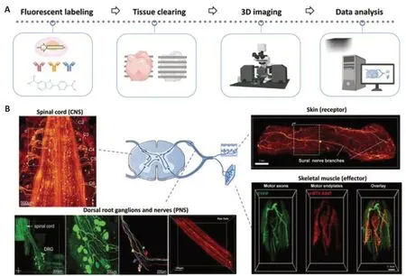
It is worth mentioning that the clearing methods and applications outlined above relate toex vivoimaging of dissected and fixed tissues or organs. The proposal ofin vivooptical clearing methods opened up a new field of research, in particular the skull optical clearing techniques, which were first proposed by our group and underwent several iterations in recent years (Li et al., 2022). The development of long-term clearing cranial and intervertebral windows enablesin vivoimaging of the cortex and spinal cord through the intact skull and intervertebral spaces, and provides alternative approaches for monitoring the dynamics of axonal response to injury or treatment. Thesein vivoclearing methods not only facilitate dynamic monitoring by circumventing the need for complex and elaborate surgical operations but also permit observation of tissue physiology and pathology without introducing immunological artifacts, likely to offer novel insights into nerve injury and repair.Tissue optical clearing in conjunction with advanced labeling, imaging, and data processing techniques holds great promise for the precise and accurate morphological analysis of nerve tissues and target organs (Figure 1), which will predictably aid the advancement of experimental neural regeneration studies and potentially the discovery of clinical intervention strategies.

Figure 1|Overview of tissue optical clearing scheme for 3D imaging and its applications in neural tissues and target organs.
This work was supported by the National Natural Science Foundation of China (Grant Nos. 61860206009 (to DZ), 81870934 (to DZ), and 81961138015 (to TY)), Innovation Project of Optics Valley Laboratory (Grant No. OVL2021BG011) (to DZ), and the Innovation Fund of WNLO (to DZ).
#Both authors contributed equally to this work.
*Correspondence to:Dan Zhu, PhD, dawnzh@mail.hust.edu.cn.
https://orcid.org/0000-0002-3640-4758(Dan Zhu)
Date of submission:August 26, 2022
Date of decision:November 21, 2022
Date of acceptance:December 1, 2022
Date of web publication:January 5, 2023
https://doi.org/10.4103/1673-5374.363827
How to cite this article:Yu T, Xu J, Zhu D (2023) Tissue optical clearing for neural regeneration research. Neural Regen Res 18(9):1940-1941.
Open access statement:This is an open access journal, and articles are distributed under the terms of the Creative Commons AttributionNonCommercial-ShareAlike 4.0 License, which allows others to remix, tweak, and build upon the work non-commercially, as long as appropriate credit is given and the new creations are licensed under the identical terms.
- 中国神经再生研究(英文版)的其它文章
- Mesenchymal stem cells, extracellular vesicles, and transcranial magnetic stimulation for ferroptosis after spinal cord injury
- Inducing prion protein shedding as a neuroprotective and regenerative approach in pathological conditions of the brain: from theory to facts
- Use of mesenchymal stem cell therapy in COVID-19 related strokes
- Brain organoids are new tool for drug screening of neurological diseases
- Emerging roles of astrocytes in blood-brain barrier disruption upon amyloid-beta insults in Alzheimer’s disease
- External anal sphincter electromyography in multiple system atrophy: implications for diagnosis, clinical correlations, and novel insights into prognosis

