
Synthesis and characterization of fluorescence active G4-quartet and direct evaluation of self-assembly impact on emission
2022-09-15 03:10YingHeMengjiLiuShunTengLukszWojtsGungxinGuXiodongShi
Chinese Chemical Letters 2022年9期
Ying He, Mengji Liu, Shun Teng, Luksz Wojts, Gungxin Gu, Xiodong Shi,∗
a Department of Chemistry, University of South Florida, Tampa, FL 33620, United States
b Department of Materials Science, Fudan University, Shanghai 200438, China
ABSTRACT Fluorescence (FL) active 8-aryl guanosine derivatives were prepared and applied for cation mediated selfassembly to form the H-bonded G8-quadruplexes.The p-cyano (p-CN) and 8-anthracene (8-An) substituted guanosines were identified to give the strongest fluorescence with the formation of G8-octamers(G8) both in solution (NMR) and solid state (X-ray).This well-defined G8-octamer system has provided the first direct evidence on the self-assembled G-quadruplex fluorescence emission with aggregationinduced emission (AIE), which could be applied as the foundation for FL molecular probe design toward G-quadruplex recognition.
Keywords:Self-assembly H-bonding G4-quartet Fluorescence Aggregation induced emission
The G-quadruplex refers to the cylindrical structure based on the stacking of planar G4-quartets (G4), which are the H-bond assemblies from four guanine units [1–3].As a well-known nucleic acid secondary structure, G-quadruplex has received tremendous attention over the past two decades [4–9].Considering that G4-quartet is a planar structure containing four aromatic purines,efforts have been made to the design of planar aromatic compounds as potential receptors to interact with G4-quartet throughπ-πinteractions [10,11].As shown in Scheme 1, one example of G-quadruplex binding/recognition was the application of external chromophore (Thioflavin T, ThT) to interact with the G4-quartet plane [12].This strategy has been successfully applied to the development of G-quadruplex receptor and fluorescence sensors [12–16].Although both terminal and internal binding have been proposed in the past, the influence of photoemission upon the formation of H-bonded self-assembly remains unclear, mainly due to the lack of well-defined and stable G-quadruplex systems as the direct fluorophore to evaluate photoluminescent (PL) activities [17–23].
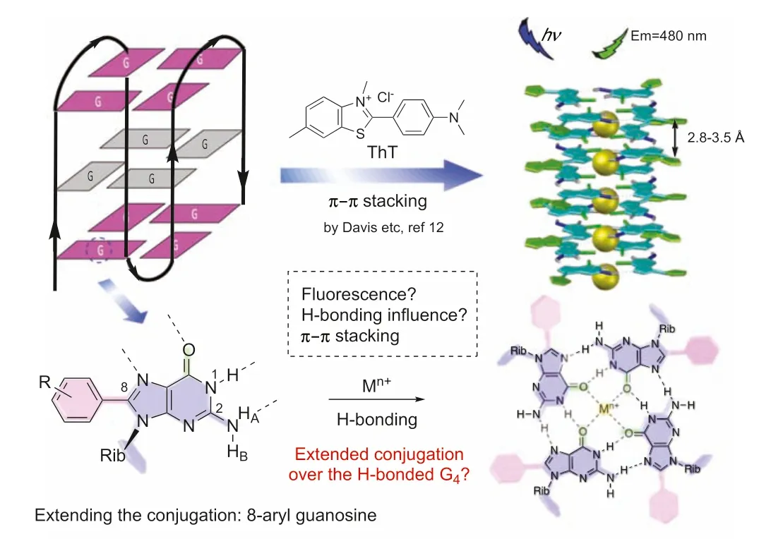
Scheme 1.G-quadruplex as fluorescence sensors.
With the interest in understanding how H-bond aggregation influences purine conjugation and G4-quartet fluorescence, in this work, we prepared a series of fluorescence active 8-aryl guanosine and evaluated their FL emission upon self-assembly.Fluorescence active G-quadruplexes were successfully prepared and characterized both in solution and solid state.para-Cyano (p-CN) and 8-anthracene (8-An) substituted guanosine analogs were identified to maintain the FL emission while holding the G4-quartet assembly.Interesting photoluminescent activity was observed between monomeric guanosine and G4-assembly, confirming the ligandcontrolled G4emission for the potentialπ-πstacking system,which provides an important foundation for future FL molecular probe design.
Recently, our group developed a well-defined guanosine G8-octamer system through the application of conformationally defined guanosine building blocks [24,25].The key to conformational control is the introduction of steric hindrance both on guanine(C8-aryl) and ribose (2′,3′-isopropylidene), which allows the G4-quartet to adopt a tight “bowl-shape” conformation and leads to the “bottom-to-bottom” stacked G8as the discrete species in solution and solid state.This dual modification is critical for the formation of well-defined G-quadruplex as modification on either C-8 or ribose alone resulted in the formation of mixtures of various stacking isomers (Fig.1A, see details in Supporting information).
To systematically study the fluorescence difference between Gmonomer and G4-quartet, a library of guanosine derivatives was prepared (Fig.1B) with variations on both 8-aryl and ribose.It is known that the introduction of 8-aryl group on purine could introduce fluorescence emission [11,21,26,27].To explore the FL activity of these guanosine monomers, we first evaluated the photoactivity of these compounds in dimethyl sulfoxide (DMSO).The results are summarized in Table 1.
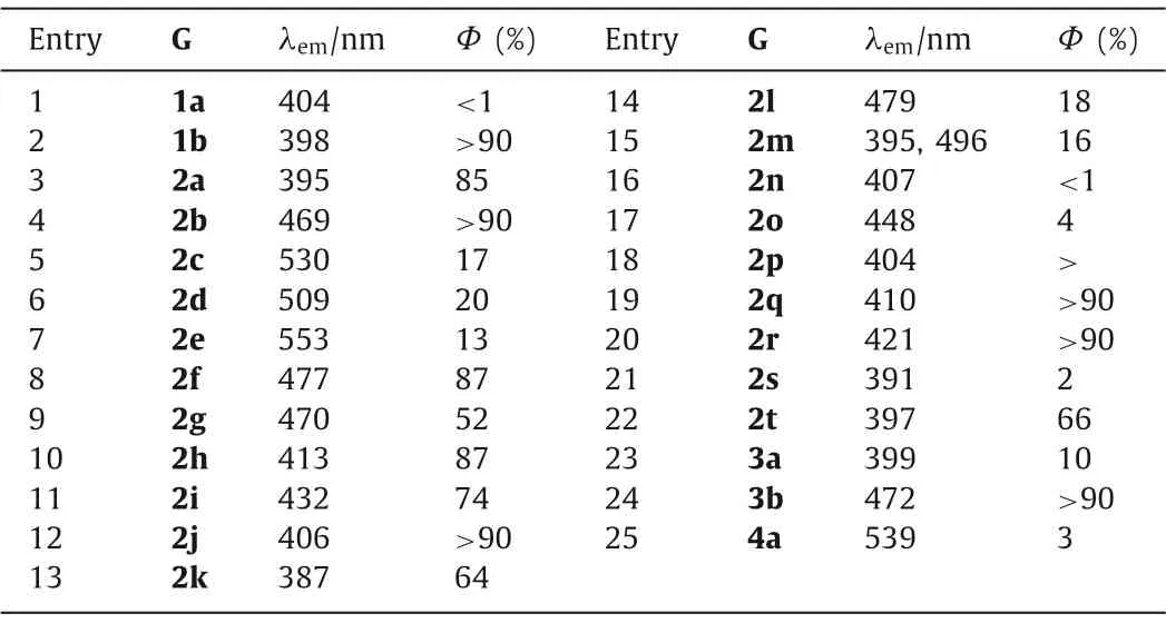
Table 1 Optical properties of monomeric guanosine in DMSO.a
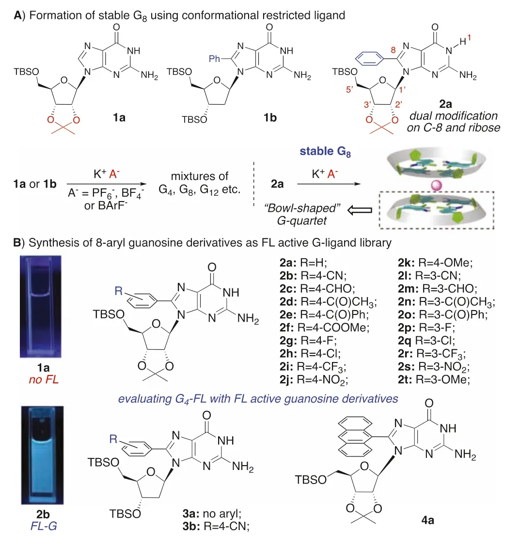
Fig.1.Exploring π-conjugation with FL active guanosines.
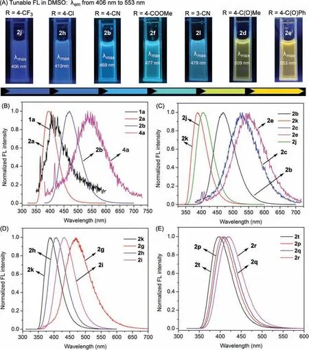
Fig.2.(A) Tuning the fluorescence with 8-aryl substituents in DMSO solution under irradiation of 365 nm UV light (1.0 mmol/L).(B) FL emission of guanosine 1a, 2a, 2b,and 4a in DMSO.(C) Normalized FL emission of para-substituted 8Ar-G, including compound 2b, 2k, 2c, 2e, or 2j in DMSO.(D) Normalized FL emission of para-substituted 8Ar-G, including compound 2g, 2h, 2i, or 2k in DMSO.(E) Normalized FL emission of meta-substituted 8Ar-G, including compound 2t, 2p, 2q, or 2r in DMSO.
As expected, a wide range of FL emission was achieved with these modified guanosine derivatives (Fig.2A).With purine as the electron rich aromatic ring, incorporation of electron-withdrawing groups (EWG) on 8-aryl should facilitate a red-shift of the emission.To our delight, compared to guanosine 1a and 2a, 8-(4-cyano)phenyl guanosine 2b exhibited a significant red-shift with strong fluorescence emission maxima at 469 nm with a bright blue-green light in DMSO (Fig.2B).Other 8-aryl guanosines withpara-substituted EWGs were also examined and compared.Overall,guanosine 2c, 2d, 2e, and 2f exhibited obvious redshifted emission(λmax>470 nm) compared to other guanosines though with comparatively lower quantum yields.It is noteworthy that the emissionλmaxof 8-aryl guanosines with carbonyl substituents is longer compared to 2g, 2i, and 2j with stronger electron withdrawing abilities.The red-shift emission achieved byp-CN, andp-carbonyl substitution can be attributed to an extended conjugated structure with smaller energy difference between the highest occupied molecular orbital (HOMO) and the lowest unoccupied molecular orbital (LUMO) [28].As shown in Fig.2C, compound 2e demonstrated the longest FL emission maxima (λmax=553 nm) likely due to the further extended conjugation by benzyl group, though with a compromise of lower emission efficiency (Φ=13%).Installing the electron donating group (EDG) at theparaposition of 8-aryl(2k,λmax=387 nm) led to a notably blue-shifted emission (up to 160 nm).For compounds with no substitution’s conjugations, including 2k, 2g, 2h, and 2i, theλmaxincreases with the increase of electron withdrawing ability.This observation highlights the effective emission control by tuning the electronic property on the 8-aryl group.These results also suggest that electronic effect and conjugation are the synergistic factors to influence the fluorescence performance on guanine.
Tuning electronic density of the 8-aryl group with variousmeta-substituted analogues gave a similar trend but with reduced impact (<24 nm difference) on the emission (Fig.2E).Most EWG atmeta-substitution resulted in a blue-shift and lower quantum yields, compared topara-substituted derivatives (2m-2o,λmax=395–448 nm, Fig.S14 in Supporting information).
To better examine the influence of electronic effect on the FL property, compounds with substituents possessing simple electronic effect (2k, 2g, 2h, 2i and 2t, 2p, 2q, 2r) were examined(Figs.2D and E).The Hammett correlation was applied to evaluate the substituent effects on 8-aryl guanosine absorption and fluorescence spectra (see Supporting information for detail).These results confirmed that electron density difference on the 8-phenyl ring and guanine can influence the luminesce as we proposed.
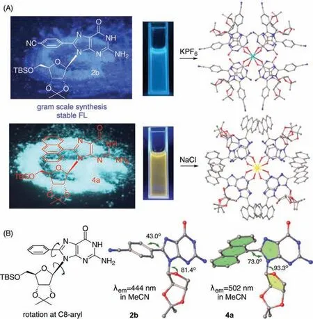
Fig.3.Crystal structures of 2b and 4a G8-octamer.(A) FL of monomeric 2b and 4a in the solid state under irradiation of 365 nm UV light (left); top view of crystal structures of G8-octamer formed by 2b and 4a through self-assembly with KPF6 and NaCl respectively (right); (B) Crystal structures showing large dihedral angles of 2b and 4a.
The influence of the sugar ribose on the fluorescence was also evaluated.Guanosine 1b, 3a, and 3b were synthesized and the absorption and emission spectra were collected (Fig.S13 in Supporting information).As expected, the FL emission data of 2′-deoxyguanosine demonstrated a very similar emission trend as 2′-oxy-guanosine, suggesting the strong influence of purine over ribose for the resulting FL emission.The 8-anthracene substituted guanosine 4a was synthesized with the expectation to extend purine conjugation.Interestingly FL red shift (λmax=539 nm in DMSO) was received as shown in Fig.2B.Switching the solvent to less polar dichloroethane (DCE) led to a blue shift (λmax=476 nm)with significantly improved intensity (see Supporting information for the optical activity of all other compounds in DCE).
With the FL emission of guanosine monomers tested, we continue to explore the fluorescence upon self-assembly in non-polar organic solvents.Treating 2b and 4a with K+or Na+in either CDCl3or CD3CN gave a complex with one set of1H NMR signals (Figs.S2 and S3 in Supporting information).The guanosine to cation ratio of these complexes is 8:1, indicating the formation of octamers.Fortunately, X-ray single crystal structures of[(2b)8K]+PF6−and [(4a)8Na]+Cl–were successfully determined,which confirmed the formation of H-bonded G8-octamers (Fig.3).
The X-ray crystal structures of [(2a)8K]+PF6−revealed the twisted conformation between 8-aryl and purine with a dihedral angle of 43°(Fig.3B).A larger dihedral angle (73°) was observed with 8-An substituted guanosine complex [(4a)8Na]+Cl–, resulting in the stronger steric hindrance between the modified purine and ribose to adopt asynconformation.These crystal data suggest the twisted intermolecular charge transfer (TICT) mechanism for the observed FL emission.
With the conditions for both G-monomer (in DMSO) and G8complexes confirmed, their FL emissions were compared.As shown in Fig.4, a blue shift emission (around 30 nm) was observed with G8complexes relative to monomer for both 2b and 4a.This result strongly suggested that no extended conjugation was formed in the H-bond linked poly-purine G4-quartet aggregation (would lead to the red shift) due to the interrupted p-conjugation.To the best of knowledge, this is the first direct evidence for FL emission evaluation upon G4-quartet formation over monomer, though in different solvents (CH3CNvs.DMSO).The1H NMR titration (mixture of CD3CN and DMSO–d6) experiments were also conducted to ensure that the supramolecular equilibrium was between G-monomer and G8-octomer without observation of intermediate aggregations as shown in Fig.4B.
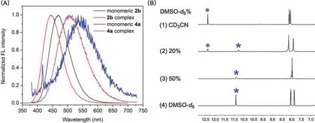
Fig.4.(A) FL emission of monomeric 2b and 4a, [(2b)8 K]+PF6−and [(4a)8Na]+Cl–.(B) 1H NMR titration experiment of [(2b)8 K]+PF6– complex in (1) CD3CN, (2) 20% of DMSO–d6 and CD3CN solvent mixture, (3) 50% of DMSO–d6 and CD3CN solvent mixture, (4) DMSO–d6.The characteristic proton NH(1) on guanosine was marked by dots and stars.The dot represents the NH(1) proton of complexes while the star represents NH(1) proton of monomers.
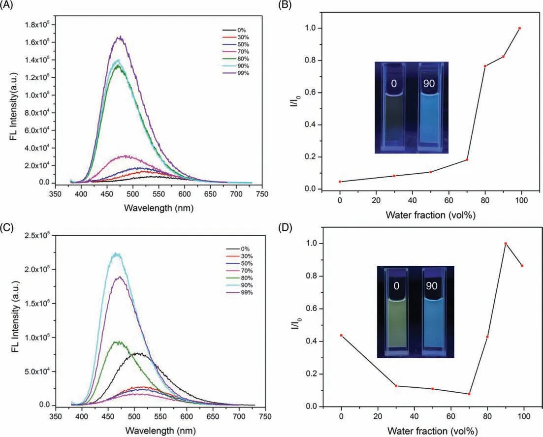
Fig.5.The titration PL spectra of (A) 4a (10 μmol/L) in DMSO/water mixtures and (C) [(4a)8Na]+Cl– complex in CH3CN/water mixtures with increasing water fractions (fw).Plots of emission intensity of (B) 4a and (D) [(4a)8Na]+Cl– complex versus the water mixtures.
The twisted conformations of 2b and 4a from the crystal structures raised our interest to know the fluorescence of these guanosine ligands in the solid state.With the 8-aryl guanosines synthesized in gram scale of these guanosine derivatives, their FL emissions were explored under solid state.Interestingly, strong fluorescence was observed for most of these compounds in the solid state (Fig.3A, see Supporting information for detail).This strong solid-state emission initiated our interest in exploring their potential aggregation-induced emission (AIE) in solution [29–31].The pioneering work of applying the AIE property for the “turn-on” Gquadruplex probe was reported by Benzhong Tang and co-workers[32].In their studies, an AIE active salts tetraphenylethenes (TPE),which is a non-emissive dye, was applied and showed surprisingly high luminescent emission after binding to the G4-quartet by electrostatic interaction.However, no FL-active G4-quartet has been reported with potential AIE property.To explore the potential AIE property with these FL-active guanosine analogs, compounds 1a, 2b, 3b, and 4a were applied into the solvent-titration experiments.As shown in Figs.5A and B, addition of water into the 4a in DMSO solution gave the drastic increase of FL emission (as of 70%), reflecting a typical AIE phenomenon in solution.Similar AIE effective was observed with the [(4a)8Na]+Cl–complex in CH3CN(Figs.5C and D), which likely attributed to the complex dissociation in protic solvent.Notably, all other tested guanosine derivative did not show this AIE property (Figs.S38-S40 in Supporting information), suggested the unique photoactivity of guanosine 4a.Detailed mechanistic studies and application of these special AIE guanosine ligands are currently under investigation in our lab.
In this study, we provided the first direct evaluation of FL activity of guanosine upon formation of G4-quartet.The introduction of various aryl groups on purine C-8 position results in tunable FL emission with strong blue to yellow light emission and large Stokes shift (up to 190 nm).The direct confirmation of guanosine monomer and G-quadruplex FL emission revealed no extended conjugation within G4-quartet.Interesting AIE property was observed with thep-anthracene guanosine, suggesting the promising applications of this new FL system in chemical and biological research.
Declaration of competing interest
The authors declare that they have no known competing financial interests or personal relationships that could have appeared to influence the work reported in this paper.
Acknowledgments
We are grateful to the National Natural Science Foundation (No.CHE-1665122), the National Institutes of Health (No.1R01GM120240–01) for financial support.
Supplementary materials
Supplementary material associated with this article can be found, in the online version, at doi:10.1016/j.cclet.2022.03.007.
 Chinese Chemical Letters2022年9期
- Chinese Chemical Letters的其它文章
- A review on recent advances in hydrogen peroxide electrochemical sensors for applications in cell detection
- Rational design of nanocarriers for mitochondria-targeted drug delivery
- Emerging landscapes of nanosystems based on pre-metastatic microenvironment for cancer theranostics
- Radiotherapy assisted with biomaterials to trigger antitumor immunity
- Development of environment-insensitive and highly emissive BODIPYs via installation of N,N’-dialkylsubstituted amide at meso position
- Programmed polymersomes with spatio-temporal delivery of antigen and dual-adjuvants for efficient dendritic cells-based cancer immunotherapy