
Effect of tetrahedral DNA nanostructures on LPS-induced neuroinflammation in mice
2022-06-20 06:20XioYngFnZhngYueDuWeitongCuiYikiDouYunfengLinZhiheZhoXiohong
Chinese Chemical Letters 2022年4期
Xio Yng,Fn Zhng,Yue Du,Weitong Cui,Yiki Dou,Yunfeng Lin,d,*,Zhihe Zho,*,Xiohong M,**
a Psychiatric Laboratory and Mental Health Center,West China Hospital of Sichuan University,Chengdu 610041,China
b State Key Laboratory of Oral Diseases,National Clinical Research Center for Oral Diseases,West China Hospital of Stomatology,Sichuan University,Chengdu 610041,China
c Physical Examination Center,West China Hospital,Sichuan University,Chengdu 610041,China
d College of Biomedical Engineering,Sichuan University,Chengdu 610041,China
1 These authors contributed equally to this work.
ABSTRACT Neuroinflammation plays a significant role in inducing depression-like behavior.Tetrahedral DNA nanostructures(TDNs)are molecules that exhibit anti-inflammatory properties and can effectively penetrate the blood-brain barrier.Thus,researchers have hypothesized that TDNs regulate the secretion of proinflammatory cytokines and consequently alleviate depression-like behavior.To test this hypothesis,we investigated the effect of TDNs on the depression-like behavior of C57 mice induced by lipopolysaccharide(LPS).We performed open-field,tail suspension,and sucrose preference tests on LPS-and LPS/TDNtreated mice.The results indicated that the injection of TDNs into LPS-treated mice resulted in increased velocity,center zone duration,frequency to the center zone,and sucrose preference,and decreased immobility time.Immunofluorescence results indicated that peripheral administration of LPS in the mice activated inflammation,which culminated in distinct depression-like behavior.However,TDNs effectively alleviated the inflammation and depression-like behavior through the reduction of the expression levels of proinflammatory cytokines,such as interleukin-1β and tumor necrosis factor-α in the brain.Additionally,TDNs normalized the expression level of microglia cell activation markers,such as ionized calcium binding adaptor molecule 1,in the hippocampus of mice.These results indicated that TDNs attenuated the LPS-induced secretion of inflammatory factors and consequently alleviated depression-like behavior.
Keywords:Tetrahedral DNA nanostructures Lipopolysaccharide Depression-like behavior Proinflammatory cytokine Microglia cell
Depression is a significantly prevalent and debilitating psychiatric disorder and the second leading cause of mental disability[1].Approximately one-third of depression do not respond to firstline treatment consisting of sequential antidepressant treatment[2].The inconsistency in clinical efficacy may be attributed to the heterogeneity and diverse etiology of the disorder.Thus,in recent years,researchers have focused on the development of more effective antidepressants for depression.
Previous studies have reported that depression is associated with neuroinflammatory responses due to the abnormal activation of the inflammatory response system(IRS)[3–5].Preclinical studies have reported that neuroinflammation plays a vital role in inducing depression symptoms through the secretion of various proinflammatory cytokines[6].Among these cytokines,the expression levels of interleukin-1β(IL-1β)and tumor necrosis factor-α(TNF-α)have been reported to be significantly high in brain samples of mice and humans with depression[7],which suggests that neuroinflammation is significantly associated with depression[8].
Furthermore,the administration of lipopolysaccharide(LPS)in mice has been reported to induce depression symptoms through microglia activation and proinflammatory cytokine expression in the peripheral blood and brain[9,10].Microglia are significant immune cells located in the central nervous system(CNS),and under abnormal conditions,may be activated to secrete proinflammatory factors[11].Microglia activation and the consequent release of various proinflammatory cytokines owing to neuroinflammation induced by LPS play significant roles in neuroprotection[7,12–14].However,cytokine dysregulation may lead to neuroinflammationrelated disorders such as depression[7,15,16].
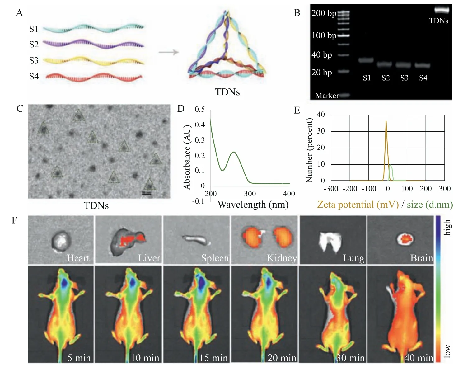
Fig.1.(A)Schematic diagram of the structure of TDNs.(B)PAGE results indicating the presence of ssDNA(S1–S4).(C)TEM image of TDNs.(D)Absorbance spectra of TDNs.(E)Zeta potential and hydrodynamic size distribution of TDNs.(F) In vivo distribution of Cy5-TDNs in BALB/c nude male mice.
Among several strategies to alleviate depression symptoms,the regulation of neuroinflammatory responses is a promising method.Thus,drug development aimed at the regulation of inflammatory responses could be significant in the treatment of depression symptoms and consequent alleviation of depression.
Nanostructures have exhibited broad application prospects in the biomedical field[17–25],such as TDNs[26–31].For the treatment of depression,tetrahedral DNA nanostructures(TDNs),which are fabricated by the self-assembly of four single-stranded DNAs(ssDNAs,Table S1 in Supporting information),are of considerable significance[23,32].TNDs serve as small molecules that can effi-ciently penetrate the blood-brain barrier(BBB)[23,33]and have been reported to regulate the mRNA expression of antioxidative enzymes,resulting in the attenuation of the release of IL-1βand TNF-αinduced by LPS[34].Thus,TDNs exhibit favorable antiinflammatory and neuroprotective properties for their application as novel antidepressants[23,35].
Therefore,in this study,we analyzed the behaviors of LPS-and LPS/TDN-treated mice and examined the effect of TDNs on the neuroinflammation response in LPS-treated mice in an attempt to verify the hypothesis that TDNs alleviate depression symptoms through the regulation of proinflammatory cytokines.
In this study,8-week-old C57 male mice(weight 20–25 g)were purchased from DOSSY(Chengdu,China)and group-housed in sets of five per cage under standard conditions(approximately 25 °C and 12/12 h light/dark cycle)with food and water.All procedures and protocols were approved by the Ethics Committee of the West China Hospital,Sichuan University.For the preparation and characterization of TDNs,four ssDNAs were obtained from Takara Bio Inc.,and TDNs were synthesized based on a previously reported procedure[32].Details of the synthesis are mentioned in Supporting information(as shown in Table S1).
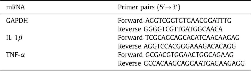
Table 1 Primer sequences of qPCR related genes.
For the animal experiment,depression-like behavior was induced in the mice through the injection of LPS,which is a common method of inducing acute depression-like behavior in mice[36,37].The experimental mice were randomly assigned to 3 groups of 10 mice each and injected with normal saline(NS,NaCl)+buffer,LPS(0.83 mg/kg)+buffer,and LPS+TDNs(1000 nmol/100 μL),respectively.NS and LPS were administered intraperitoneally(10 mL/kg),followed by injection of the buffer and TDNs into the tail vein(100 μL/mice)0.5 h and 6 h after the LPS administration,respectively.Behavioral tests were performed on the mice 24 h after the NS and LPS administration.The mice were anesthetized,and half of them received systemic blood perfusion with 0.9% NaCl and 4%paraformaldehyde(PFA)prior to their sacrifice,and their brain tissues were preserved in 4% PFA.The other half were sacrificed,and their blood samples and hippocampal tissues were stored at-80 °C for further use.
With regard to the behavioral tests,based on previously reported procedures[14,38,39],open-field test(OFT),tail suspension test(TST),and sucrose preference test(SPT)were performed.The distance covered by the mice,center zone duration,frequency to the center zone,velocity of the mice,and immobility time were measured using the Noldus EthoVision XT(version 12.0).The sucrose preference ratio(SPR)(%)was calculated using the equation SPR =[(sucrose consumption)/(water and sucrose consumption)]× 100%[15].Details of the test procedures are provided in Supporting information.
The composition of the synthesized TDNs was examined using polyacrylamide gel electrophoresis(PAGE).Absorption spectroscopy was carried out to examine the absorbance of TDNs.The zeta potential and hydrodynamic size of the TDNs were measured using a Zetasizer Nano ZS90.The distribution of TDNs in the various organs of the mice,including the brain,heart,liver,kidneys,and lungs,was analyzed using anin vivoimaging system(Caliper Life Sciences,IVIS Spectrum,USA).Nude mice were anesthetized with isoflurane,and then 100 μL of Cy5-TDNs was injected into the tail veins of the mice.The distribution of TDNs was analyzed at 5,10,15,20,30,and 40 min.
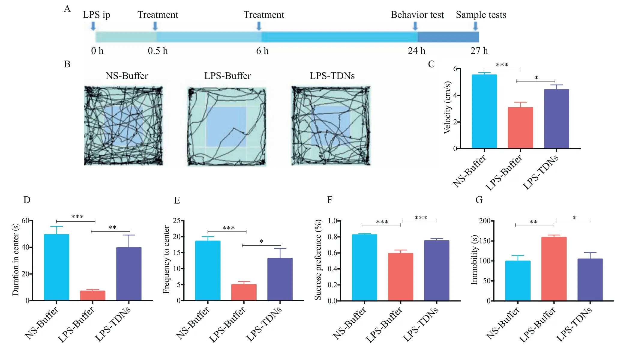
Fig.2.Effect of TDNs on LPS-induced depression-like behavior(n = 10 and *P <0.05,**P <0.01,***P <0.001).(A)Schematic of the experimental design.(B)Representative tracks in the OFT.(C)Velocities of the mice.(D)Center zone duration.(E)Frequency to the center zone.(F)Sucrose preference ratios.(G)Immobility times.
To examine the effects of LPS and TDNs on the gene expression of proinflammatory cytokines,such as IL-1βand TNF-α,qPCR was performed.Total RNA was extracted from the brain tissues of the mice using an RNeasy Plus Mini Kit.The extracted RNA samples were reverse-transcribed using a reverse transcription-polymerase chain reaction(RT-PCR)kit;the samples were heated at 94 °C for 3 min,followed by 40 cycles of 5 s at 94 °C and of 34 s at 60 °C.The cycle threshold(Ct)values of the samples were analyzed and quantitative results were obtained using the 2–ΔCt method.Primer sequences of the genes encoding IL-1β,TNF-α,and GAPDH are listed in Table 1.
The brain tissues frozen at-80 °C were sectioned at 8–12 μm.Then,phosphate buffer saline(PBS)was used to wash the sectioned and the PFA-treated brain tissues thrice for 5 min each at room temperature(approximately 25 °C)using a shaker.Then,the samples were incubated with 3% bovine albumin for 30 min and washed again using PBS.Then,they were incubated with solutions of the primary antibody of rabbit Iba-1 in PBS(concentration of 1:2000)at 4 °C for 24 h and then washed with PBS.Subsequently,the samples were incubated with solutions of the corresponding secondary antibody in PBS(concentration of 1:200)(Servicebio)at 37 °C for 50 min under dark conditions and then washed with PBS.Next,the samples were incubated with 4′,6-diamidino-2-phenylindole(DAPI)for 10 min followed by incubation with a spontaneous fluorescence quenching agent for 5 min and then washed in tap water for 10 min.Prior to obtaining the fluorescence microscopy images,the samples were covered with an antifade mounting medium.Under excitation,the nuclei stained by DAPI were represented in blue,whereas the positive expression of IBA-1 was in red.The images were obtained by a Nikon confocal laser microscope(Japan).
For the statistical analysis,one-way analysis of variance(ANOVA)was carried out using the GraphPad Prism 6 software(GraphPad,USA)to analyze the variation in the results of the three groups.The data were presented as the mean ± standard deviation.APvalue<0.05(*)was considered statistically significant.
As shown in Fig.1A,TDNs consist of four ssDNAs(Table S1)and every ssDNA forms a triangle whose sides are shared with the other three triangles.PAGE results indicated that four ssDNAs were effectively self-assembled to form TDNs(Fig.1B).From the TEM image,the size of TDNs was estimated to be<20 nm(Fig.1C).The absorbance spectra of the synthesized TDNs indicated an absorbance of approximately 260 nm(Fig.1D).The zeta potential and hydrodynamic size of TDNs were approximately-9.1 mV and 10 nm(Fig.1E),respectively.These results indicated that smallsized TDNs with special spatial structures were successfully synthesized.The fluorescence signals generated by thein vivoimaging system indicated that TDNs penetrated the BBB and accumulated in the brain(Fig.1F).Thirty minutes after their injection into the tail vein,TDNs were predominantly concentrated in the brain tissue,whereas small amounts were distributed in the kidneys and liver.These results confirmed the ability of TDNs to rapidly penetrate the BBB and accumulate in the brain.Furthermore,they indicated that TDNs could remain in other organs,such as the kidneys and liver,without undergoing degradation.
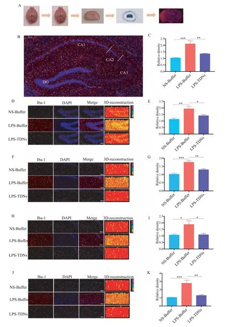
Fig.3.Effect of TDNs on the expression of Iba-1 induced by LPS(n = 5 and *P <0.05,**P <0.01,***P <0.001).(A)Schematic of the experimental design.(B)Structure map of the hippocampus.(C)Expression level of Iba-1 in the entire hippocampus.(D)Immunofluorescence of Iba-1 in DG of the hippocampus.(E)Expression level of Iba-1 in DG.(F)Immunofluorescence of Iba-1 in CA1 of the hippocampus.(G)Expression of Iba-1 in CA1.(H)Immunofluorescence of Iba-1 in CA2 of the hippocampus.(I)Expression level of Iba-1 in CA2.(J)Immunofluorescence of Iba-1 in the cortex.(K)Expression level of Iba-1 in the cortex.
To identify the effect of releasing LPS-induced depression-like behaviorsviaregulating inflammation response,as shown in Fig.2,LPS successfully induced depression-like behavior in the mice,which is consistent with previously reported studies[14,38].The OFT results indicated that the velocity,center zone duration,and frequency to the center zone of the group injected with LPS were significantly lower compared with those of the control group(NStreated group)(Figs.2B–E).
However,compared with the LPS-treated group,the group of mice treated with TDNs exhibited higher velocity,center zone duration,and frequency to the center zone.The sucrose preference ratio of the LPS-treated group was lower(less than 50%)than that of the control group,and the sucrose preference ratio of the TDN-treated group was higher than that of the LPS-treated group(Fig.2F).The TST results indicated that the immobility time of the LPS-treated group was considerably longer than that of the control group,and the immobility time of the TDN-treated group was shorter than that of the LPS-treated group(Fig.2G).
Immunofluorescence was also used to examine the localization,qualitative expression,and quantitative levels of ionized calciumbinding adaptor molecule 1(Iba-1)in the subregions of the hippocampus,including the dentate gyrus(DG),cornu ammonis 1,2,and 3(CA1,CA2,and CA3),and cortex(Fig.3A).The immunofluorescence results indicated that the expression levels of Iba-1 in DG,CA1,CA2,CA3,and the cortex of the mice significantly increased owing to the injection of LPS.However,after the mice were treated with TDNs,the abnormally increased expression levels of Iba-1 significantly reduced(Figs.3B–K).Also,histological assessment of the biocompatibility found that there is no obvious toxicity(Fig.4A).Additionally,the expression levels of IL-1βand TNF-αin the peripheral blood and brain tissues of the LPS-treated mice were abnormally high,and after the mice were treated with TDNs,the expression levels normalized(Figs.4B–E).These results were consistent with the results of the behavioral tests and support the antiinflammatory effects of TDNsviaregulating inflammation signaling pathway in mice.
On the basis of the above results and discussion,TDNs could reduce the expression levels of proinflammatory cytokines,such as IL-1βand TNF-α,in the peripheral blood and brain tissues,which alleviated the LPS-induced neuroinflammation and depression-like behavior.Furthermore,TDNs significantly normalized the inflammation signaling and downstream components,such as the activation markers of microglial cells(Iba-1),in the hippocampus of the mice.Thus,our results verified the hypothesis that TDNs alleviate depression-like behavior through the regulation of proinflammatory cytokines.
The immunofluorescence results indicated that the injection of LPS in the mice resulted in increased expression levels of inflammatory cytokines.LPS has been widely used to induce central neuroinflammation[40,41]which is associated with the increased expression of proinflammatory cytokines and abnormal microglia cell activation[42].LPS administration in mice has been reported to result in peripheral immune activation,which stimulates the activation of microglia cells and accelerates the production and transportation of a few cytokines to the CNS[43,44].However,the excessive activation of microglial cells could damage the surrounding neural tissues in the brain and result in a gradual loss of neurons.Thus,increased production of proinflammatory cytokines and enhanced activation of microglia and Iba-1 owing to LPS administration typically leads to excessive neuroinflammation that is associated with psychiatric disorders[11].Furthermore,previous studies have reported that individuals who committed suicide exhibited highly activated microglia cells in their brains[43,45].Therefore,based on previous reported research and the findings of our study,we speculated that excessive cytokine levels and neuroinflammation in individuals may induce synaptic changes that could lead to depression-like behavior[8,46–48].According to the current results,we deduced that the LPS-induced distinct depression-like behavior could be normalized by TDNs.
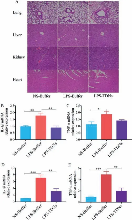
Fig.4.qPCR analysis of mRNA expression levels(n = 5 and *P <0.05,**P <0.01,***P <0.001).(A)Histological assessment of the biocompatibility.(B)Expression levels of IL-1β mRNA in the plasma.(C)Expression levels of TNF-α mRNA in the plasma.(D)Expression levels of IL-1β mRNA in the hippocampus.(E)Expression levels of TNF-α mRNA in the hippocampus.
In summary,we investigated the effect of TDNs on the depression-like behavior of C57 mice in this study.The results of the behavioral tests indicated that TDNs could alleviate the LPSinduced depression symptoms in mice.The fluorescence signals generated by thein vivoimaging system indicated that TDNs successfully penetrated the BBB and accumulated in the brain.The immunofluorescence results indicated that TDNs normalized the increased expression levels of Iba-1,IL-1β,and TNF-αthat were induced by LPS.However,there are some limitations:Acute LPSinduced model may limit the possibility of exploring longer effect of TDNs on depressive model.Also,the current findings need to be validated and replicated on different depressive model.In spite of the limitations,these results indicated that TDNs can effectively regulate the inflammatory response and consequently alleviate the LPS-induced neuroinflammation and depression symptoms in mice.Furthermore,TDNs exhibit high capability for their potential application as effective antidepressants for the treatment of depression.
Declaration of competing interest
The authors declare that they have no known competing financial interests or personal relationships that could have appeared to influence the work reported in this paper.
Acknowledgments
This research was supported by the National Key R&D Program of China(No.2019YFA0110600),the National Natural Science Foundation of China(Nos.82001432,81970916),the China Postdoctoral Science Foundation(Nos.2020TQ0213,2020M683319),and the West China Hospital Postdoctoral Science Foundation(No.2020HXBH104).Furthermore,we thank Dr.C.H.Li for his aid with the TEM image collection(Analysis &Testing Center,Sichuan University).
Supplementary materials
Supplementary material associated with this article can be found,in the online version,at doi:10.1016/j.cclet.2021.10.029.
 Chinese Chemical Letters2022年4期
- Chinese Chemical Letters的其它文章
- Key progresses of MOE key laboratory of macromolecular synthesis and functionalization in 2020
- Small nanoparticles bring big prospect:The synthesis,modification,photoluminescence and sensing applications of carbon dots
- Cell membrane-coated nanoparticles for immunotherapy
- Diketopyrrolopyrrole-derived organic small molecular dyes for tumor phototheranostics
- Exosome based miRNA delivery strategy for disease treatment
- Recent advances in targeted stimuli-responsive nano-based drug delivery systems combating atherosclerosis