
Recent progress on microfluidic biosensors for rapid detection of pathogenic bacteria
2022-06-20 06:18GaowaXingWeifeiZhangNanLiQiaoshengPuJinMingLin
Chinese Chemical Letters 2022年4期
Gaowa Xing,Weifei Zhang,Nan Li,Qiaosheng Pu,Jin-Ming Lin,*
a College of Chemistry and Chemical Engineering,Lanzhou University,Lanzhou 730000,China
b Beijing Key Laboratory of Microanalytical Methods and Instrumentation,Department of Chemistry,Tsinghua University,Beijing 100084,China
c Division of Chemical Metrology and Analytical Science,National Institute of Metrology,Beijing 100029,China
ABSTRACT Rapid on-site detection of pathogenic bacteria with high sensitivity and specificity is becoming an urgent need in public health assurance,medical diagnostics,environmental monitoring,and food safety fields.Despite being reliable and widely used,the existing methods of bacteria detection are cumbersome and time-consuming,which is not conducive to field detection.Microfluidic lab-on-a-chip technology has provided a detective tool for various analytes,due to its miniaturization,portability and low reagent consumption.Within this progress report,advances in the bacteria detection using microfluidic biosensors were discussed.Typical methods for pathogenic bacteria capture,separation and detection were introduced respectively in the first part.Then key applications of microfluidic biosensor-based rapid bacteria detection were presented.Finally,we made a conclusion and discussed possible research prospects in aspects of microfluidic biosensors for rapid detection of pathogenic bacteria.
Keywords:Microfluidic chip Biosensors Pathogenic bacteria Rapid detection
1.Introduction
Millions of people in the world got infected by contaminated food and water by pathogenic bacteria each year,resulting in various diseases against human health.For example,foodborne pathogens(mostly bacteria such asEscherichia coli,Salmonella enterica,orListeria monocytogenes)are estimated to cause 600 million food-related illnesses and 420,000 deaths annually[1].These bacteria are usually highly infectious and only a few dozen colony forming units(CFUs)or lower can cause disease.Therefore,early and sensitive detection of pathogenic bacteria is crucial to manage these problems and avoid outbreaks.Traditional detection methods rely on Gram staining,culturing and biochemical analysis,which have made great contributions to the detection of pathogenic bacteria.However,they often take 2 or 3 days or longer,making them difficult to deal with sudden public health and safety incidents[2,3].Currently,the most commonly used methods for microbial detection include flow cytometry[4,5],polymerase chain reaction(PCR)[6,7],enzyme-linked immunosorbent assay(ELISA)[8,9]and so on.These methods presented the advantages of high sensitivity,specificity and selectivity and short detection time.However,these detection technologies were expensive and required skilled personnel for sample preparation and detection,which is not suitable for on-site applications.Hence,detecting viable pathogenic bacteria cells with simple,fast,cheap,and on-site analytic strategies becomes urgent in public health assurance,medical diagnostics,environmental monitoring,and food safety fields.
Microfluidic chip refers to fluid flow and heat and mass transfer scaled down to the micrometer or nanometer scale,and many operations including specimen preparation(sampling and capture),reagent manipulation,biological recognition and detection can be successfully miniaturized into this platform,which is often called“lab-on-a-chip”(since 1979)[10].Microfluidic system is always applied as the biosensor for the detection of specific targets by converting a biomolecular recognition event into measurable physical or chemical signal[11].The integrated microfluidic biosensor permits low sample and reagent consumption,flexible liquid manipulation and process integration with reduced detection time.A broad range of characterization methods based on electrical,magnetic,and optical techniques can be readily incorporated into microfluidic biosensor devices,enabling rapid identification and detection within minutes.In addition,based on range of inherent features,microfluidic biosensors have received enormous attention due to their potential use in detecting pathogenic bacteria in many fields such as clinical diagnostics,food safety and environmental analysis[12–14].
In this review,advances in the detection of bacteria using microfluidic biosensors were discussed.First,the strategies of bacteria identification,separation and detection were briefly overviewed.The second part was devoted to introduce several detection applications in clinical diagnostics,food safety,and environmental monitoring.The results achieved were also summarized and future perspectives were proposed in the last part.
2.Microfluidic biosensors for the detection of pathogenic bacteria
Microfluidic biosensors could combine a series of individual functions,such as sample transfer,target capture,reagent mixing and separation,biological/chemical reaction and detection(signal output),into a single complex microfluidic chip system.The most important aspect was the design of the microfluidic chip with suitable materials and structure.The definition of microfluidic chips has developed over time.Various materials have been designed and manufactured for microfluidic chips at the 2D and 3D levels including silicon[15],glass[16],quartz[17],polymethyl methacrylate(PMMA)[18],hydrogel[19],polydimethylsiloxane(PDMS)[20–23],paper[24,25],cloth[26,27]and wood[28].Recently,the 3D printing technology was used for 3D-printed microfluidic device fabrication to detect microorganisms[29].Melt-extruded microcapillary film(MCF)with several parallel capillaries have also been used for microbial detection which could be easily and costeffectively turned into multiplex bioassay strips suitable for microfluidic chip fabrication[30].
These detection platforms were generally combined with other equipment for data acquisition,signal processing and monitoring.They might be combined with the handheld instruments to achieve device miniaturization.Note that the selection of the microfluidic device should be associated with the target analytes and the process of identification,separation and detection,so as to form a suitable microfluidic biosensor for the detection of pathogenic bacteria.
2.1.Capturing strategies for target pathogenic bacteria
Specific capture probes are dominant to microfluidic biosensors for improving the detection selectivity of target bacteria from complex samples and many strategies have been performed as bacteria-specific capture units.Antibody-antigen interactions,aptamer binding,bacteriophage biological recognition,antibiotics/antimicrobial peptide capture,and other target recognition interactions were commonly applied in microfluidic biosensors before the bacteria detection.
2.1.1.Antibody-antigen interactions
Antibody-antigen interactions are the most popular capture elements used in microfluidic biosensors to achieve the specific capture of bacteria from various complex samples[19,29,31].In most microfluidic biosensors,antibody-antigen sandwich is widely used to capture whole bacterial cells through a variety of bacterial extracellular receptors without cell lysis or release of enzyme makers for further signal transduction and amplification,such as ELISA.Generally,the capturing probes and the signal output molecules for detection are introduced in the antibody-antigen sandwich reactions.Magnetic nanoparticles(MNPs)modified with the target bacteria antibodies forming immunomagnetic beads were usually used for bacteria capture.Kimet al.realized bacteria identification based on magnetic capture particles(CPs),and europium-fluorescence labeled particles(LPs)functionalized with antibodies(Fig.1a)[18].After capturing the bacteria through antibody-antigen interactions,the fluorescence signal of the bacterial immune complexes was acquired for quantitative analysis ofVibrio parahaemolyticusfrom 0.1 mL pure broth culture samples within 30 min at single cell level.Meanwhile,Caiet al.described a similar strategy of visual microfluidic immunosensor for the determination ofSalmonella typhimurium(S.typhimurium)in chicken samples[31].The visual travelling distance of the red dye driven by the oxygen pressure generated from the catalyzed bacterial immune complexes by hydrogen peroxide was measured for bacteria content.
2.1.2.Aptamer binding
Although antibodies showed good specificity and selectivity,the stability was low and the price was usually high.In recent years,aptamers are widely used in the recognition of analytes.Compared with antibodies,they are easier to synthesize and modify with higher purity,smaller molecular weight(~12–30 kDa)and lower cost,and have better thermo/pH stability for easy storage[32,33].Suet al.have replaced the antibodies with aptamer for dual aptamer assays to detectAcinetobacter baumannii(AB)[34].MNPs coated withAB-specific aptamers were used to capture bacteria and the quantum dots(QD)bound to a second aptamer were utilized to quantify the amounts of bacteria with LED induced fluorescence module integrated into the device.Only 10 μL of sample and reagents were required with the detection limit of 100 CFU/reaction in 30 min(Fig.1b).Moreover,the sensitivity could be further improved by many approaches,such as the rolling circle amplification(RCA)technique.Liet al.modified the inner surfaces of the microfluidic channels with RCA products that featured repeating tandem capturing aptamers specific forE.coliO157:H7 cells[35].They demonstrated that compared with the unit-aptamer approach,this modality significantly improved the capturing efficiency at a wide range of flow rate.The capturing performance exhibited in the microchannels for the spiking in different food matrices(iced tea,bottled water,orange juice and milk)were similar to that in PBS.Furthermore,the combination of antibody and aptamer was used to form an antibody-target-aptamer sandwich for the identification of cells[36],which provided a certain reference for the identification of bacteria.
2.1.3.Bacteriophage biology recognition
Bacteriophage(phage)can attack specific bacteria and infect them using their metabolic machinery[37].They may further be used as bio-recognition element alternatives to the currently available molecular probes in the development of highly specific,stable,sensitive,selective,rapid,easily available,cost-effective and reliable platforms for pathogenic bacteria detection.Hussainet al.have reviewed about bacteriophage biology with various recognition sites,receptor-binding proteins on the surfaces of tailed phages,and methodologies for phage-based bacterial detection[38].They discussed the strategies of phage-based bacterial detection including phage-induced bacterial lysis,phages immobilized on a transducer surface,fluorescently labelled phages,phageconjugated quantum dots,and recombinant reporter phages.Dowet al.reported a work about bacteriophage-based identification of three wellknown pathogen species by luminescence assay after acoustophoresis separation in blood,and the limit of detection of this work was 6 bacteria,which was 33-fold improvement compared with the unpurified sample(Fig.1c)[39].
2.1.4.Antibiotics
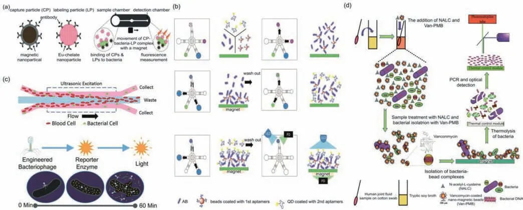
Fig.1.(a)Magnetic capture particles(CPs)and europium-fluorescent labeling particles(LPs)functionalized with antibodies were used to identify Vibrio parahaemolyticusa at the single cell level.Reproduced with permission[18].Copyright 2020,Royal Society of Chemistry.(b)Dual aptamer assays which first aptamer was modified with MNPs to capture bacteria and a second aptamer was bounded to QD to be as fluorescence signal detect AB.Reproduced with permission[34].Copyright 2020,Elsevier.(c)A system of bacteriophage-based luminescence assay.Reproduced with permission[39].Copyright 2018,the Royal Society of Chemistry.(d)The bioprobes including vancomycin for GPB and magainin for GNB detected and identified periprosthetic joint infection-associated bacteria performed on an integrated microfluidic system.Reproduced with permission[40].Copyright 2019,the Royal Society of Chemistry.
Antibiotics could interact specifically with bacterial cell walls so as to be used as pathogenic bacteria capture probes.Vancomycin could bind to the amine group of the peptidoglycan on the cell wall of the bacteria through the carbonyl group to form a strong hydrogen bond.Recently,it has been found that vancomycin-modified nanomaterials have been used to capture gram-positive(GPB)and gram-negative(GNB)bacteria.Liuet al.developed a microfluidic system to detect four bacteria associated with periprosthetic joint infections(Staphylococcus aureus(S.aureus),methicillin-resistantS.aureus,E.coli,andAB)integrating sample treatment,bacterial isolation,bacterial lysis,PCR and optical detection[40].The vancomycin-coated MNPs were used to capture the four target bacteria and enhance isolation.After separation,a universal 16S ribosomal ribonucleic acid PCR primer set and four species-specific primer sets were used for PCR-based detection and identification of the four bacteria.The whole experiment took less than 90 min with the limit of detection below 100 CFU/mL(Fig.1d).Kimet al.first demonstrated portable dual antibiotics(as bioprobes)conjugated graphene micro pattern fieldeffect transistor(ABX-GMFET)combined with a microfluidic chip for on-site detection of the presence of GPB and GNB bacteria in the samples[41].The bioprobes including vancomycin for GPB and magainin for GNB were attached to ABX-GMFET based on charge or chemical moiety interaction between the bioprobes and target bacteria.The limit of detection was 10° CFU/mL(1–9 CFU/mL).
2.1.5.Other target recognition interactions
Antimicrobial peptides(AMPs)with high stability and low cost have been studied in the detection of bacteria by Qiaoet al.[42].Maet al.have reviewed DNAzyme biosensors for the detection of pathogenic bacteria where DNAzyme was used as a molecular recognition element(RNA-cleaving DNAzyme)or as a reporter element(peroxidase mimicking DNAzyme)[43].Miet al.summarized lectins as a promising alternative recognition element in bacterial detection biosensors[44].These strategies of capturing bacteria could be introduced and integrated in the microfluidic biosensor system.
2.2.Separation of pathogenic bacteria
In many microfluidic biosensors for bacteria detection,the operations of target bacteria identification and signals output were performed simultaneously.However,sometimes it is necessary to separate the target bacteria after capture and then proceed to the next detection,especially in the complex samples.To solve this problem,MNPs-based separation,inertial microfluidic sorting and other separating methods have been used in the microfluidic biosensors.
2.2.1.MNPs-based separation
The MNPs-based separation was widely used for the enrichment and separation of pathogenic bacteria cells in the microfluidic biosensors[19,29,31,45–48].The bacteria could be fixed on the surface of magnetic nanoparticles and easily separated under the effect of an external magnetic field according to its magnetic characteristics.Lin’s group have reported a series of magnetic separation works for pathogen separation in the detection process[46–48].The magnetic bacteria based on immune reaction between the immune MNPs and the target bacteria and the polystyrene microspheres(PSs)modified with the detection antibodies and the catalases were captured by the external magnetic field[46,47].After separated by a magnetic field,the MNP-bacteria-PS complex modified with enzymes would produce the detection signals to quantitatively analyze bacteria in food(Fig.2a)[46].They also developed another magnetic bacteria complex by MNPs and ZnO-capped mesoporous silica nanoparticles(MSNs)forSalmonelladetection[48].The detection signal of the microfluidic biosensor was the fluorescence changes from acid-responsive curcumin modified on the MSNs which was also used for signal amplification.
2.2.2.Inertial microfluidic sorting
The inertial microfluidic sorting device based on the size differences between bacteria and blood cells were used with external field-free operation to detect bacteria from the blood sample.Huanget al.reviewed the theoretics and applications of inertial microfluidics for focusing,concentrating,isolating,or separating various bioparticles such as blood components,circulating tumor cells and bacteria[49].Luet al.designed a unique microfluidic channel to enhance inertial lift force at the microsquare zone and produced localized secondary Dean flow drag force in addition to global Dean flow drag force,realizing the separation of bacteria with the size between 5.5 μm and 6.0 μm at high recovery ratio(Fig.2b)[50].This inertial sorting device was used to purify two species ofCandida(Cornus glabrataandCandida albicans)fromCandida-spiked blood samples for enhanced molecular diagnosis of bloodstreamCandidainfection.
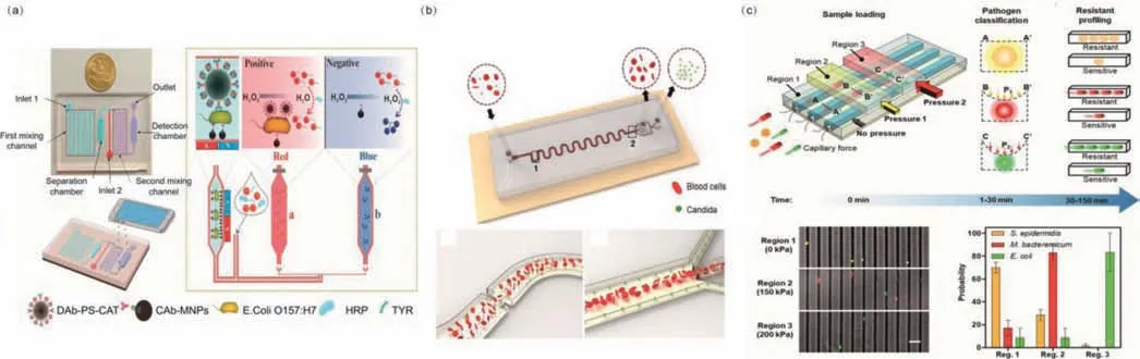
Fig.2.(a)MNPs-based separation for rapid detection of E.coli O157:H7 by smart phone imaging colorimetric photos.Reproduced with permission[46].Copyright 2019,Elsevier.(b)Separation between blood cells and Candida cells through the channel by inertial microfluidic sorting.Reproduced with permission[50].Copyright 2020,American Chemical Society.(c)Bacteria are trapped in different regions of the channels and classified according to the applied pressure.Reproduced with permission[51].Copyright 2019,National Academy of Sciences.
2.2.3.Other separation methods
Size differences between bacteria and blood cells were also used in the acoustic separation.Dowet al.described a plastic microfluidics based on the size differences between bacteria and blood cells for rapid detection of both GPB and GNB from blood[39].They revealed a 33-fold improvement in limit of detection compared with the unpurified sample.Liet al.reported an adaptable microfluidic system for rapid pathogen classification and antimicrobial susceptibility testing(AST)at the single-cell level[51].Particularly,an adaptable microchannel with tunable pneumatic valves physically trapped bacteria and classified the bacterial species according to their physical size and shape within 3 min(Fig.2c).By monitoring their growth in the presence of antibiotics at the single-cell level,AST could be finished in 30 min compared with days required for standard procedures.
2.3.Detection technology
The microfluidic biosensors were generally combined with other equipment for data acquisition and readout,signal processing and monitoring.The detection technology could employ various optical techniques,electrochemical detection,mass spectrometry methods(MS),and so on.On the other hand,these microfluidic systems might combine with the handheld instruments to realize the device miniaturization such as smartphone,personal glucose meters(PGMs)for the point of care test(POCT).
2.3.1.Optical-based detection method
The existing optical detection methods used in the microfluidic biosensors were mainly fluorescence,chemiluminescence,Raman spectroscopy and colorimetric methods,among which fluorescence and colorimetry methods were commonly used.
Fluorescent analytical strategies,offering wild response ranges and excellent sensitivity,have great potential to become an ideal quantitative detection method in the microfluidic biosensors for bacteria detection.Functionalized nanoparticles,such as AuNPs and graphene oxide were widely used as acceptor and donor fluorophores rather than traditional organic dyes with the disadvantages of narrow absorption range,wide emission range and photobleaching.Zhenget al.developed a sensitive optical biosensor based on porous gold@platinum nanocatalysts(Au@PtNCs)and a passive 3D microfluidic chip for fast detection ofS.typhimuriumin chicken samples[52].The targetS.typhimuriumcells were first separated by the immune MNPs in the 3D-fluidic chip and polyclonal antibody-coated Au@PtNCs were labeled onto the target cells as signal output to catalyze H2O2-TMB.The color absorbance was measured at 652 nm to calculate the bacterial amount within 10 min.The potential application to detect other bacteria could be realize by changing the corresponding antibodies.Ahnet al.developed a single-step fluorescence detection method of foodborne pathogens on paper chip device-based recombinase polymerase amplification(RPA)[53].The reaction zone was loaded with RPA reagent and fluorescent probes,and the RPA reaction was initiated by adding pathogen DNAs into an injection hole at 37 °C for 20 min.This paper chip device detectedE.coli,S.aureusandS.typhimuriumsimultaneously with detection limit of 102CFU/mL(Fig.3a).
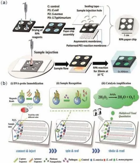
Fig.3.(a)Paper chip for multiple foodborne pathogens detection simultaneously by fluorescence on single-step.Reproduced with permission[53].Copyright 2018,American Chemical Society.(b)Multiplexed instrument-free V-Chip for visual color quantitative detection of multiple pathogens.Reproduced with permission[59].Copyright 2018,American Chemical Society.
Chemiluminescence(CL)without excitation light source will terminate the noise source and background emission.Beside its selectivity,it is highly sensitive for trace analysis.Due to the above advantages,microfluidic chemiluminescence has been used in the analysis of bacteria[27,54].Shanget al.developed a cloth-based CL biosensor for the first time to determine target DNA ofListeria monocytogenes(L.monocytogenes)with high sensitivity[27].The integration of desirable hydrophobic barrier fabricated by sequential wax screen-printing and gravity/capillary flow onto the flow channel of the cloth-based device made the biosensor easily fabricated and associated with a flow CL.The hemin/Gquadruplex DNAzyme was used for amplifying the signals of luminol/H2O2-based CL system,and the signal was recorded by a low-cost CCD.Electrochemiluminescence(ECL)combining CL and electrochemistry is advantageous because it can increase sensitivity and widen the dynamic range inherited from conventional CL.Liuet al.have reported a low-cost,label-free,and wireless paperbased bipolar electrode ECL(pBPE-ECL)analysis system for the rapid and sensitive genetic detection ofL.monocytogenes.The simple and rapid label-free pBPE-ECL distinguishedL.monocytogenesfromSalmonella,E.coliO157:H7,andS.aureus,and had the potential in point of care applications for pathogen detection[55].
The colorimetric detection methods were especially favored by researchers allowing direct observation of the results with naked eye.The key challenge was to transform the bio-recognition signal into color variations.Chromogenic substrates and enzymes were two main tools for colorimetric methods.Meanwhile,TMB coloration assays based on catalysis assay[56,57],and AuNPs-based immunochromatography assay were often used in the visual detection systems[46].Wanget al.identified three common nosocomial and antibioticresistant bacteria:AB,E.coli,and multidrugresistantS.aureussuccessfully[56].The biotin-labeled aptamers for the three bacteria were first bound to nitrocellulose membranes housed within the microfluidic chip channel and incubated with bacteria.TMB-streptavidin(blue color)reaction was next exploited upon binding of secondary aptamers to primary ones,thereby permitting bacterial detection.In other works,HCR and LAMP techniques were used to amplify the signal for improving the sensitivity[57].Trinhet al.fabricated a foldable microdevice with colorimetric detection for the monitoring of multiplex foodborne pathogens[58].They used 2-hydroxyethyl agarose loaded with LAMP reagents and silver nitrate deposited in the reaction and detection chambers to maintain the reagent activity for at least 45 days.Before folding the reaction and detection chambers,a heater-based thin graphene associated with a handheld battery was utilized to provide a constant temperature for amplification within 30 min.The colorimetric strategy was used for simple visual read-out of the results on-site.This method was successful in the identification ofSalmonella spp.andE.coliwith high sensitivity and selectivity.Furthermore,volumetric bar-chart chip(VChip)was developed for visual quantitative instrument-free detection of multiple pathogens[59,60].Weiet al.first reported using V-Chip for quantitative detection of multiple pathogens simultaneously with high sensitivity without specialized instruments.Three major foodborne pathogens includingSalmonella enterica,E.coliandL.monocytogeneswere specifically quantified in apple juice with the LOD about 10 CFU/mL,and the visual detection time was within 10 min(Fig.3b)[59].
Surface-enhanced Raman spectroscopy(SERS),with the advantages of nondestructive and on-line qualitative analysis of multicomponent samples,could be used to directly obtain molecular fingerprint information.It has therefore become an effective technique for bacterial detection[61,62].Mühliget al.developed a closed droplet-based lab-on-a-chip device for the differentiation of six species ofmycobacteriausing SERS[61].The obtained SERS spectra was dominated by the cell-wall component mycolic acid without extraction or further treatment of the sample,and the successful identification of nontuberculous mycobacteria and mycobacteria tuberculosis complex species was achieved within 1 h.Witkowskaet al.investigated the detection ofPorphyromonas gingivaliswhich was the keystone pathogen implicated in the development of gum disease(periodontitis)by SERS[62].Bacteria could easily adsorb to silver-coated magnetic nanoparticles(Fe2O3@AgNPs),which is possible to magnetically separate the investigated bacteria from other components of a specimen using the microfluidic chip.The bacteria-NP complex could be magnetically attracted to the Si/Ag SERS platform by external magnetic field to obtain additional enhancement of the signal.
2.3.2.Electrochemical method
Electrochemical biosensors have enabled sample preparationfree detection of pathogens in various matrices,which might be suitable forin siturapid detection of pathogens[26,63–65].Cesewskiet al.have overviewed electrochemical biosensors for pathogen detection and discussed the transduction,biorecognition and measurement formats associated with electrochemical biosensors[64],which could be referenced in the microfluidic biosensors.In Jiang’s work,EC cloth-based DNA sensors(ECDSs)was developed for the first time to detect genomic DNA fromL.monocytogenes,without the need for cumbersome chip fabrication and high-cost peripheral facilities[26].The EC signal was amplified by modifying nanocomposite and double linear hybridization chain reaction.Additionally,the ECDSs had an acceptable storage stability and high selectivity with low cost($0.045 per sensor).The genomic DNA fromL.monocytogeneswas detected by the ECDSs coupled with simple enzyme digestion,and the detection limit was 0.039 ng/μL(Fig.4a).Yuet al.developed an ultrasensitive biosensor for fast detection ofSalmonellausing self-assembled MNP chains for continuous-flow separation ofSalmonellafrom largevolume sample and linear scan voltammetry for sensitive detection[65].The separation was finished in 3D spiral channel using magnetic bacteria which was combined with urease coated gold nanopaticles.After the catalysate transferred into microfluidic chip with the thin-film Ag/AgCl reference electrode array for linear scan voltammetric measurement,exhibiting detection limit of 101CFU/mL forSalmonelladetection within 1 h.
2.3.3.Mass spectrometry
Matrix-assisted laser desorption ionization-time of flight mass spectrometry(MALDI-TOF MS)has been regarded as a fast and reliable method for bacterial identification.It could provide unique fingerprint mass spectra for identifying bacteria by comparing with the reference spectra in the database.Liet al.developed a fast bacterial identification method in urine samples by capturing bacteria on a microchannel silicon nanowire microfluidic chip,followed by MALDI-TOF MS detection[66].The library of reference spectra was built based on 15 spectra acquired from five chips afterE.colienrichment from suspensions in PBS buffer(0.5 mL,4 × 108CFU/mL).Bacteria with a concentration of 106CFU/mL without culture and 103CFU/mL after culture for 6 h in urine samples were directly identified by comparing with the reference spectra after enrichment(Fig.4b).
2.3.4.Multi-detection technology
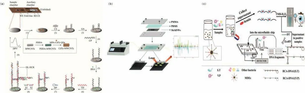
Fig.4.(a)Electrochemical cloth-based DNA sensors for genomic DNA from L.monocytogenes detection.Reproduced with permission[26].Copyright 2020,American Chemistry Society.(b)MALDI-TOF MS identification of bacteria captured by the microchannel silicon nanowire microfluidic chip from liquid samples.Reproduced with permission[66].Copyright 2021,Royal Society of Chemistry.(c)The universal dual-mode aptasensor for simultaneous determination of different bacteria based on naked eyes and microfluidic-chip together with magnetic DNA encoded probes.Reproduced with permission[67].Copyright 2021,Elsevier.
The combination of detection methods was used to overcome the shortcomings of a single detection method thereby improving detection sensitivity[48,67].Huanget al.developed an acidresponsive microfluidic biosensor for detection ofS.typhimuriumin colorimetric and fluorescent model in spiked chicken samples[48].The curcumin(CUR)as signal reporter and ZnO-capped mesoporous silica nanoparticles for signal amplification were developed.Acetic acid was introduced to release CUR from the complexes for the first time,and both the color and fluorescence intensity changes were measured to determine the concentration ofS.typhimurium.The detection limits were calculated to be 63 CFU/mL for colorimetric measurement and 40 CFU/mL for fluorescent measurement.Yuet al.developed a universal dual-mode aptasensor based on naked eyes and a microfluidic chip for simultaneous onsite assay ofS.typhimuriumandVibrio parahaemolyticusin food[67].The dual-mode aptasensor could quickly screen positive samples by naked eyes,and perform quantitative detection of the bacteria in positive samples using DNA fragments with different lengths in the microfluidic chip(Fig.4c).And the whole operation was finished within 3 min.
2.3.5.Point-of-care testing detection
Point-of-care testing(POCT)for pathogen detection has been reported in recent years using thermometers,smartphones,personal glucose meters(PGMs),etc.These miniaturized sensors are gradually being used in combination with microfluidic systems to detect pathogenic bacteria as a new detection method.The reviews of strategies of the miniaturized biosensors for POCT and the microfluidic devices with simplified signal readout have been published[68,69].The current POCT detection for pathogen bacteria based on microfluidic biosensors were summarized in Table 1[46,70–81].
Table 1 The POCT detection of pathogen bacteria in microfluidic biosensor.
3.Application of microfluidic biosensors for rapid pathogenic bacteria detection
The microfluidic biosensors for detecting pathogenic bacteria cells with high sensitivity and specificity in a short period of time on site have been widely used in the fields of public health assurance,clinical diagnostics,environmental monitoring,and food safety.
3.1.Clinical diagnostics
Bacterial could cause infections in different parts of the human body from skin and cellulitis to invasive infections,such as tuberculosis,sepsis,nephritis and cystitis.The rapid and sensitive detection of bacteria is a prerequisite for the treatment of these diseases.The bacteria in urine[12,73]and blood[24,29,39,49,51]have been detected mostly by microfluidic biosensors.Zhouet al.firstly developed a low-cost photothermal biosensing method for the quantitative genetic detection ofMycobacterium tuberculosisDNA on a paper hybrid device using a thermometer[80].The DNA capture probes were simply immobilized on paper through a one-step surface modification process.After DNA sandwich hybridization,oligonucleotide-functionalized AuNPs were introduced on paper and then catalyzed the oxidation reaction of 3,3′,5,5′-tetramethylbenzidine(TMB).The produced oxidized TMB,acting as a strong photothermal agent used for the photothermal biosensing of MTB DNA under 808 nm laser irradiation.The detection limit of this work was 39 nmol/L(or 0.58 μg/mL)and without color interference issues compared to conventional colorimetric methods(Fig.5a).Kimet al.reported an unpowered microfluidic sample preparation device in on-site molecular diagnosis which did not require expertise and electrical devices[82].The device integrated a plasma separation chip and a microfluidic chip for nucleic acid extraction and purification.This unpowered sample pretreatment method was precise and reliable even at low concentration down to 103CFU/mL ofE.coliin blood samples.
3.2.Food safety
Foodborne bacterial pathogens exist in various kinds of foods,and may cause the incidence of foodborne diseases which are the main source of health problems around the world.Hence,it is necessary to find a way for detecting the foodborne pathogenic microorganisms in different food products.Several reviews about microfluidic devices for foodborne pathogens detection have been published in recent years[83,84].Rapid screening of pathogenic bacteria contaminated foods is essential to prevent food poisoning including seafood[19],milk[25–27,35,47,53,61,66,67,71,76,81],juice[29,72],chicken[31,46,48,52,57,76,85],vegetables[74],etc.Qiet al.developed a microfluidic biosensor for rapid,sensitive and automatic detection ofSalmonellausing metal-organic framework(MOF)used for mimicking peroxidase activity to amplify biological signals,and Raspberry Pi coupled with self-developed App for analyzing color images[85].In this study,the immune MNPs were introduced to capture the target bacteria and labeled with the immune MOFs to form MNB-Salmonella-MOF complexes.After catalyzing colorless o-phenylenediamine and H2O2,the system produced yellow 2,3-diaminophenazine(DAP)(Fig.5b).Raspberry Pi App was employed to measure the color for bacterial concentration calculating presenting the detection limit of 14 CFU/mL.This microfluidic biosensor was applied for detection ofS.typhimuriumspiked in chicken meats and the recoveries for different concentrations of spiked samples ranged from 107.44% to 118.75%.
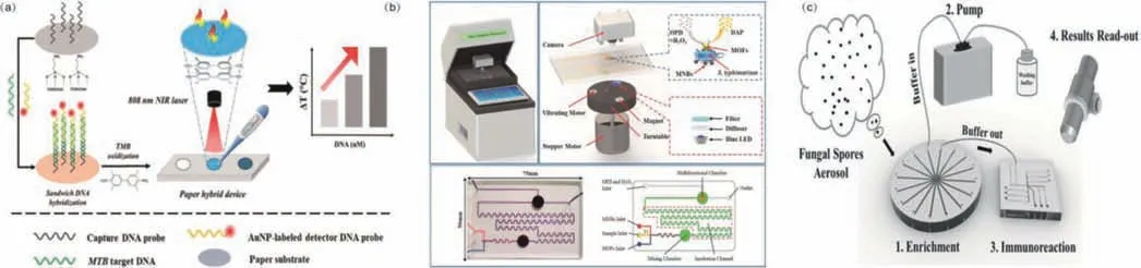
Fig.5.(a)Photothermal genetic detection of Mycobacterium tuberculosis using a thermometer.Reproduced with permission[80].Copyright 2020,American Chemistry Society.(b)A microfluidic biosensor for rapid and automatic detection of Salmonella in chicken meats.Reproduced with permission[85].Copyright 2021,Elsevier.(c)Microfluidic system for rapid detection of airborne pathogenic Fungal Spores.Reproduced with permission[87].Copyright 2018,American Chemistry Society
3.3.Environmental monitoring
Microorganisms can easily be found in a variety of environments,including soil,air and water.Many of these organisms are potentially harmful to animals and humans causing infectious diseases.Rapid capture and detection of pathogens are essential for disease prevention and public safety.Unlike traditional methods,microfluidic biosensor systems have been used for rapid detection of microorganisms in the environment[14,54,77,86,87].Sui’s group first reported the airborne bacteria detection using microfluidic system[86].They presented a microfluidic system that could capture and enrich airborne pathogens and perform high-throughput LAMP analysis,thereby realizing the detection of five species of bacteria.The results could be detected by naked eye and the detection limit could down to approximately 24 cells per reaction.Later they published another work by integrating a microfluidic system which was capable of enrichment and high-throughput detection ofAspergillus nigerbased on immunofluorescence analysis[87].The microfluidic system integrated sampling and detection to avoid additional sample concentration step,and the whole analysis time was less than 3 h including 1 h of enrichment and 1 h for target detection(Fig.5c).The detection limit was 20 spores(300 spores m-3of the concerned targets in air).Kimet al.have assayedE.coliK12,S.typhimurium,andS.aureusin environmental water samples,which represented GPB,GNB,rod-and roundshaped bacteria[77].They used protamine as a guanidinium rich polymerthe to capture bacteria without the need of specific antibodies or primers.Samples and the protamine conjugated fluorescent particles were sequentially loaded to the paper microfluidic chips,and a smartphone was used as a fluorescence microscope.This paper-based microfluidic biosensor assisted with smartphone was fabricated to detect all three bacteria(P <0.05)with a detection limit of 101–102CFU/mL.
4.Conclusions and future perspectives
This paper provided a review of microfluidic biosensor development for pathogenic bacteria detection over the recent years.Various target capturing and separation strategies,detection methods and applications were introduced.The design of microfluidic chip should consider the biological recognition elements which directly interact with specific target and the transducer components for signal output.Antibody immune recognition and aptamer recognition were widely used in the biomolecular recognition strategies combined with microfluidic chips,making it easy to realize target capture and high detection sensitivity.The separation of bacteria was not necessary for detection,although the immunomagnetic bead separation technology was the most common separation modality.In addition,research on chip structure to realize the separation and enrichment of bacteria without external force was also needed.The transducer components for signal output were usually miniaturized with handheld instruments using optical or electrochemical methods for pathogenic bacteria detection.Attractively,the smartphone has been widely used for detection in microfluidic systems with built-in cameras and internal microprocessors which could carry out image processing without external computers and share real-time results when necessary.
In the end,the establishment of fast,highly specific and highly sensitive detection methods will become the focus of future research.In addition,appropriate microfluidic biosensors should also be capable of integrated,automated and multiplex detection.Meanwhile,significant challenges still need to be addressed over years to translate the developed microfluidic biosensors for commercial use in POCT.With the development of microfluidic droplet preparation technology[88,89],the rapid detection of bacteria in microfluidic droplets provides a possible development direction.The strategies for cell analysis in the microfluidic system also provide a certain reference for the identification,separation,and detection of pathogenic bacteria[90,91].
Declaration of competing interest
The authors declare no conflict of interest.
Acknowledgments
This work was supported by Research and Development Program in Key Areas of Guangdong Province,China(No.2019B020209009)and the National Natural Science Foundation of China(Nos.21727814,81872829 and 21621003).
 Chinese Chemical Letters2022年4期
- Chinese Chemical Letters的其它文章
- Key progresses of MOE key laboratory of macromolecular synthesis and functionalization in 2020
- Small nanoparticles bring big prospect:The synthesis,modification,photoluminescence and sensing applications of carbon dots
- Cell membrane-coated nanoparticles for immunotherapy
- Diketopyrrolopyrrole-derived organic small molecular dyes for tumor phototheranostics
- Exosome based miRNA delivery strategy for disease treatment
- Recent advances in targeted stimuli-responsive nano-based drug delivery systems combating atherosclerosis