The promise of neuroprotection by dietary restriction in glaucoma
Rossella Russo , Carlo Nucci, Annagrazia Adornetto
Abstract Glaucoma, a progressive age-related optic neuropathy characterized by the death of retinal ganglion cells, is the most common neurodegenerative cause of irreversible blindness worldwide. The therapeutic management of glaucoma, which is limited to lowering intraocular pressure, is still a challenge since visual loss progresses in a significant percentage of treated patients. Restricted dietary regimens have received considerable attention as adjuvant strategy for attenuating or delaying the progression of neurodegenerative diseases. Here we discuss the literature exploring the effects of modified eating patterns on retinal aging and resistance to stressor stimuli.
Key Words: aging; caloric restriction; fasting; glaucoma; neurodegeneration; retina; retinal ganglion cells
Introduction
Glaucoma is the most common neurodegerative cause of irreversible blindness worldwide.
The global prevalence for the disease is 3–5% for people aged 40–80 and it has been estimated that the number of affected people will reach about 111.8 million by 2040 (Tham et al 2014). Despite the currently available therapies, focusing on reducing intraocular pressure (IOP),the therapeutic management remains challenging since the disease may progress even when target IOP values are achieved. The partial inefficacy of IOP-lowering therapies,and the neurodegenerative nature of glaucoma have driven the research toward IOP-independent approaches focusing on neuroprotective strategies. Studies on dietary restriction(either as reduced calorie intake or intermittent fasting)support the notion that metabolic and energy balance aretightly linked with aging and neurodegeneration (Mattson et al., 2012; Pani, 2015). Indeed, the regulated limited access to food activates mechanisms (i.e. autophagy, mitochondrial biogenesis, neuronal plasticity, reduced oxidative stress and inflammation) that translate into protective adaptive responses improving neuronal health and survival (Lin et al.,2014; Bagherniya et al., 2018; Gabandé-Rodríguez et al., 2019;Popov et al., 2020).
Here we discuss the effects of caloric restricted regimens on retinal aging and neurodegeneration focusing on the available evidence from animal models of glaucoma.
Search Strategy and Seletion Criteria
Literature search for studies published up to 2020 was performed on PubMed, Web of Science and Google Scholar databases using the following keywords: glaucoma, retinal aging, retinal neurodegeneration, caloric restriction, fasting,restricted diet, dietary restriction, caloric restriction mimetic and combinations of the above terms.
Glaucoma: an Age-Related Neurodegenerative Disease
Glaucoma is an umbrella term used for a group of progressive optic neuropathies sharing common structural alterations of the optic nerve head and characterized by the progressive loss of retinal ganglion cells (RGCs) (Quigley, 2011). The neurodegenerative process is associated with typical pattern of visual field defects which are initially localized in the paracentral areas and become wider and more numerous as the disease progresses (Nucci et al., 2016). Primary open angle glaucoma, the form accounting for three-quarter of all glaucoma cases, is basically painless and asymptomatic and about 50% of the patients are diagnosed in an advanced stage, when significant and irreversible RGC loss has occurred(Kapetanakis et al., 2016).
The etiopathogenesis of the optic neuropathy is complex and includes genetic and environmental factors, which are only partially defined. Along with the presence of elevated or unstable intraocular pressure (IOP), age is the main risk factor and the prevalence of the disease exponentially increases with population aging (Tham et al., 2014). The therapeutic management of glaucoma, which relies on drugs or surgical procedures aiming at lowering IOP, is a therapeutic challenge. Indeed, despite the treatments, in approximately 10% of glaucoma patients vision loss progresses even when“target” IOP values are achieved (Cohen and Pasquale,2014). Furthermore, not all patients with ocular hypertension develop the neuropathy and normal tension glaucoma, a subtype of the disease accounting for one-third of all open angle glaucomas, occurs in patients with IOP values falling in the physiological range (Mallick et al., 2016).
Although it is clear that additional, IOP-independent and still unidentified factors are involved in the pathogenesis of the disease, the apoptotic death of RGCs is the road end of a cascade of molecular events (i.e., excitotoxicity, alteration of neurotrophic support, hypoxic/ischemic events, oxidative stress, mitochondrial damage, chronic inflammation,autoimmunity, autophagy dysregulation, etc.) altering neuronal homeostasis and creating a hostile environment for neuronal survival (Zhang et al., 2016; Thomas et al., 2017;Russo et al., 2018). The mechanisms underlying the RGC degeneration are similar to those engaged by other neuronal subtypes vulnerable in neurodegenerative disorders, such as Parkinson’s and Alzheimer’s disease, and also recur in the process of cell senescence (Mirzaei et al., 2017; Matlach et al.,2018).
The Effects of Dietary Restriction on Retinal Aging and Glaucoma-Related Neuronal Loss
Over the past few decades the research on calorie restriction has rapidly expanded and changing of caloric intake and meal timing has been identified as a reproducible environmental intervention to extend life-span and improve health in several species, from yeast to rodent and primates (Fontana and Partridge, 2015). In particular, the effects of two different eating patterns, caloric restriction and fasting, have been studied as possible strategies to sustain healthy aging and modify the risk and progression of age-related chronic diseases (Belsky et al., 2017; Das et al., 2017).
Caloric restriction is a dietary regimen based on the reduction(between 10 and 40%) of daily caloric consumption without affecting the intake of essential nutrients (i.e. vitamins and minerals). At variance, fasting imposes the ingestion of limited or no food and may or may not include caloric restriction during the non-fasting periods. Several regimens in terms oftime, length and type can be applied for caloric restrictions,and fasting can be part of a caloric-restricted diet (Longo and Mattson, 2014).
Data from the literature also show that caloric restriction is the most effective non-pharmacological intervention able to increase resistance to stress and offering protection in pathological conditions like diabetes, cancer, cardiovascular and neurodegenerative diseases (Baumeier et al., 2015; Noyan et al., 2015; Ntsapi and Loos, 2016; Kopeina et al., 2017).Indeed, restricted caloric regimens prevents age-related neuronal loss and attenuates or delays neurodegeneration in animal models of Parkinson’s and Alzheimer’s diseases,as well as in acute neuronal injury induced by focal stroke or cervical spinal cord lesion (Plunet et al., 2008; Ran et al., 2015;Bayliss et al., 2016). Despite the amount of scientific reports supporting the beneficial results of caloric restricted regimens in pathological states affecting the nervous system (Gillette-Guyonnet et al., 2013), evidence regarding their effects on the onset and progression of retinal neurodegenerative diseases,and in particular glaucoma, are still limited. Nevertheless,data available in the literature suggest that, in animal models,modify the eating pattern could improve the resistance of RGCs to stressor stimuli, delay retinal aging and support retinal neurons’ viability (Kawashima et al., 2013; Guo et al.,2016; Adornetto et al., 2020).
Aging is associated with a decline in retinal cell densities, a reduction of retinal layers thickness (Obin et al., 2000) and an age-dependent inherited loss of RGCs (Kawai et al., 2001).In mammals, a total loss of approximately 35–40% has been estimated over the lifetime (Harman et al., 2000). Obin et al. (2000) firstly observed that a reduction of caloric intake by 40% preserved retinal cell densities and thickness and modulated aging in the sensory neurons of 30-month-old rats.Similarly, an age-related decline in all three nuclear layers of the neural retina (outer nuclear layer, inner nuclear layer and ganglion cell layer) was reported in Brown Norway rats by Li et al. (2003). This was attenuated by a progressive restriction of dietary regimen initiated at 14 weeks of age and maintained throughout the animals’ life (Li et al., 2003).
Kaway et al. (2001) analyzed the loss of peripheral RGCs following exposure to transient elevation of IOP in old (2-years old) versus young (2-month old) rats subjected to ad libidum access to food or caloric restriction regimen (achieved by providing food three days per week for three months) (Kawai et al., 2001). The study revealed that RGCs from old rats show an increased susceptibility to ischemia reperfusion injury as compared to young ones and caloric restriction protected animals from both ages against the loss of RGCs in the peripheral retina (Kawai et al., 2001).
A similar finding was reported by Kong and colleagues (2012)who demonstrated a clear age-related decline in the ability of optic nerve and retina to withstand an acute IOP stress (Kong et al., 2012). Indeed, following a transient elevation of IOP,18 months old mice with ad libidum access to food showed increased retinal vulnerability, with greater functional loss and poorer recovery, as compared to 3-months old mice. This negative effects of aging on retinal function were significantly reduced in old mice subjected to a dietary restricted regimen(limited access to food for 24 hours every other day) between 12 and 18 months of age (Kong et al., 2012).
More recently our group has shown that 48 hours of fasting significantly reduced the RGC loss induced by acute ocular hypertension; the neuroprotection afforded by the restricted caloric regimen was associated with the suppression of mTOR activity and the activation of autophagy (Russo et al.,2018). Using EAAC1 (excitatory carrier 1)-deficient mice,an animal model of normal tension glaucoma, Guo and colleagues showed that seven weeks of every-other-day fasting suppressed RGC degeneration and ameliorated visual impairment by upregulating the expression of neurotrophic factors and reducing oxidative stress levels (Guo et al., 2016).Beside supporting neuronal survival, caloric restriction may also contain the retinal glial cell activation in response to detrimental insults inhibiting the release of pro-inflammatory factors. Indeed the prevention of ischemia-induced retinal damage observed in old rats subjected to a caloric restriction regimen was associated with suppression of reactive gliosis(Kim et al., 2004).
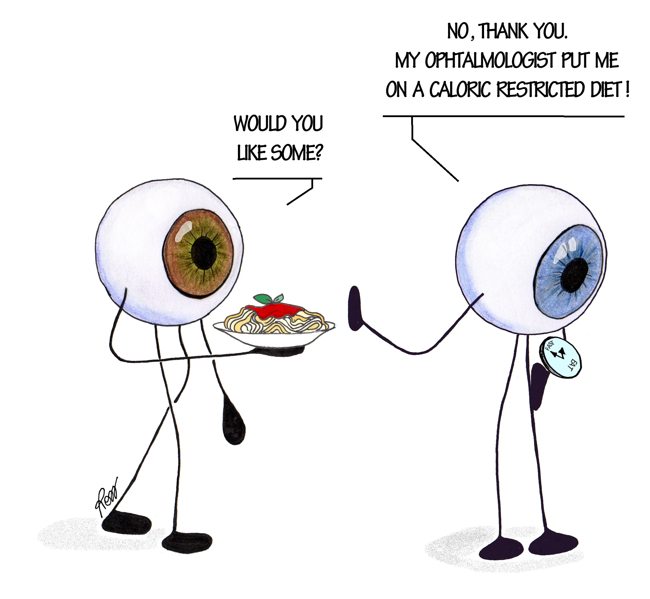
While the data on the effects of caloric restricted dietary regimens seem promising in preclinical animal studies, to the best of our knowledge there are no reports regarding its effect on patients with glaucoma (Figure 1). However, it is worth noting that in a retrospective cohort study the risk of developing primary open angle glaucoma was reduced in diabetic patients taking the hypoglycemic drug metformin but not in patients assuming other diabetes medicationswith similar efficacy on glycemic control. Since metformin is considered a drug mimicking the health promoting effects of caloric restriction it can be conceivable to hypothesize that the reduced risk of developing glaucoma may involve the activation of longevity pathways similar to those involved in the protective effects exerted by restricted caloric regimen (Lin et al., 2015).

Figure 1|Will the knowledge of the beneficial effects of calorie restriction(mainly derived from animal studies) be applicable to patients affected by glaucoma?
The positive effects of calorie restriction may rely on several mechanisms including induction of autophagy, improvement of mitochondrial function, activation of sirtuins, reduced oxidative stress and anti-inflammatory effects (Madeo et al., 2019). Furthermore, some of the metabolic adaptations induced by these dietary regimens in neuronal tissues may not stem from local effects but can be mediated by circulating factors present in the sera (Amigo et al., 2017).
Conclusion
To date, the molecular basis of the dietary restrictionmediated effects in the aging retina and after exposure to detrimental insults remain elusive, although an overlapping with those observed in other tissues can be speculated.
In-depth studies aimed at identifying the genetic and physiological mediators of caloric restriction beneficial effects in the retina are fundamental for developing the research in this field. Indeed, the adherence to a long-term or early life starting caloric restriction regimen in humans is questionable and basically impractical and would not meet the compliance of patients. The alternative approach is represented by the search and identification of caloric restriction mimetics:substances/drugs that mimic, at the molecular level, the beneficial physiologic responses of caloric restriction without the need to reduce calorie intake (i.e., metformin, resveratrol,rapamycin) (Madeo et al., 2019).
Further studies are warranted to strengthen the hypothesis that modified dietary regimens or caloric restriction mimetics represent potential adjuvant approaches to be associated with the currently used therapies (Figure 1), for preventing or delaying glaucomatous neurodegeneration and other agerelated ophthalmic disorders.
Author contributions:RR, AA and CN wrote and revised the manuscript; RR conceived and drew the figure. All authors approved the final version of the manuscript.
Conflicts of interest:The authors declare no conflicts of interest.
Financial support:None.
Copyright license agreement:The Copyright License Agreement has been signed by all authors before publication.
Plagiarism check:Checked twice by iThenticate.
Peer review:Externally peer reviewed.
Open access statement:This is an open access journal, and articles are distributed under the terms of the Creative Commons Attribution-NonCommercial-ShareAlike 4.0 License, which allows others to remix, tweak,and build upon the work non-commercially, as long as appropriate credit is given and the new creations are licensed under the identical terms.
Open peer reviewer:Georgios D. Panos, Nottingham University, UK.
- 中国神经再生研究(英文版)的其它文章
- Genes for RNA-binding proteins involved in neuralspecific functions and diseases are downregulated in Rubinstein-Taybi iNeurons
- Research advances on how metformin improves memory impairment in “chemobrain”
- Dendritic spine density changes and homeostatic synaptic scaling: a meta-analysis of animal studies
- Optogenetic activation of intracellular signaling based on light-inducible protein-protein homo-interactions
- Presenilin mutations and their impact on neuronal differentiation in Alzheimer’s disease
- Growth differentiation factor 5: a neurotrophic factor with neuroprotective potential in Parkinson’s disease

