Zuogui Wan(左归丸)improves trabecular bone microarchitecture in ovariectomy-induced osteoporosis rats by regulating orexin-A and orexin receptors
LIU Feixiang,TAN Feng,FAN Qiaoling,TONG Weiwei,TENG Zhanli,YE Sumin,LI Xing,ZHANG Mingyue,CHAI Yi,MAI Chongying
LIU Feixiang,TAN Feng,FAN Qiaoling,TONG Weiwei,TENG Zhanli,YE Sumin,LI Xing,ZHANG Mingyue,CHAI Yi,MAI Chongying,School of Chinese Medicine,School of Integrated Chinese and Western Medicine,Nanjing University of Traditional Medicine,Nanjing 210023,China
Abstract OBJECTIVE:To investigate the protective effects of Zuogui Wan (左归丸,ZGW) on bone loss induced by ovariectomy (OVX) and its mechanism via orexin-A and orexin receptors in the osteoporosis rat model.METHODS:Fifty Sprague-Dawley female rats were randomly divided into sham-operated (sham)group and four OVX subgroups.Rats subjected to sham and OVX were treated with the vehicle(OVX,1 mL/100 g weight, n= 10),17β-estradiol(E2,50 μg·kg-1·d-1),and ZGW at the doses of 2.3(ZGW-L)and 4.6(ZGW-H)g/kg/day lyophilized powder daily for 3 months,respectively.The serum biochemical parameters of 17β-estrogen (17β-E2),tartrate-resistant acid phosphatase (TRACP-5b) and bone alkaline phosphatase (BALP) were measured by enzyme-linked immunosorbent assay.Hematoxylin-eosin staining was used to detect the changes in the morphological structure in bones.Microcomputed tomography was used to evaluate the bone mineral density and microarchitecture of the distal femur.The gene or protein expression of orexin-A,orexin receptor 1 (OX1R),orexin receptor 2 (OX2R),osteoprotegerin(OPG)and receptor activator of nuclear factor-κ B ligand(RANKL)were assayed by either quantitative polymerase chain reaction or Western blot analysis.RESULTS:Compared with the OVX group,ZGW could reduce the serum level of TRACP-5b and increased the serum levels of BALP and17β-E2 (P <0.01).Meanwhile,ZGW could prevent bone loss and improved bone trabecular microarchitecture by increasing the trabeculae structure thickness and trabecular number,and arranging the trabeculae structure properly.Compared with the OVX group,it was upregulated for the orexin-A and OX2R mRNA or protein expression from the hypothalamus and tibiae,and OPG in the tibiae of ZGW groups (P < 0.01, < 0.05),while downregulated for the OX1R mRNA and protein expression in the tibiae and hypothalamus and RANKL from the tibiae(P<0.01).CONCLUSION:ZGW exhibited a protective effect for PMOP that may be mediated via orexin-A and orexin receptors regulation.
Keywords:osteoporosis,postmenopausal;orexin receptors;orexins;Zuogui Wan
INTRODUCTION
Postmenopausal osteoporosis (PMOP),is a generalized bone disease caused by estrogen deficiency and characterized by the alterations in the bone trabecular microarchitecture,decrease in bone mass,and increase in bone fragility.1According to a research issued by the French statutory health insurance system for salary workers in 2013,approximately 135 000 female patients aged over 50 years old that are admitted in the hospitals in France sustain fractures,which caused substantia economic losses of more than €770 million.2The agents of stimulating bone formation or inhibiting bone resorption,such as estrogen,bisphosphonates,fluoride,teriparatide,and calcitriol,are commonly applied for PMOP treatment and play essential roles in clinical settings.3,4However,the side effects of these agents,including atypical femoral fractures,endometrorrhagia,stroke,et al.,affect the quality of the life of patients.5Therefore,necessary precautions should be taken to reduce the cost and risk of future fractures.
Zuogui Wan (左归丸,ZGW),which was formulated by Zhang Jingyue,a famous physician in Ming Dynasty and first recorded in Jingyue Quanshu,could be used to strengthen bone.6Our previous study also indicated that ZGW exhibits an osteoprotective effect by correcting bone metabolism imbalanceviaRANKL/OPG regulation mediated by β2AR in ovariectomized(OVX) rats.7However,there is lacking information on the precise mechanism underlying tonifying kidney to increase BMD.
Orexin-A and-B are neuropeptides,produced laterally in the hypothalamus,which are associated with sleep,food intake,and bone metabolism regulation.8The action is regulated by two G-protein-coupled receptors,namely,orexin receptors 1 (OX1R) and 2 (OX2R).9The lack of orexins in humans and rats can cause narcolepsy,anorexia,and obesity,which are also important risk factors for the increased incidence of low bone mass and osteoporotic fractures in PMOP.10Orexins regulate bone remodeling bidirectionally through OX1R and OX2R in brain and bone marrow tissues and maintain bone mass balance through their interaction,respectively.11
In this study,we hypothesized that orexin system regulation may be attributed to the mechanism and effect of ZGW in improving bone microstructure and correcting bone metabolism imbalance.
MATERIALS AND METHODS
ZGW lyophilized powder preparation
Eight Chinese medicines in ZGW,including Dihuang(Radix Rehmanniae)240 g,Shanyao(Rhizoma Dioscore-ae Oppositae) 120 g,Shanzhuyu (Fructus Macrocarpii)120 g,Gouqizi (Fructus Lycii) 120 g,Niuxi (Radix Achyranthis Bidentatae) 90 g,Tusizi (Semen Cuscutae)120 g,Guijiajiao(Colla Carapacis et Plastri)120 g,and Lujiaojiao (Colla Cornus Cervi) 120 g were supplied by the affiliated hospital of Nanjing University of Chinese Medicine.The method of preparing ZGW lyophilized powder was adopted from Liu et al.8 In brief,all drugs except Guijiajiao(Colla Carapacis et Plastri)and Lujiaojiao (Colla Cornus Cervi) were mixed,dried,crushed to 80 mesh,and boiled 2 times in 10.5 L of water for 1 h each.The two gelatins were melted and added to the decoction.The concentrated liquid was rapidly frozen and dried in a flask with liquid nitrogen and then lyophilized under −50 ℃by freeze dryers with a product yield of 51.2%.
Animals
These 4-month-old female Sprague-Dawley rats [(313± 20) g]were purchased from the Animal Breeding Center of Qinglong mountain,Jiangning district,Jiangsu,China with the certification No.SCXK (SU)2017-0001 (n=50).The Institutional Animal Care and Use Committee of Nanjing University of Chinese Medicine approved the experimental animal facility and research protocols(Agreement No.201803A002).
Modeling and grouping
Rats were subjected to OVX or sham and left untreated for 4 weeks to develop significant osteopenia.The specific operation methods referred to the literature described by Yuanet al.12These OVX rats were randomly divided into 4 separate groups by random number table method as follows (n=10 per group):OVX with 17β-estradiol(17β-E2)dissolved in absolute ethyl alcohol and then diluted with water (OVX-E2,50 μg·kg-1·d-1),OVX model group with water as vehicle(OVX,1 mL water/100 g per day),OVX with high-dose ZGW lyophilized powder dissolved in water (ZGW-H,4.6 g·kg-1·d-1),and OVX with low-dose ZGW lyophilized powder dissolved in water(ZGW-L,2.3 g·kg-1·d-1) once a day for 12 weeks.Sham group rats were treated with water (n=10).The intake dose for animals was derived from the clinical human dose.After 3 months of administration,animals were anaesthetized with 3% pentobarbital solution for blood collection and then sacrificed via bloodletting.Tibiae,bilateral femora,and hypothalamus were extracted and preserved for further experiments.
Drugs and reagents
Enzyme-linked immunosorbent assay (ELISA) kits of rat 17β-E2,bone alkaline phosphatase(BALP)and tartrate-resistant acid phosphatase 5b (TRAP-5b) were provided by Enzyme-linked Biotechnology Co.,Ltd.(Shanghai,China).17β-E2 was purchased from Sigma Co.,Ltd.(St.Louis,MO,USA).The Tissue/cell RNA rapid extraction kit was supplied by Yi Fei Xue Biotechnology Co.,Ltd.(Nanjing,China).HifairTMII 1 Strand cDNA Synthesis SuperMix for quantitative polymerase chain reaction (qPCR),HieffTM qPCR SYBR GREEN Master Mix,Cy3-AffiniPure goat anti-mouse IgG (H+L) and Cy3-AffiniPure goat anti-rabbit IgG (H+L) were purchased from Yi Sheng Biotechnology Co.,Ltd.(Shanghai,China).Rabbit anti-OPG,mouse monoclonal actin and anti-RANKL were provided by Abcam,Co.,Ltd.(Cambridge,UK).Rabbit anti-OX1R and rabbit anti-OX2R antibodies were purchased from Bioss Biotechnology Co.,Ltd.(Beijing,China).
Serum biochemical parameters
The serum17β-E2,BALP and TRACP-5b concentrations were measured according to the commercial instructions in ELISA kits.The serum biochemical parameters were analyzed by the enzyme-linked immune detector (EnSpire,Perkin Elmer Instrument Co.,Ltd.,Waltham,MA,USA).
Histological examination of distal femurs
The right distal femurs were immobilized with 10%formalin and decalcified by ethylenediamine tetraacetic acid solution for 4 weeks.Then,all samples were embedded in paraffin and sectioned into 4 μm thick specimens for routine hematoxylin-eosin (HE) staining.The pathological changes in the stained femurs were photographed and viewed under ×100 magnification by using fluorescence-inverted microscope camera system(DMI3000B,Leica,Weztlar,Germany).
BMD and micro-computed tomography(µCT)
The computed tomography images of the bone mineral density and trabecular bone microarchitecture of the left distal femur were acquired and measured using Bruker μCT (SkyScan 1176,Bruker,Co.,Ltd.,Karlsruhe,Germany).The detailed scan method and parameter set-up in SkyScan 1176 were obtained in our previous study.7Those methods of the BMD measuredviaμCT by using Bruker μCT,the volume investigated by 3D models for structure thickness and separation distribution in CTVox and analyzed the boneviaμCT general information were performed from Yuanet al.12The following direct trabecular metric parameters,including trabecular number (Tb.N),bone volume/tissue volume (BV/TV),trabecular thickness (Tb.Th),trabecular separation (Tb.Sp),and bone surface/volume ratio(BS/BV),were calculated and analyzed as described by Stephenset al.13
Quantitative real-time PCR assay
Total RNA was extracted from the right tibiae plateau and hypothalamus in accordance with the commercial instructions of the tissue/cell RNA rapid extraction kit.cDNA was synthesized in accordance with the commercial protocol of HifairTM Ⅱ1 Strand cDNA Synthesis SuperMix for qPCR.qPCR was analyzed on the cDNA samples via real-time PCR (ABI 7500,Applied Biosystems Inc.,Waltham,MA,USA) in accordance with the commercial instructions of HieffTM qPCR SYBR GREEN Master Mix,with the amplification procedure of 1 cycle for 95 ℃for 5 min,followed by 40 cycles of 10 s of denaturation at 95 ℃,20 s of annealing at 60 ℃,20 s of extension at 72 ℃,and then followed 1 cycle,with the three stages of dissolution curve for 15 s at 95 ℃,60 s at 60 ℃,and 15 s at 95 ℃.The primer sequences are listed in Table 1.GAPDH was used as the internal control.All primers were purchased from Shenggong Co.,Ltd.(China).Data were analyzed and calculated by the 2-ΔΔCttechnique.

Table 1 Primer sequences for RT-PCR analysis
Western blot
The total protein from the tibiae plateau and hypothalamus were extracted by using protein extraction buffer.Approximately 20 μg of each protein samples were separated by 10% SDS-PAGE.Afterward,the samples were transferred into 0.45 μm polyvinylidene fluoride membranes.After 1 h of blocking with 3% BSA solution,bands were incubated with the first antibodies,including mouse monoclonal actin (1∶1000),rabbit anti-OX1R(1∶1000),rabbit anti-RANKL(1∶1000),rabbit anti-OX2R (1∶1000),and rabbit anti-OPG (1∶1000).Then bands were incubated with secondary antibodies,as follows:Cy3-AffiniPure goat anti-mouse IgG(H+L)(1∶2000)or Cy3-AffiniPure goat anti-rabbit IgG (H+L) (1:2000).Membranes were visualized with the Enhanced ECL Chemiluminescent Substrate Kit and then exposed to X-film by gel documentation system (Imagequant LAS 4000,GE Co.,Ltd.,Boston,MA,USA).Image J software (National Institutes of Health,Bethesda,MD,USA) was used to detect and analyze the sum density of membranes.
Statistical analyses
All data were expressed as means ± standard deviation(±s).One-way analysis of variance followed by post hoc analysis using Student-Newman-Keuls test (more than two groups),and Student'st-test(two-group comparison) were used to calculate and analyze all results.Statistical analysis was performed with the SPSS 19.0 software(IBM SPSS,Armonk,NY,USA).P<0.05 was considered to indicate statistically significant difference.
RESULTS
Biochemical assay
In the OVX group,the serum BALP and 17β-E2 levels were lower (P <0.01) and the serum TRACP-5b level was higher (P <0.01) than those of the sham group(Table 2).After 12 weeks of treatment with ZGW or E2,the serum BALP and 17β-E2 levels were higher(P<0.01)and the serum TRACP-5b concentration was lower(P<0.01)than those of the OVX group(Table 2).
Histopathological analysis
In the OVX group,the HE staining of the distal femur showed irregular trabecular arrangement,thinned and incomplete trabecular structure,a considerable amount of empty bone lacunae,disorganized distribution,and widened intertrabecular spaces compared with the sham group (Figure 1).After 12 weeks treatment with ZGW,the bone histomorphology improved compared with that in the OVX group and showed slightly ordered arrangement of the trabeculae,increased bone trabecula in terms of number and width,slight thinning of the trabeculae,and decreased empty bone lacunae(Figure 1).
µCT evaluation
The local BMD of the distal femur was lower in the OVX group (P <0.01) than that in the sham group.After 3 months of treatment with either ZGW or E2,the local BMD levels were significantly higher (P <0.05,P<0.01)than those in the OVX group(Table 3).The μCT analysis of left distal femur trabecular indicat-ed that the rat trabecular bone microarchitecture was decreased after OVX.After 12 weeks of ZGW administration,both trabecular volume and structure thickness were improved,as shown in the 3D models compared with the OVX group (Figure 2).The analysis of the representative sample data illustrated that Tb.Th,Tb.N,and BV/TV levels were decreased in the OVX group (P <0.01),and BS/BV and Tb.Sp levels were significantly increased (P <0.01) compared with those in the sham group (Table 3).In the ZGW groups,Tb.N,BV/TV,and Tb.Th levels were increased (P <0.01),and BV/TV and Tb.Sp levels were decreased(P <0.01) compared with the control vehicle-treated rats(Table 3).
Table 2 Effect of ZGW on biochemical parameters in serum17β-E2,BALP and TRACP-5b(±s)

Table 2 Effect of ZGW on biochemical parameters in serum17β-E2,BALP and TRACP-5b(±s)
Notes:sham group was treated with water.OVX group(OVX,1 mL water/100 g per day),OVX-E2 group(OVX-E2,50 μg·kg-1·d-1),OVX-ZGW(H)group(ZGW-H,4.6 g·kg-1·d-1),and OVX-ZGW(L)group(ZGW-L,2.3 g·kg-1·d-1)were treated once a day for 12 weeks.ZGW:Zuogui Wan;17β-E2:17β-estradiol;BALP:bone alkaline phosphatase;TRAP-5b:tartrate-resistant acid phosphatase 5b;OVX:ovariectomy;OVX-E2:ovariectomy-17β-E2;OVX-ZGW(H):ovariectomy with high-dose ZGW lyophilized powder;OVX-ZGW(L):ovariectomy with low-dose ZGW lyophilized powder.Groups of sham and OVX were treated with water(1 mL water/100 g per day);OVX-E2 was treated with 17β-E2(50 μg·kg-1·d-1);Groups of OVX-ZGW(L)and OVX-ZGW(L)were treated with ZGW lyophilized powder(2.3,4.6 g·kg-1·d-1)for 12 weeks.aP<0.01,compared to sham;bP<0.01,compared to OVX.

Figure 1 Histopathological changes in different groups
Table 3 SelectiveµCT parameters of the left distal femoral trabecular(±s)

Table 3 SelectiveµCT parameters of the left distal femoral trabecular(±s)
Notes:BMD:bone mineral density;Tb.Th:trabecular thickness;BV/TV:bone volume/tissue volume;Tb.N:trabecular number;BS/BV:bone surface/volume ratio;Tb.Sp:trabecular separation;OVX:ovariectomy OVX-E2:ovariectomy-17β-E2;OVX-ZGW(H):ovariectomy with high-dose ZGW lyophilized powder;OVX-ZGW(L):ovariectomy with low-dose ZGW lyophilized powder;ZGW:Zuogui Wan.aP<0.01,compared to sham;bP<0.05,cP<0.01,compared to OVX.
qPCR analysis
Compared with the sham group,it was decreased for the orexin-A and OX2R mRNA expression levels in the rat tibiae and hypothalamus and OPG in the tibiae of the OVX group (P <0.01),but increased for the OX1R and RANKL mRNA expression levels (P <0.01) (Table 4).After 12 weeks of treatment with ZGW,the orexin-A and OX2R mRNA expression levels of the rat tibiae and hypothalamus and OPG in the tibiae were upregulated (P <0.05,<0.01),and OX1R and RANKL mRNA expression levels were significantly downregulated compared with the OVX group (P <0.01)(Table 4).
Table 4 Effects of ZGW on orexin-A,OX1R,OPG,RANKL and OX2R signaling in the rat hypothalamus and tibiae(±s)

Table 4 Effects of ZGW on orexin-A,OX1R,OPG,RANKL and OX2R signaling in the rat hypothalamus and tibiae(±s)
Notes:OVX:ovariectomy OVX-E2:ovariectomy-17β-E2;OVX-ZGW(H):ovariectomy with high-dose ZGW lyophilized powder;OVX-ZGW(L):ovariectomy with low-dose ZGW lyophilized powder;ZGW:Zuogui Wan;OX1R:orexin receptors 1,OX2R:orexin receptors 2,OPG:steoprotegerin,RANKL:receptor activator of nuclear factor-κ B ligand.aP <0.01,compared to sham;bP <0.05,cP <0.01,compared to OVX.
Protein analysis
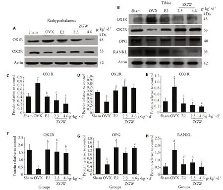
The OX1R protein expression level in the rat tibiae and hypothalamus and RANKL in the tibiae of the OVX group were highly upregulated (P <0.01),and those of OPG and OX2R were downregulated compared with the sham group (P <0.05,<0.01) (Figure 3).After 12 weeks of administration with either ZGW or E2,the OPG and OX2R protein expression levels in the rat tibiae or hypothalamus were significantly upregulated (P <0.05,<0.01),and those of OX1R and RANKL were significantly downregulated compared with the OVX group(P<0.01)(Figure 3).

Figure 2 Micro-CT generated 3D models of the left distal femur trabecular in different groups

Figure 3 Production of proteins in the rathypothalamus and tibiae with Western blot
DISCUSSION
ZGW,with the function of tonifying kidney and essence,benefiting marrow,and strengthening bone,is commonly used in the treatment of PMOP and the improvement of the PMOP-induced symptoms.14Combining our results with those of the previous researches,we proposed that ZGW may play an important role in bone formation by the central regulation of orexin-A and its receptors.
As a steroid hormone,estrogen primarily modulates osteoclast proliferation and development.The treatment with estrogen can improve the symptoms of bone loss,dyssomnia,and depression,which are caused by the deficiency of estrogen in postmenopausal women.15Osteoclasts and osteoblasts play critical roles in regulating bone remodeling and maintaining the balance of bone resorption and bone formation.With the abnormal fluctuation and decreasing of estrogen,the rate of bone formation and bone resorption are highly increased in perimenopausal women.16BALP is a specific osteoblast activity marker,while TRAP5b is the osteoclast activity marker.17-18The current research indicated that the therapeutic effect of ZGW not only inhibits bone resorption but also promotes bone formation.
According to the World Health Organization,PMOP diagnosis and fracture risk assessment should be based on BMD measurement.19However,increasing evidence has demonstrated that BMD cannot predict and estimate the fracture risk perfectly and is comprehensively unrelated to bone strength.20Given its characteristics of sensibility and visualization for the quantitative analysis of skeletal structures,μCT can clearly observe the pathological changes in the trabecular bone microarchitecture.21Dual-energy X-ray absorptiometry combined with μCT can provide highly diagnostic information about skeletal structures and bone quality.22The present study showed that 12 weeks of administration with either ZGW or E2 exerted significantly positive effects on bone morphological structures and trabecular bone microarchitecture.However,the difference in the morphological characteristics of apoptosis and the changes in the μCT parameters of BMD,Tb.Th,Tb.N,BV/TV,BS/BV,and Tb.Sp between the ZGW and E2 groups was statistically insignificant.
Orexins produced in the lateral hypothalamus plays a crucial role for people's health.Studies showed that orexin,as an important rheostat of skeletal homeostasis but previously unrecognized regulator,exerted aYin-Yangdual regulation for bone metabolism by differentially utilizing OX1R and OX2R.23Orexin-A could accelerate the differentiation and matrix mineralization of osteoblast,while orexin B could inhibit the proliferation cultured rat calvarial osteoblast-like cells.24Orexin can enhance bone formationviathe central activation of OX2R by lowering the circulating leptin level and also suppress bone formationviathe peripheral activation of OX1R by decreasing the osseous ghrelin expression.23Leptin could suppress bone formation by increasing the expression of RANKL.25The activity of RANKL/RANK signaling pathway could be inhibited when OPG binds RANKL by competing with RANK on the surface of bone-degrading osteoclasts.26Both orexin-A and OX1R are present in the brain and bone.27OX2R is expressed in brain,but whether it is present in bone remains unknown.27The dysfunction of the hypothalamic-pituitary-gonadal axis in patients with kidney deficiency can lead to unbalanced bone formation and absorption and damage the bone microstructure.Therefore,to explore whether the central control dominating over the peripheral regulation is involved in the underlying mechanism of ZGW in treating PMOP,we examined the mRNA or protein expression of OX1R,orexin-A,OX2R,OPG and RANKL in the rat hypothalamus and tibiae.Our study showed that OX2R was expressed in the tibiae of OVX rats.ZGW could improve bone mass by significantly increasing the orexin-A,OX2R and OPG protein or mRNA expression levels and significantly downregulating the OX1R and RANKL protein and mRNA expression levels in the hypothalamus or tibiae.
However,certain insufficiencies still existed in this study.The ZGW mechanism underlying the occurrence of PMOP was illustrated through the regulation of orexin-A and orexin receptors,which was only an aspect.The comprehensive evaluation of the effect and mechanism of ZGW against osteoporosis requires an in-depth study.
In conclusion,the results of this study illustrated that the regulation of orexin-A and orexin receptors played a role and contributed to the mechanism of ZGW underlying the increase in the bone mass in the OVX rat model.ZGW may be a potential herbal alternative for PMOP treatment.
 Journal of Traditional Chinese Medicine2021年6期
- Journal of Traditional Chinese Medicine的其它文章
- Editorial Board Listing
- In vivo anti-diarrheal activity of jujube honey on castor oil-induced diarrhea in mice
- Chemical characters and protective effect of Baqi Lingmao formula(巴芪灵猫方)on experimental liver injury
- Effectiveness of herb-partitioned moxibustion combined with electroacupuncture on polycystic ovary syndrome in patients with symptom pattern of kidney deficiency and phlegm-dampness
- Traditional Chinese Medicine enhances absorption of lung lesions in corona virus disease 2019 patients
- Shumian capsule(舒眠胶囊)improves symptoms of sleep mood disorder in convalescent patients of Corona Virus Disease 2019
