
A Case of Congenital Syringocystadenoma Papilliferum
2021-08-04 07:15:18LianshengZHONGMeilianLIUXingqiangGAO
Liansheng ZHONG ,Meilian LIU ,Xingqiang GAO
1 Department of Dermatology,Xiamen Children’s Hospital,Xiamen,Fujian 361006,China
2 Department of Pathology,Xiamen Children’s Hospital,Xiamen,Fujian 361006,China
3 Department of Otolaryngology and Head and Neck Surgery,Xiamen Children’s Hospital,Xiamen,Fujian 361006,China
ABSTRACT Syringocystadenoma papilliferum (SCAP) is a rare benign skin tumor that develops from the apocrine or eccrine glands.Here,we report the case of a 2-year-old girl who presented with an asymptomatic,slowly growing lesion on her scalp.On physical examination,a 2 cm × 1 cm red papillary plaque with a crusted surface was noted on her scalp.Histopathological examination revealed papillomatous expansion of the epidermis and cystic invaginations extending from the epidermis down to the deep dermis.The cystic structures were lined by papillae with two layers of columnar epithelium,which showed decapitation secretion.Based on the above clinicopathological findings,a diagnosis of congenital scalp SCAP was made.
KEY WORDS Syringocystadenoma papilliferum;Congenital;Apocrine gland;Eccrine gland
INTRODUCTION
Syringocystadenoma papilliferum (SCAP),also known as nevus syringadenomatosus papilliferus,is a rare benign skin tumor that develops from the apocrine or eccrine glands[1].It is commonly seen on the scalp in association with nevus sebaceous.Approximately 50% of the lesions are present in infancy or at birth[2].They are commonly located on the head and neck but can also occur on the trunk and extremities[3].Herein,we describe a case of congenital SCAP on the scalp of a 2-year-old girl.
CASE PRESENTATION
A 2-year-old girl presented with an asymptomatic,slowly growing lesion on her scalp that had been present since birth.Her parents noticed a mung bean-sized red papule on her scalp at birth,and the lesion gradually increased in size.Occasionally,erosion and exudation can occur on scratching.Her family history was unremarkable,and her general health was good.On physical examination,a 2 cm × 1 cm red papillary plaque with a crusted surface was noted on the scalp (Fig.1A and 1B).A cutaneous appendage neoplasm of the scalp was suspected,and the lesion was excised under general anesthesia.Histopathological examination revealed papillomatous expansion of the epidermis and cystic invaginations extending from the epidermis down to the deep dermis.The cystic structures were lined by papillae with two layers of columnar epithelium,which showed decapitation secretion,and the underlying stroma of the papillary projections was rich in plasma cells (Fig.1C and 1D).Based on the above clinicopathological findings,a diagnosis of congenital scalp SCAP was made.No recurrence was observed during the follow-up of 6 months after excision (Fig.2).
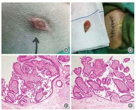
Fig.1 (A-B) Physical examination revealed a 2 cm × 1 cm red papillary plaque with crusted surface on the scalp.(C-D) Histological studies revealed papillomatous expansion of the epidermis,and there were cystic invaginations extending from the epidermis down to the deep dermis.The cystic structures were lined by papillae that had two layers of columnar epithelium,which showed decapitation secretion;the underlying stroma of the papillary projections was rich in plasma cells.Hematoxylin and eosin staining:× 200 magnification.
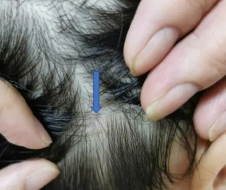
Fig.2 No recurrence 6 months after excision
DISCUSSION
SCAP is a rare benign skin adnexal tumor that develops from an apocrine or eccrine gland that has hamartomatous characteristics and was first described by Pinkus in 1954[4].SCAP usually occurs after birth or in infants and young children.Although the disease often appears at an early age,very few cases have been reported in children,which may be because the disease has no obvious symptoms of itching and pain,and the low rate of pathological biopsies performed in children.There are three clinical types of SCAP:plaque type,linear type,and solitary nodular type.The plaque type is more common on the scalp and appears as a hairless area that may become larger,nodular,verrucous,or crusted at puberty and is often associated with nevus sebaceous.Approximately 75% of cases of SCAP occurs on the scalp,20% on the trunk,and 5% on the extremities,especially on the lower extremities.In addition,some rare sites,such as the nipple,scrotum,and lip,have also been reported[5].SCAP can be accompanied by other skin tumors,such as sebaceous nevus,apocrine adenoma,ductal eccrine carcinoma,basal cell carcinoma,and trichoepithelioma.Approximately one-third of the skin SCAPs are accompanied by sebaceous nevus[6],and 5%-19% of sebaceous nevi are associated with SCAP[7].Characteristic histopathological changes of this disease include an obviously enlarged cystic cavity and papillary structures protruding toward the cystic cavity.The cyst wall is composed of two layers of cells.The inner layer consists of tall columnar cells with apocrine cells,and the outer layer consists of cubic cells.The stroma is abundant with edema,plasma cells are visible,and dilated apocrine glands are seen below the tumor cell masses.SCAP has typical histological characteristics and is not difficult to distinguish from other skin diseases such as seborrheic dermatitis,clear cell sweat adenoma,and papillary sweat adenoma.Surgical excision is the first-choice treatment because of the possibility of malignant transformation.Successful treatment with Mohs’ micrographic surgery and laser excision has also been reported[8].In this patient,the lesion was excised under general anesthesia and she recovered well with no recurrence after 6 months of follow-up.
ETHICS DECLARATIONS
Ethics Approval and Consent to Participate
The need for ethical approval was waived as this study was a case report.Written informed consent was obtained from the patient’s parents.
Consent for Publication
All the authors have consented to the publication of this article.
Competing Interests
The authors declare that they have no competing interests.The authors state that the views expressed in the article are their own and not the official position of the institution or funder.
 Chinese Journal of Plastic and Reconstructive Surgery2021年2期
- Chinese Journal of Plastic and Reconstructive Surgery的其它文章
- The Practice of China’s Cosmetic Medicine Dated Back to 3 800-4 800 Years Ago
- Progress in Implant-Based Breast Reconstruction:What Do We Know?
- Electric Field:A Key Signal in Wound Healing
- Mechanisms and Management of Postparalysis Facial Synkinesis
- Looped,Broad,and Deep Buried Suturing Technique for Wound Closure
- A Novel Composite Skin Graft Technique with Fat Derivatives