Hemodynamic changes in arterial flow velocities throughout the first six months of pregnancy in buffalo heifers by Doppler ultrasonography
Elshymaa A.Abdelnaby
Theriogenology Department, Faculty of Veterinary Medicine, Cairo University, Giza, Egypt
ABSTRACT
KEYWORDS: Uterine; Doppler; Pregnancy; Ovarian artery;Umbilical cord
1.Introduction
During the first and the second trimester of the critical gestation period, functional and structural changes occur in the utero-ovarian vascular system depending upon the amount of fetal nutritional requirements; the ovarian and middle uterine arteries, the two major blood vessels adapt to these hemodynamic changes to ensure adequate blood supply to the developing placenta/fetus in addition to the functionality of the corpus luteum[1,2].The mechanism responsible for the maintenance of a corpus luteum during pregnancy and in particular the way in which the embryo influences this process, are not clearly understood but may involve a local effect of the conceptus on uterine blood flow.Therefore,function and perfusion of uterine arteries as well as uterine branch of ovarian blood flow is a vital determinant of fetal growth[3].In medicine, there have been several studies to assess uterine blood flow in cows[4,5], mares[6], bitchs[7], queens[8]and rabbits[9], in addition to some studies, use Doppler to evaluated women uterine vascularization[10,11], However, there was a very little information about the ovarian vascular perfusion changes in buffalos during pregnancy.
The utero-ovarian system sustains the most important adaptation during the period of gestation.This includes an increase in known vessel area under the pregnancy stress factors and compliance by the action of the trophoblastic maternal cells invasion.Moreover, the placenta is developed and maintained throughout pregnancy, which is a low resistance organ that facilitates the transport of nutrients and minerals required by the foetus, via the umbilical cord[12].The applicability of Doppler technique in blood flow studies of uterine and umbilical arteries has been considered as a tool where the maternal and fetal unit normality can be detected[13].Additionally,the uterine artery Doppler abnormal parameters as Doppler indices and peak with end velocities indicated an intrauterine growth restriction and abnormal pregnancy leading to fetal death or fetus born with abnormalities; Doppler technique can be a useful diagnostic tool to determine any abnormalities in the fetus before the date of birth perinatal morbidity, and abnormal delivery or fetal death[14].In addition to any maternal disorder indicated by uterine arteries blood flow leading to inadequate blood supply as well as placental attribution[15], the abnormality in uterine artery Doppler indicated by Doppler indices like a mean pulsatility index was increased with alteration of end-diastolic notch[16].Doppler ultrasound scanning is an effective preventive and diagnostic tool for any fetal or maternal abnormalities[14].
The objective of the present study was to examine blood flow velocity changes in the ovarian and middle uterine arteries in heifers at the first six months of pregnancy to confirm the base-line information on parameters that can be used to assess the progress of pregnancy in buffalo heifers.
2.Materials and methods
2.1.Animals and management
This study was conducted on 15 healthy, cycling, Egyptian buffalo heifers.All animals were examined in each month of gestation twice at an organized dairy farm in the Faculty of Veterinary Medicine,Cairo University (30.0276 °N, 31.2101 °E) and maintained under uniform conditions of feeding and management and were kept in an indoor paddock individually with a natural light and temperature at morning and with artificial lightening at night.Animals maintenance requirements of nutrition consist of concentrated ration and tracemineralized salts with free access to water all over the day.They were housed in semi-intensive housing systems.Body condition score of the heifers ranged from 3.5 to 4.0 on a scale of 5.0.The age of heifers ranged from 15-19 months, weighing 300-400 kg [mean(350±50) kg].Heifers would come into puberty early and cycle 2-3 times then achieve the target of an average age at first calving of 25-30 months.Pregnancy was established with high-quality semen by natural mating of only one sire.From farm records, all animals with no apparent clinical disease were examined weekly along three successive estrous cycles normally, then twice per month during pregnancy.The ovarian and middle uterine arteries ipsilateral and contralateral to the fetus side were scanned by the same operator at the same time of day on early morning (07:00-10:00 a.m.).The ovarian artery emerged from dorsal aorta, while the middle uterine artery was a branch of the internal iliac artery which was located cranial to the external iliac artery[17].
2.2.Doppler examination and Doppler parameters
Doppler measurements were performed by using Exago transportable ultrasound device (ECM Co., Angouleme, France)in pulsed-wave mode.A 7.5 MHz linear probe Doppler equipped with linear-array transducer [Power: Max (100%) PRF: 3.3-5 kHz Doppler angle: 30-60 Brightness: 70%]was used with the color and spectral modes.The parameters used to assess blood flow were determined by using automatic options on the device and the images were stored in the device.
The Doppler indices and other parameters that the device displayed for the waveform by applying the automatic pulsed wave mode were resistance index (RI), pulsatility index (PI), peak systolic velocity(PSV), end-diastolic velocity (EDV), and blood flow rate (R) in beats per minute (bpm).After activating the B mode of the known artery, color Doppler mode was used to assess the known ovarian and middle uterine arteries by red or blue color according to the blood flow direction.The spectral Doppler mode was activated and the specific gate entered in the known artery and all Doppler indices with blood flow velocity waveforms were calculated.To avoid any artifacts, the gate size must be suitable to the artery size.The operator manually adjusted the gate location, gate size, and other spectral Doppler imaging control parameters.All animals were scanned without the use of anesthesia.Averages of three successive waves were required to develop measurements during one cardiac cycle.The averages of all parameters were calculated until the six months of pregnancy.The RI as well as PI indicated the vascular perfusion to the particular organ and tissues[17-20].As pregnancy progressed, the ovarian arteries ipsilateral or contralateral to the side of a fetus could not be evaluated through their origin from the great abdominal aorta[21], hence, its branch called uterine branch of the ovarian artery that supply the uterus cranially was evaluated in addition to umbilical artery evaluated.A mean PI >1.55 was defined to be abnormal[22].
2.3.Statistical analysis
All data were recorded in an excel spreadsheet and statistical analysis was taken by using SPSS version 16 Chicago, IL,USA[23].Numeric data for all the parameters were expressed as mean±standard deviation (mean±SD).Pearson's correlation coefficient was calculated to ascertain the correlation between blood flow parameters.Statistical analysis was performed by using t-test and P-value <0.01 was considered to be statistically significant.Repeated measures of analysis of variance for measurements were taken over time on the same animal during the first 6 months of pregnancy.
2.4.Ethics statement
This study was approved by the Institutional Animal Care and Use Committee of Faculty of Science, Cairo University, Egypt (approval number: Vet CU23012020433).
3.Results
3.1.Doppler indices of uterine branch of ovarian arteries and middle uterine arteries
With the respect to all Doppler indices and parameters of both ipsilateral and contralateral ovarian and middle uterine arteries,there was a variation within animals of a particular gestation in the first and second trimester.RI of the ipsilateral uterine branch of ovarian arteries was positively significantly correlated with PI(r=0.62, P<0.01).Since RI and PI were highly correlated, RI was used for comparison with other parameters.RI was negatively correlated with all other Doppler parameters (PSV: r=-0.25,P<0.001, EDV: r=-0.76, P<0.001, and R: r=-0.85, P<0.001).There was a positive correlation between PSV and EDV (r=0.38,P<0.01), PSV and R (r=0.35, P<0.01), and EDV and R (r=0.59,P<0.01).Contralateral RI of the uterine branch of ovarian arteries and uterine artery was positively correlated with PI (r=0.39,P<0.01) and negatively correlated with other parameters (PSV:r=-0.54, P<0.001, EDV: r=-0.98, P<0.001, and R: r=-0.55,P<0.01).
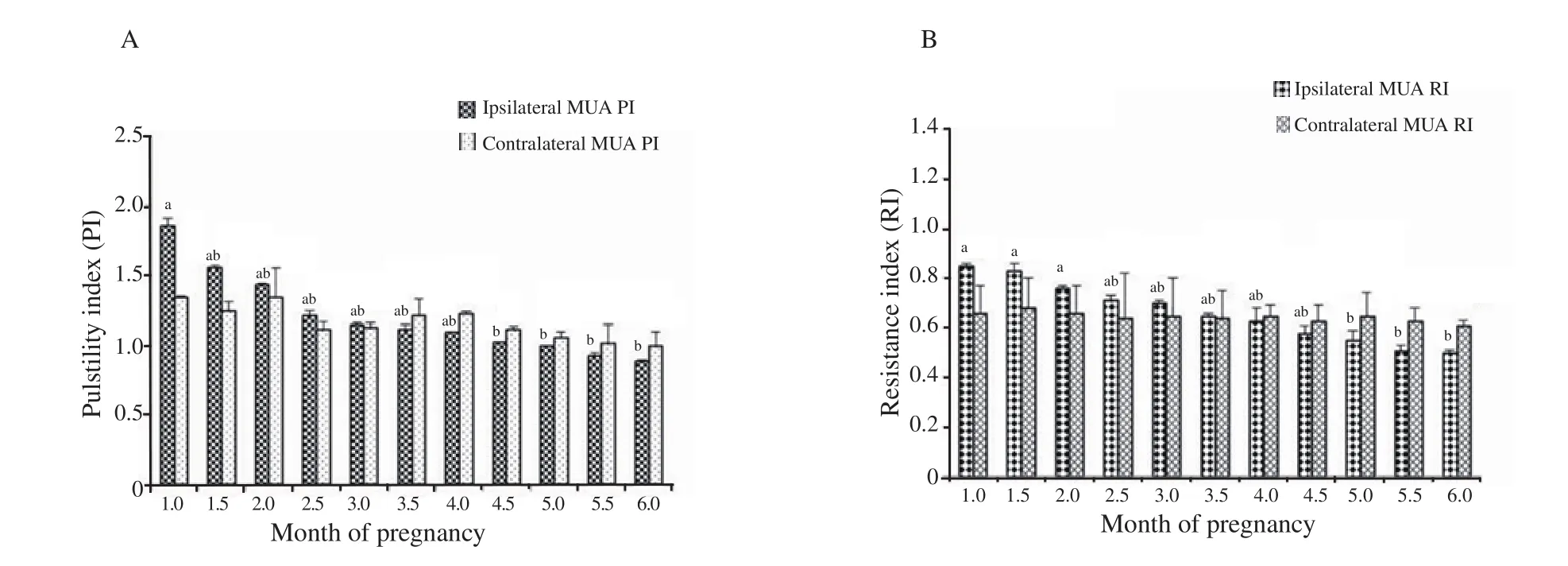
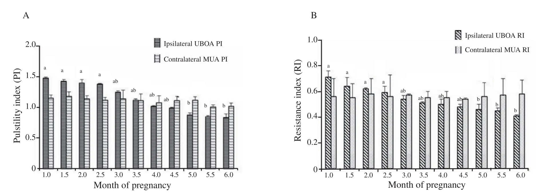
The two Doppler indices values, RI and PI, also together showed a pattern of decline in the middle uterine artery ipsilateral to the fetus compared with contralateral one (P<0.01) (Figure 1) and the RI showed a continuous decrease during the first six months of gestation.In addition, the mean RI and PI values showed a linear decrease in the uterine branch of ovarian artery ipsilateral to the fetus side compared with contralateral artery at the first and second trimester of pregnancy (P<0.01) (Figure 2); the RI showed a continuous decrease during the first 6 months of gestation and remained fairly constant or slightly higher thereafter (P<0.01).

Figure 1.Pulsatility (PI) and resistance index (RI) of the middle uterine arteries (MUA) ipsilateral and contralateral to the fetus at the first and second trimester of pregnancy.a: significantly higher than the successive value in the different month, P<0.01; b: ipsilateral MUA PI vs.contralateral MUA PI at the same month, P<0.01.

Figure 2.Pulsatility (PI) and resistance index (RI) of the uterine branch of the ovarian arteries (UBOA) ipsilateral and contralateral to the fetus at the first and second trimester of pregnancy.a: significantly higher than the successive value in the different month, P<0.01; b: ipsilateral MUA PI vs.contralateral MUA PI at the same month, P<0.01.
3.2.Doppler velocities of uterine branch of ovarian arteries and middle uterine arteries
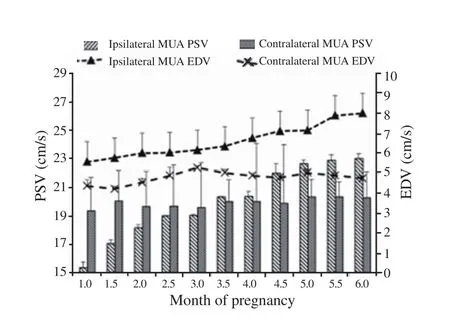
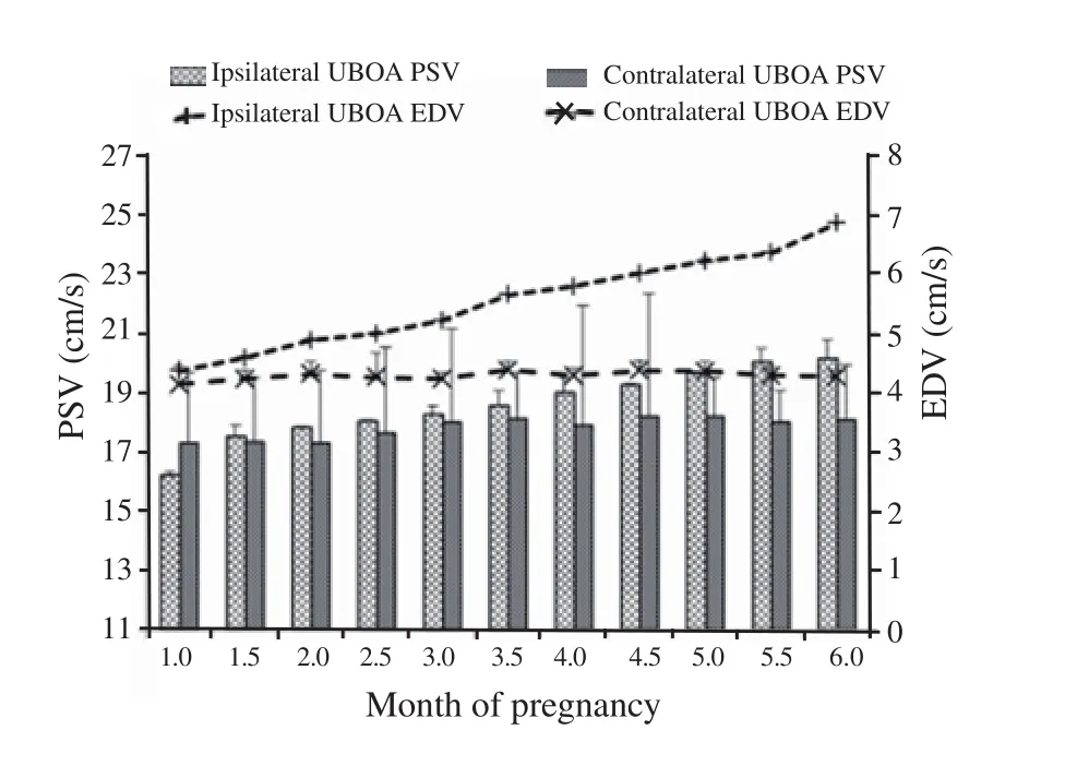
The PSV and EDV of the middle uterine artery ipsilateral to the fetus side increased linearly till the six months of pregnancy(Figure 3), while the PSV and EDV in the other contralateral side remained on a constant level (19.36±1.85 to 20.25±1.24 for PSV and 4.36±0.01 to 4.75±0.01 for EDV) throughout the first and second trimester of the pregnancy.The same went for Doppler parameters of the ipsilateral uterine branch of ovarian artery, as both blood flow velocities showed a pattern of increase in the ipsilateral side compared with the contralateral one (Figure 4).
The R in the ipsilateral uterine branch of ovarian artery was lower compared with the contralateral artery in most of the months of gestation in the first and second trimester (Table 1).The R in the middle uterine artery ipsilateral to the fetus side was lower than that in the contralateral artery side.
3.3.Normal and abnormal Doppler parameters of the middle uterine artery, uterine branch of ovarian artery and umbilical artery between 4 weeks 0 day (1 month) and 24 weeks 6 days (6 months)
Of the 15 evaluated heifers, 5(33.3%) had abnormal uterine artery Doppler findings leading to fetal death, which had a mean PI of>1.55.The uterine branch of ovarian artery, middle uterine artery,and umbilical artery of Doppler measurements between 4 weeks 0 day (1 month) and 24 weeks 6 days (6 months) did help in prediction of any abnormalities in five pregnant animals in comparison to those with normal results shown in Table 2.Ipsilateral RI and PI of uterine branch of ovarian artery were higher in the abnormal Doppler reading results than the normal one.The same for uterine branch of ovarian artery was done in the middle uterine artery and umbilical artery ipsilateral to the fetus side; all RIs and PIs increased abnormally when compared with the normal reading.Nevertheless,all PSVs and EDVs of uterine, ovarian and umbilical arteries showed a significant abnormal decrease (P<0.01)when compared with normal animals without any pregnancy abnormalities (Table 2).

Figure 3.Peak systolic velocity (PSV) and end diastolic velocity (EDV)of the middle uterine arteries (MUA) ipsilateral and contralateral to the fetus at the first and second trimester of pregnancy.

Figure 4.Peak systolic velocity (PSV) and end diastolic velocity (EDV)of the uterine branch of the ovarian arteries (UBOA) ipsilateral and contralateral to the fetus at the first and second trimester of pregnancy.

Table 1.Blood flow rate in the ovarian and middle uterine arteries ipsilateral and contralateral to the fetus in the first six months of gestation.

Table 2.Abnormal and normal Doppler measurement results of uterine branch of ovarian artery, middle uterine artery, and umbilical artery from 4 weeks 0 day (1 month) to 24 weeks 6 days (6 months) of gestation.
4.Discussion
A mature healthy calf after gestational months in buffalos is the most favorable goal for dairy farmers, producers and professionals'enterprise.With this attainment to be carried out, every heifer must undergo pregnancy to term, to deliver a normal mature calf.Maintenance of a successful pregnancy involves interactions between conceptus and the dam of surrounding environment to get rid of any embryonic abnormities or lethality during organogenesis as abnormal vascular development in domestic and small ruminants[24,25].Maternal adaptations in the vascular perfusion observed during the pregnancy in normal cases include decreases in uterine and systemic vascular resistance with dramatic increases in uterine blood flow and several changes of cardiac parameters in humans[12,26,27]or the same parameters not changed in both ovarian and uterine arteries[28].
Although the growth and development of new vessels in addition to the rebuilding of existing present major vessels during the first months of pregnancy participates in the increased blood supply and blood flow of the ovarian and uterine arteries, the period that the blood flow reaches a maximum level occurs after the completion of new vessel growth, which indicates the maintenance of vasodilation in existing or newly developed vessels is fateful[27,29].In this study,as the period of gestation developed progressively, there were clear reductions in Doppler indices in both ovarian and uterine arteries (RI and PI) with a linear decrease in the value in the ipsilateral artery compared with the contralateral artery which is indicative to greater perfusion to the gravid horn with least resistance by increasing the two velocities of the blood flow parameters in both ovarian and uterine arteries.This reduction is essential for continuous maternal vascular perfusion through the placenta and for the critical supply of oxygen as well as nutrients[30].Contrast to our report, no significant difference was observed between the right and left uterine arteries,or between the right and left ovarian arteries in the value of each vascular index in women with hypo-estrogenic amenorrhea[31].Thus,the uterine and ovarian arteries of one side were selected for further analysis.
There was a considerable increase in PSV and EDV in the ipsilateral ovarian and uterine arteries as compared with the contralateral artery with a linear rapidly increase as the pregnancy progress to meet the growing fetus demands and these findings were similar to those in previous studies[4].This change is essential for the fetal chance of survivability to improves fetus body condition and development.In the studies of luteal blood flow assessment during the first trimester of pregnancy, PSV and EDV did not change, and the PI was decreased in some studies[10]but not in others[3,32].
Similarly, a recent study[33]had reported the predictive role of the uterine artery PI, RI, and PSV indices measurement on the day of human chorionic gonadotropin injection in the success rate of invitro fertilization procedure.They found that only mean uterine artery PI and RI were significantly lower in pregnant women with successful in-vitro fertilization than those with an unsuccessful procedure.As well as the PSV of ipsilateral uterine artery was higher in pregnant women, but the differences were not statistically significant.The rate of the flow in ipsilateral ovarian artery was lower compared with the contralateral arteryas the blood flow rate determine the blood flow velocity in the important vessels.Uterine artery blood flow is affected by a rate/bpm which may change according to the vessel diameter.During pregnancy, the diameter of the main uterine artery approximately doubles in size in humans[34].The best predictor of different abnormal findings by Doppler was the two Doppler indices together (PI and RI) and bilateral notching in addition to velocities as peak systolic related to maximum contraction and end diastolic related to maximum relaxation.Similar to our findings, for determination of risk at pregnancy in abnormal uterine artery, Doppler images and study of the importance of abnormal uterine artery were determined by Doppler new technology with pulsed wave mode at middle stage of gestation[16].
In our study, a PI of >1.55, an excellent predictor was used and 33.3% of the heifers developed fetal death, with abnormal uterine artery ipsilateral to fetus and umbilical artery Doppler reading results.Another report[35]evaluated the middle uterine artery Doppler changes in the second stage of pregnancy in women, for determination of risk this stage, and concluded that many cases of pre-eclampsia were detected with abnormal PI and many adverse pregnancy outcomes were suffered from abnormal uterine artery Doppler indices.The study limitation is the applicability of color Doppler to evaluate specific artery blood supply, which needs a stable state in the animal body without any movement, but the main disadvantage is a quick fetal movement inside the uterus, as the accurate detection of the genital arteries with their branching requires the constant and correct positioning of the ultrasound probe.In conclusion, this study has provided the hemodynamics changes in uterine and umbilical arteries waveform velocities that take place during the first six months of pregnancy in buffalo heifers in the developing placenta/fetus.
Conflict of interest statement
There are no conflicts of interest to declare.
Acknowledgements
The author thanks Dr.Refaat Ragab, professor of College Farm for animals' availability.
 Asian Pacific Journal of Reproduction2020年4期
- Asian Pacific Journal of Reproduction的其它文章
- Prospects of diagnostic and prognostic biomarkers of pyometra in canine
- Seasonal changes in sperm parameters, testicular histology and circulating levels of reproductive hormones in the male African straw-colored fruit bat (Eidolon helvum)
- Genistein improves the vaginal epithelium thickness in a rat model of vaginal atrophy through modulation of hormone and heat shock protein 70 levels
- Process of becoming a mother in women receiving donated egg: Based on the grounded theory
- Food insecurity and other possible factors contributing to low birth weight: A case control study in Addis Ababa, Ethiopia
- Effects of nitric oxide on reproductive organs and related physiological processes
