Anti-proliferative potential of sodium thiosulfate against HT 29 human colon cancer cells with augmented effect in the presence of mitochondrial electron transport chain inhibitors
Bhavana Sivakumar, Sri Rahavi Boovarahan, Gino A Kurian
Vascular Biology Lab, School of Chemical and Biotechnology, SASTRA Deemed University, Thanjavur, India
ABSTRACT
KEYWORDS: Colorectal cancer; Sodium thiosulfate; HT-29;IEC6; Mitochondria; Rotenone; Clonogenic assay
1.Introduction
Colorectal cancer is becoming one of the emergent cancers and according to the Global Cancer Observatory, the number of new cases in 2018 was around 18 495 118[1].The incidence rate of colorectal cancer is linked to people’s nutritional status and its transition is followed by rapid change in the economy and society.The proportion of colorectal cancer cases in the world has increased in individuals younger than 50 years of age, primarily associated with dietary habits and sedentary lifestyle (which generally induces inflammatory response) along with smoking and alcohol consumption[2].Under chronic inflammatory conditions,the colon polyps develop as the aberrant crypt foci which leads to malignant tumors[3].Chemotherapeutic agents play a key role in the management of colorectal cancer and are becoming an indispensable part of the treatment in these days[4].
Many sulfur-based drugs like thiophene derivatives which include sulfonamide, isoxazole, benzothiazole, quinoline and anthracene are very promising agents in the treatment of colon cancer[5].In addition, sulfur-based naturally occurring products are widely used in the traditional system of medicines in India (Ayurveda) and China.The sulfur-containing compound in garlic namely, allicin is found to be effective in the management of gastric cancer because of its pro-apoptotic, anti-proliferative, and anti-helicobacter activities[6].However, sulfur-based compound efficiency may be linked to its bioavailability as the half-life of many of these compounds are low and the possible cure depends on its concentration in the cancer environment.Documents from the literature have shown that hydrogen sulfide (H2S), a sulfur-based endogenous molecule,possesses both pro and anti-proliferative effects on colon cancer cells[7], thereby putting a hold on the bioavailability issue.By using the slow-releasing H2S donor, GYY4137, investigators have shown that H2S is specifically targeted to induce cancer cell deathviaactivating caspase systems[8], but without making an apprehension on its cellular toxicity on normal cells.
Unlike H2S, sodium thiosulfate (a metabolite of H2S), a sulfurbased compound has high tissue tolerance even up to 250 mg/mL(according to Food and Drug Administration reports).A number of studies have shown that thiosulfate possesses antioxidant potential,calcium chelation effect and can also modulate mitochondrial activity[9] and thereby regulate different signaling pathways that involve NFκB and PI3K[10].However, the anti-proliferative effect of thiosulfate has not been widely recognized except for a few investigators who have shown the detoxifying effect of platinum-based chemotherapeutic drugs[11].Also, recent research developments have shown that one of the major etiology of cancer lies within mitochondrial DNA mutations and many investigators consider cancer as a type of mitochondrial metabolic disease[12,13]The treatment with sodium thiosulfate leads to mitochondrial functional modulation due to its interference with NF-κB and PI3K signaling pathways that converge into the mitochondria, an effective target in the treatment of cancer.Hence, more studies are required to explore its potential to act as an anti-proliferative agent, which is addressed in this study.
2.Materials and methods
2.1.Chemicals and reagents
All the chemicals and reagents used in this study were purchased from Sigma-Aldrich (St.Louis, USA).
2.2.Cell culture
The human colon cancer cell line (HT-29) and intestinal epithelial cells (IEC6) were purchased from National Centre for Cell Science,Pune.The cells were maintained in Dulbecco’s Modified Eagle Medium (DMEM, Invitrogen, USA) and 10% fetal bovine serum(FBS) and 1% penicillin-streptomycin were used as supplements.5% CO2humidified incubator at 37 ℃ was used for the incubation of the cells which were later passaged after it reached its 70%confluence.
2.3.Cell viability tests
Cells (HT-29 and IEC6) were plated in a 96-well plate of 1×10⁵cell density and treated with different concentrations of sodium thiosulfate (0.5 mM, 1 mM,10 mM, 20 mM, 40 mM, 60 mM, 80 mM) for 24 h.The cell viability was measuredviacrystal violet and MTT assays.
2.3.1.Crystal violet assay
Cell viability was assessed using the crystal violet assay according to the protocol described[14].Paraformaldehyde at 4% was used to fix HT-29 and IEC6 cells and was then incubated for 30 min at room temperature.The plate was washed twice with water in order to remove the fixative.The cells were stained with 0.5% crystal violet and incubated for 20 min.To dissolve the dye, 200 μL methanol was added and gently shook for 20 min.The absorbance was measured at 570 nm.
2.3.2.3-(4, 5-Dimethylthiazol-2-yl)-2, 5-diphenyltetrazolium bromide (MTT) assay
The MTT assay was done to assess the cell viability and the ability of mitochondrial malate dehydrogenase enzyme in viable cells to reduce yellow-colored MTT to form purple-colored formazan, which was measured[15].A total of 10 μL MTT solution was added to each well containing IEC6 and HT-29 cells.The cells were incubated at 37 ℃ for 4 h.Then 150 μL of 0.1% acidic isopropanol was used to dissolve the dye.The absorbance was measured using a multimode reader at 540 nm.
2.3.3.Clonogenic assay
Cells (HT-29 and IEC6) were plated in a 35 mm culture dish at 0.3×10⁶cell density and treated with different concentrations of sodium thiosulfate (0.5 mM, 1 mM, 10 mM, 20 mM, 40 mM, 60 mM) for 15 d.
The clonogenic assay or colony formation assay determines the ability of a cell to grow in a colony that determines its proliferation capacity.IEC6 and HT-29 cells were plated at different concentrations (0.5 mM, 1 mM, 10 mM, 20 mM, 40 mM, 60 mM) in order to form colonies in 1-3 weeks[16].The colonies were fixed with glutaraldehyde (6.0% v/v), further stained with crystal violet (0.5%w/v) and counted.Cell viability was measured by crystal violet assay.
2.4.Inhibitor studies
HT-29 was plated in a 96-well plate of 1×10⁵cell density and treated with 1 μM sodium azide and 1 μM rotenone [mitochondrial electron transport chain (ETC) inhibitors], 300 μM diazoxide and 25 μM glibenclamide (KATPchannel opener and closer), H2S inhibitors 0.2 mM DL-propargyl glycine (PAG) and 0.2 mM aminooxy acetic acid (AOA) for 24 h followed by a combination with sodium thiosulfate at different concentrations (0.1 mM, 0.5 mM, 1 mM, 25 mM).
2.4.1.Effect of sodium thiosulfate in the presence of mitochondrial ETC inhibitors
To evaluate whether sodium thiosulfate modulates its cytotoxic action in HT-29 through mitochondria, we used ETC inhibitors in the presence and absence of sodium thiosulfate.Rotenone is a naturally occurring mitochondrial complex Ⅰ inhibitor that affects two reversible conformational states of complex Ⅰ, which leads to the activation and inactivation of the complex.Similarly, selective reduction of complex Ⅳ activity was mediated by administrating the cells with sodium azide.Therefore, the HT-29 cells were first treated with 1 μM sodium azide (mitochondrial complex Ⅳ inhibitor) for 24 h and different concentrations of sodium thiosulfate (0.1 mM,0.5 mM, 1 mM, 25 mM).Similarly, the cells were treated with 1 μM rotenone (mitochondrial complex Ⅰ inhibitor) followed by different concentrations of sodium thiosulfate (0.1 mM, 0.5 mM, 1 mM, 25 mM) in order to understand the cytoprotective action which was measured by crystal violet assay.
2.4.2.Effect of sodium thiosulfate in the presence of KATP channel modulators
KATPchannel of HT-29 cells was modulated by using diazoxide(300 μM acts as KATPchannel opener) and glibenclamide (25 μM acts as KATPchannel closer).Prior to sodium thiosulfate treatment,HT-29 cells were incubated with KATPchannel modulators for 24 h.At the end of the experiment, cytotoxicity was determined by crystal violet assay.
2.4.3.Effect of sodium thiosulfate in the presence of endogenous H2S inhibitors
The influential role of endogenous H2S enzyme in the antiproliferative effect of sodium thiosulfate was evaluated by using H2S biosynthetic enzyme inhibitors like DL-PAG and AOA.Prior to sodium thiosulfate administration, the cells were treated with PAG and AOA for 24 h and the cytotoxicity effect was evaluated by crystal violet assay.
2.5.Statistical analysis
All values were expressed as mean ± SD.The data were subjected to ANOVA for comparison of differences between groups using GraphPad Prism 7 software.P<0.05 was considered statistically significant.
3.Results
3.1.Sodium thiosulfate induces dose-dependent decline in survival of HT-29 colon cancer cells
HT-29 and IEC6 cells were cultured in the presence or absence of sodium thiosulfate at various concentrations (0.5 mM, 1 mM,10 mM, 20 mM, 40 mM, 60 mM, and 80 mM) for 24 h and cell viability was measured (Figure 1).Exposure to 20 mM sodium thiosulfate was found to be toxic to HT-29, but IEC6 cells could withstand the 20 mM sodium thiosulfate and sodium thiosulfate was toxic from 40 mM concentration.The IC50value of HT-29 was found to be 40.93 mM and 42.45 mM when evaluated by crystal violet and MTT assays, respectively.IEC6 cells showed IC50values of 45.17 mM and 47.22 mM.
In order to determine the anti-proliferative activity of sodium thiosulfate, the clonogenic assay was performed and the results are shown in Figure 2.HT-29 cells treated with 40 mM of sodium thiosulfate showed only 46% of live cells in 15 d whereas IEC6 cells showed 68% of live cells at the same concentration of sodium thiosulfate and same time period.
3.2.Sodium thiosulfate augments the cytotoxicity effect in the presence of ETC inhibitors in HT-29 colon cancer cells
Treatment with complex Ⅳ inhibitor sodium azide resulted in(28±7)% and (30±6)% cell death, estimatedviaMTT and crystal violet assays, respectively (Figure 3).However, a combination of sodium azide with different concentrations of sodium thiosulfate(0.1 mM, 0.5 mM, 1 mM, 25 mM) led to approximately (60±4)%,(65±4)%, (67±3)%, and (69±3)% cell death, respectively in MTT assay.In the case of crystal violet assay, cell death of (57±4)%,(61±4)%, (63±3)%, and (65±3)% were found at 0.1 mM, 0.5 mM,1 mM, and 25 mM, respectively.These results indicate a significant(P<0.05) cell death in both cases.
Similarly, 1 μM rotenone treatment of HT-29 cells for 24 h showed (57±4)% cell death in MTT and (55±4)% death in crystal violet assay.Conversely, the combined treatment of rotenone with different sodium thiosulfate concentrations as mentioned above led to (72±4)%, (75±4)%, (78±3)%, and (81±3)% cell death in MTT assay and (70±4)%, (72±4)%, (75±3)%, and (77±3)% in crystal violet assay, indicating a significant (P<0.05) cytotoxicity of sodium thiosulfate in the presence of rotenone (Figure 4).Hence it can be concluded that neither sodium azide nor rotenone, when given individually, can impart prominent cytotoxicity to the HT-29 cells.On the other hand, the presence of ETC inhibitors enhanced the cytotoxic effect of sodium thiosulfate on HT-29 cells.
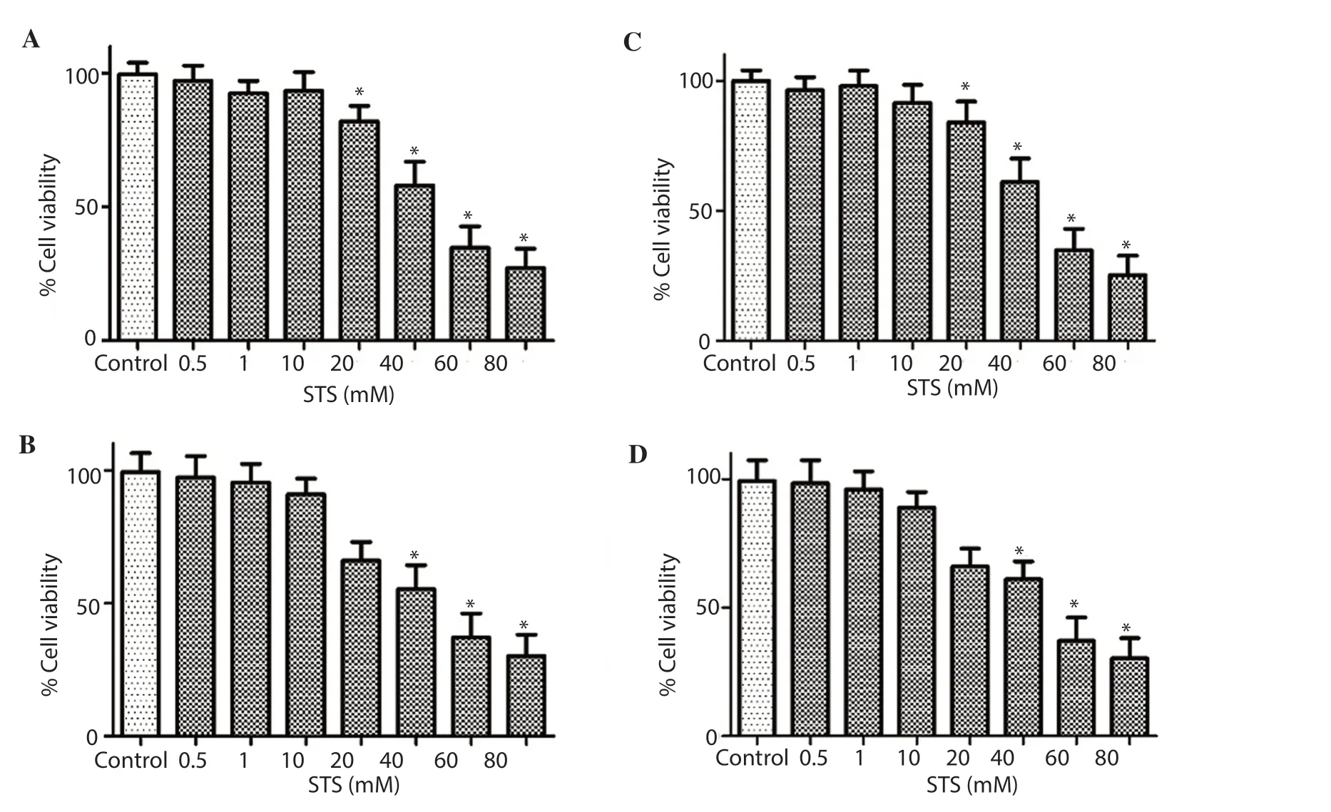
Figure 1.Cytotoxicity of sodium thiosulfate (STS) in HT-29 cells and IEC6 cells.HT-29 and IEC6 cells were cultured in the presence or absence of sodium thiosulfate at various concentrations.A: HT-29 cells measured via MTT; B: IEC6 cells measured via MTT; C: HT-29 cells measured via crystal violet assays; D: IEC6 cells measured via crystal violet assays.* indicates P<0.05 vs. control.
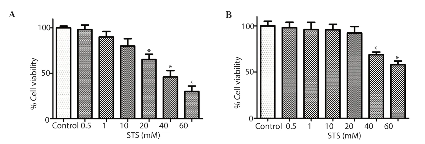
Figure 2.Clonogenic assay.The anti-proliferative capacity of sodium thiosulfate (STS) reconfirmed by clonogenic assay done on (A) HT-29 cells and (B)IEC6 cells.* indicates P<0.05 vs. control.
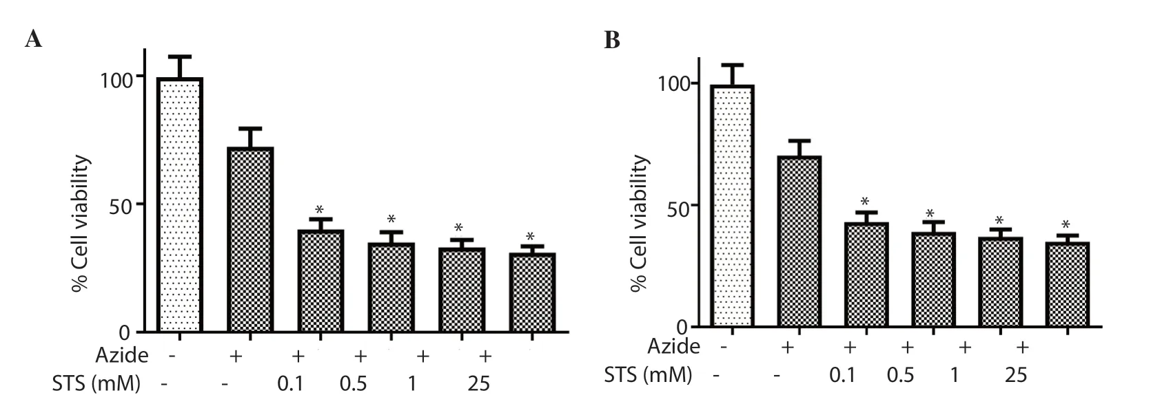
Figure 3.Effect of electron transport chain (complex Ⅳ) inhibitor, 1 µM sodium azide on the anti-proliferative effect of sodium thiosulfate (STS) in HT-29 cell line evaluated by (A) MTT assay and (B) crystal violet assay.* indicates P<0.05 vs.control.
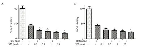
Figure 4.Effect of electron transport chain (complex Ⅰ) inhibitor, 1 µM rotenone on the anti-proliferative effect of sodium thiosulfate (STS) in HT-29 cell line evaluated by (A) MTT assay and (B) crystal violet assay.* indicates P<0.05 vs.control.

Figure 5.Effect of sodium thiosulfate (STS) on HT-29 in the presence of KATP channel modulator-diazoxide (300 µM) evaluated by (A) MTT assay and (B)crystal violet assay.No significant changes were observed.
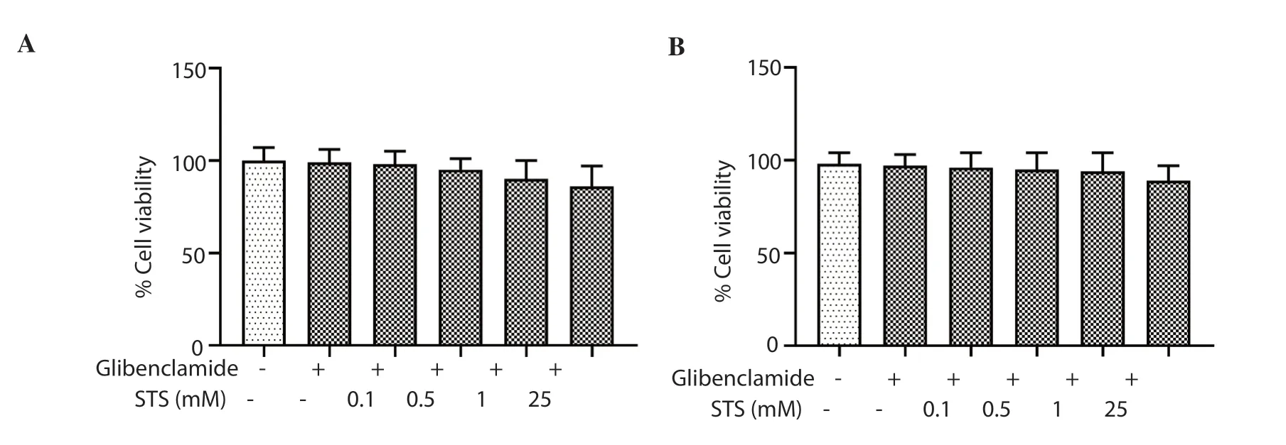
Figure 6.Effect of sodium thiosulfate (STS) on HT-29 in the presence of KATP channel modulator-glibenclamide (25 µM), evaluated by (A) MTT assay and (B)crystal violet assay.No significant changes were observed.
3.3.Sodium thiosulfate mediated cytotoxicity in HT-29 colon cancer cells is independent of mito-KATP channel
Since ETC enzymes need intact KATPchannel for its functional activity, we further assessed the effect of sodium thiosulfate on HT-29 in the presence of KATPchannel modulators.According to Figures 5 and 6, the cytotoxic effect of sodium thiosulfate was not significantly changed in the presence of 300 μM diazoxide (KATPchannel opener) and 25 μM glibenclamide (KATPchannel closer),when compared with the sodium thiosulfate group without the above-mentioned inhibitors.
3.4.Anti-proliferative effect of sodium thiosulfate was intact in HT-29 cells in the presence of DL-PAG or AOA

Figure 7.Cytotoxicity of sodium thiosulfate (STS) in the presence of endogenous H2S inhibitor- propargyl glycine (PAG) in the HT-29 cell line evaluated by (A)MTT assay and (B) crystal violet assay.No significant changes were observed.

Figure 8.Cytotoxicity of sodium thiosulfate (STS) in the presence of endogenous H2S inhibitor-aminooxy acetic acid (AOA) in HT-29 cell lines evaluated by (A)MTT assay and (B) crystal violet assay.No significant changes were observed.
Next, we evaluated whether the anti-proliferative effect of sodium thiosulfate depends on endogenous H2S, where the latter has already been proven to be a potent anticancer agent on HT-29 cells.We found that the cytotoxic effect of sodium thiosulfate on HT-29 was unaltered in the presence of DL-PAG or AOA, indicating the sodium thiosulfate mediated cytotoxicity is H2S independent (Figures 7 &8).
4.Discussion
The ambiguous mechanisms in cancer pathology make cancer therapy difficult and thus the research focusing on its eradication is still attracting greater attention of both clinicians and scientific investigators.In the present study, the anti-proliferative capacity of sodium thiosulfate was evaluated on HT-29 colorectal cancer cells and the effects were compared with IEC6 normal small intestine cells.Based on the clonogenic and cytotoxicity assays, we found that sodium thiosulfate can inhibit the growth of HT-29 cells.The IC50values of HT-29 cells were 40.93 mM and 42.45 mM by crystal violet and MTT assay whereas in IEC6 cells, the values were 45.17 mM and 47.22 mM, respectively.Moreover, we also found that the anti-proliferative effect of sodium thiosulfate was augmented in the presence of rotenone (complex Ⅰ) and sodium azide (complex Ⅳ)inhibitors.
Among the different anticancer drugs approved by the Food and Drug Administration from 2010 to 2015, two-thirds of them are heterocyclic compounds[17].Sulfur-based heterocyclic compounds have exhibited potent anticancer property primarily because of its role in co-enzyme chemistry and its ability to interact with different regulatory molecules[18].A recent clinical trial by Hauschild and his co-workers showed that dabrafenib, a well-known sulfur-based heterocyclic compound, is known to inhibit mutatedBRAFand thereby improve the survival rates inBRAFmutated colorectal cancer patients[19].Similarly, few investigators have shown that thiazole derivatives are effective against colon cancer cell lines[20].Even though sulfur-based heterocyclic compounds provide many promises in the management of colorectal cancer, they have their own limitations.Sulfur-based heterocyclic compounds work in tight regulatory control of cells, making it difficult to distinguish the delicate balance of cell metabolism in normal and cancer cells.Hence it has become an utmost necessity to identify a potential sulfur-based molecule that is endogenous in nature.This shortcoming can be addressed to a certain extent by recent findings that prove the anti-proliferative action of endogenous sulfur compounds like hydrogen sulfide[18].Accordingly, literature against cancer suggested that H2S can inhibit the growth of cancer cells[21.22].On the contrary,there are evidences which suggest the tumor-promoting activity of H2S as well[23].The controversial findings of H2S (pro and anti-cancer effects) will bring doubt in the use of H2S as a therapeutic agent in the management of colon cancer.So this calls for a new therapeutic endogenous sulfur-based drug which is safer than H2S.
Thiosulfate, one of the main metabolites of hydrogen sulfide,is reported to interact with the mitochondrial electron transport system[24], apoptosis and is believed to be a potent candidate in the management of cancer.In the present study, we found that exogenous administration of thiosulfate attenuated the proliferation capacity of HT-29 cells.Exposure to 20 mM sodium thiosulfate inhibited the growth of colon cancer epithelial cells (HT-29) while the normal intestinal epithelial cell growth (IEC6) was found to be inhibited from 40 mM sodium thiosulfate.
In agreement with our findings, Freyer and his group have shown that sodium thiosulfate can reduce hearing loss due to cisplatin in cancer patients[25].Similarly, Ramasamy and his workers demonstrated that thiosulfate sulfur transferase can act as a tumor marker for colorectal cancer as its mRNA expression level was found to be markedly declined in cancer colonocytes.Thiosulfate sulfur transferase and mercapto pyruvate sulfur transferase are the isoforms of rhodanese enzymes that catabolize intracellular hydrogen sulfide to thiosulfate[26].
Since the anti-proliferative property of sodium thiosulfate was confirmed in HT-29 cells, we evaluated whether the inhibition of HT-29 cell growth was mediatedviathiosulfate metabolites (H2S,sulphate).We used H2S metabolizing enzyme inhibitors PAG and AOA before the administration of sodium thiosulfate.These results indicate the cytotoxic effect of sodium thiosulfate on HT-29 cells was unaltered even in the presence of PAG or AOA, suggesting sodium thiosulfate mediated cytotoxicity is H2S independent.
A recent study by Brown and his coworkers showed that caspase-3 knockout in colorectal cancer cells leads to the sensitization of colon cancer cells towards chemotherapeutic agents by promoting receptor-interacting serine/threonine-protein kinase 1, procaspase 8 and ROS dependent necrosis[27].Similarly, anin silicostudy predicts the interaction of sodium thiosulfate with the cascade of the caspase systemviabinding to caspase 3, which is one of the major executors of apoptosis[28].Evidence from literature signifies the influence of dysregulated apoptosis (downregulation of caspase 9&3) system in cancer pathology[29].
Thus, the anti-proliferative activity of sodium thiosulfate may be attributed to the activation of caspase 3 (based on our previousin silicoresult[28].Since caspase-3 is widely present in mitochondria[30]and its site of action is linked with complex 1 enzyme activity, we further investigated the role of mitochondria in the anti-proliferative property of sodium thiosulfate.Accordingly, we found that the underlying mode of action of sodium thiosulfate was not mediated through the mitochondrial ETC enzyme activity.In fact, in the presence of ETC enzyme inhibitors, the anti-proliferative action of sodium thiosulfate was enhanced, thus suggesting a synergetic effect.In addition, we used mitochondrial KATPchannel inhibitors along with sodium thiosulfate to confirm its interactive effect with mitochondria.The result suggests that sodium thiosulfate action was intact even in the presence of KATPchannel inhibitors, confirming the absence of mitochondrial linked sodium thiosulfate anti-proliferative effect.Similarly, H2S mediated sodium thiosulfate anti-proliferative action was ruled out by using inhibitors of endogenous H2S metabolizing enzymes, thereby asserting the anti-proliferative effect attributed to sodium thiosulfate rather than its metabolite.
Based on the above findings, we conclude that sodium thiosulfate possesses the potential to curb HT-29 growth.Moreover, the antiproliferative activity of sodium thiosulfate can be enhanced in the presence of mitochondrial ETC inhibitors.
Conflict of interest statement
We declare that there is no conflict of interest.
Authors’ contributions
GAK desiged the work.SRB and BS analysed data, wrote and revised the article The final approval of the version to be published was done by GAK.
 Asian Pacific Journal of Tropical Biomedicine2020年7期
- Asian Pacific Journal of Tropical Biomedicine的其它文章
- Anticancer effect of Psidium guajava (Guava) leaf extracts against colorectal cancer through inhibition of angiogenesis
- Ferruginol alleviates inflammation in dextran sulfate sodium-induced colitis in mice through inhibiting COX-2, MMP-9 and NF-κB signaling
- Sesamum indicum (sesame) enhances NK anti-cancer activity, modulates Th1/Th2 balance, and suppresses macrophage inflammatory response
- Antibiofilm activity of alpha-mangostin loaded nanoparticles against Streptococcus mutans
