Ferruginol alleviates inflammation in dextran sulfate sodium-induced colitis in mice through inhibiting COX-2, MMP-9 and NF-κB signaling
Xiao-Yan Zhu, Chun-Ling Zhang, Yukiat Lin, Min-Yan Dang✉
1Department of Gastroenterology, Jinan Central Hospital Affiliated to Shandong University, Jinan, 250013, Shandong Province, China
2Department of Emergency, Taian City Central Hospital, Taian, 271000, Shandong Province, China
3Innoscience Research Sdn Bhd, Subang Jaya, 47650, Selangor, Malaysia
ABSTRACT
KEYWORDS: Ferruginol; Dextran sulfate sodium; Inflammation;Sulfasalazine; Ucerative colitis
1.Introduction
It is known that the incidence of colorectal cancer is being increased every year, and the mortality rate is the second-highest worldwide[1].Ulcerative colitis is an inflammatory bowel disease and is an important risk factor for developing colorectal cancer.Ulcerative colitis is characterized by severe stomach pain, diarrhea, bodyweight loss, and bleeding in colon (hematochezia) which rigorously worsen the quality of patients' life[2].
Nuclear factor-κB (NF-κB) is an essential mediator of the inflammatory response and the NF-κB pathway is responsible to produce inflammatory cytokines that are important for the pathological process in chronic inflammatory diseases such as ulcerative colitis and gastric ulcers[3].It is normally originated in the cytoplasm through the relative inhibitory proteins called NF-κB inhibitors (IκBs).Upon the activation, IκBs are phosphorylated with the IκB kinases complex that leads to the deprivation of IκB kinases.Consequently, the movement of NF-κB to the nucleus takes place, which stimulates inflammatory factors and additionally induces the transcription of inflammatory cytokines like inducible nitric oxide synthase (iNOS), cyclooxygenase(COX)-2, tumor necrosis factor-α (TNF-α), interleukin (IL)-1β, and IL-6[4,5].
Nowadays, a variety of drugs are available for the treatment of ulcerative colitis including steroid hormone, 5-aminosalicylic acid, anti-TNF-α, and immunosuppressive agents.However, the uses are restricted because of the administration frequency, excessive cost, deleterious side effects and high prices[2].More studies supported that natural compounds are effective in treating various cancers and inflammatory disorders[6].Ferruginol (FGL) is an active phenolic compound with partial terpenoid structure (meroterpene) derived from thePodocarpus andina(Podocarpaceae) andPersea nubigena.Ferruginol exists in plants from the Verbenaceae, Podocarpaceae, Cupressaceae,and Lamiaceae families.FGL has antifungal, antibacterial[7],cardioprotective[8], miticidal[9], antioxidative[10], antiplasmodial[11],and antimalarial activity[12].However, the anti-inflammatory effect of FGL on ulcerative colitis has not been completely studied.Hence, this investigation aimed to determine the anti-inflammatory mechanism of FGL in dextran sulfate sodium (DSS) induced ulcerative colitis mice.
2.Materials and methods
2.1.Chemicals and drugs
FGL, DSS, and sulfasalazine were obtained from Sigma Chemical Co (USA).Precise antibodies for iNOS, NF-κB p65, COX-2 and matrix metalloproteinases (MMP)-9, and β-actin were procured from Cell Signaling Technology (MA) and Santa Cruz Biotechnology(CA).The ELISA kits for IL-6, TNF-α, and IL-1β were purchased from R&D Systems Inc (Minneapolis).The other reagents and chemicals were of analytical grades.
2.2.Experimental animals
The animal experimentation procedures and handling were performed based on the experimental animal guidelines suggested by the International Animal Ethics Committee.The C57BL/6J mice(male breed) weighing about 26 g were fed with a typical laboratory diet with waterad libitum.Animals were maintained in controlled laboratory conditions [temperature: (22 ± 1) ℃; humidity: 60%-70%; light and dark cycle were 12 hours].
2.3.Induction and measurement of colitis in mice
Totally, 24 mice were used in this experiment and all mice were randomly divided into four groups with six animals in each.Mice in the normal control group had drinking water without DSS.For DSS-treated mice, the ulcerative colitis was induced by administering 2% of DSS orally through the drinking water[13].The mice in the treatment group were treated with DAA+50 mg/kg/day of FGL orally.In the positive control group, sulfasalazine (50 mg/kg/day)was used alongside with DSS[23,24].On the 8th day of treatment,the animals were killed under anesthesia.The midline laparotomy technique was employed to open the abdominal cavity; then the whole colon tissue was taken and cut longitudinally for the morphological analysis.The colon and spleen tissues were taken and weighed.
2.4.Clinical activity scores
The bodyweight, character of stool and feces occult blood were recorded by a guaiac test (Hemoccult Sensa, CA) daily.The disease activity index (DAI) was calculated as follows: (a) percentage of bodyweight loss: 0% scored 0, 1%-5% scored 1, 6%-10% scored 2, 11%-15% scored 3, above 16% scored 4, (b) character of stool:normal as 0, loose stool as 1, diarrhea condition as 2; (c) feces occult blood: negative test result as 0, positive test result as 1, gross blood in stool as 2.The sum of all the mean scores was calculated as DAI[14].
2.5.Histopathological analysis
A piece of ulcerated colon tissue was taken, processed with neutral formalin solution (10%), fixed with paraffin wax and cut into the 5-μm slice.Then the slides were stained with hematoxylin & eosin and examined under a light microscope (Olympus, Shinjuku, Tokyo,Japan) to detect tissue damage such as the severity of ulceration,hemorrhage, mucosal damage and leucocytes infiltration[15].
2.6.Measurement of myeloperoxidase (MPO) enzyme activity
The colon tissues were mixed with the 10× of phosphate buffer(pH-6.5) to reach 5% of homogenate.Then homogenized tissues were centrifuged at 14 000 rpm for 30 min at 4 ℃.Then 0.125 mL of supernatant was mixed with the 2.9 mL of reaction medium containing 0.16 mg/mL of o-dianisidine hydrochloride and 0.005% of hydrogen peroxide.It was incubated for 5 min, then the absorbance was read at 460 nm by using a spectrophotometer[16].
2.7.Determination of pro-inflammatory cytokines by ELISA method
The colon tissues were homogenized with the cell lysis buffer(pH-7.6).Then homogenate was centrifuged at 13 000 rpm for 25 min and the supernatant containing tissue lysates was collected for further estimations.The inflammatory cytokines level including IL-1β, IL-6, and TNF-α were assayed by ELISA (manufacturers of R&D systems) based on manufacturer's protocol.
2.8.Western blotting analysis
The colon tissue was cleaned thrice with saline buffer and homogenized with RIPA buffer containing the protease enzyme inhibitors for lysis.Then cell suspensions were centrifuged at 13 000 rpm for 20 min.After that, the supernatant was assayed by Lowry’s protein estimation method[17] and then by Western blotting analysis with the primary antibodies for iNOS, MMP-9, COX-2, NF-κB,and β-actin to determine the protein expression level.The ECL kit (Cell Signaling Tech, USA) was used to visualize the protein present in the membrane.The band was measured using enhanced chemiluminescence reagent kits (Santa Cruz Biotech, USA) and the intensity of the band was analyzed by ImageJ software.
2.9.Immunohistochemical analysis
For the immunohistochemical study of iNOS and COX-2 in the colon tissues, the paraffin-fixed sections in 5 μm thickness were processed by the method of Chaet al.[18].The immunostained colon sections were observed under the electron microscope (Olympus,Shinjuku, Tokyo, Japan) at 40×magnification to detect the immunesubcellular complex.
2.10.Statistical analysis
The results were described as mean±standard deviation.The differences among the groups were analyzed with one-way analysis of variance (ANOVA) followed by Duncan multiple range test by SPSS (version 19).The significance level was set as α=0.05.
2.11.Ethical statement
All the experiments were approved by Institutional Ethical Committee (IAEC: JXCJCH20181011S72), Jinan Central Hospital Affiliated to Shandong University, Jinan, Shandong Province, China.
3.Results
3.1.Effect of FGL on DAI score, bodyweight loss and character of stool
As compared to the normal control group, the significant bodyweight loss and increased DAI score were observed in DSS-treated mice (P<0.05).The DSS-treated mice exhibited diarrhea with bloody stool.After the treatment of FGL, the bodyweight was significantly increased, and the DAI score was significantly reduced,which was comparable to the effect of sulfasalazine.No mouse showed any diarrhea symptoms in the treatment groups (Figure 1).
3.2.Effect of FGL treatment on the colon weight, colon length, and spleen enlargement
The colon weight and the length were significantly decreased, and spleen weight was significantly increased in the DSS-treated group compared to the normal group (P<0.05).After the treatment of FGL,the colon weight and the length were significantly increased, and the spleen weight was significantly decreased (P<0.05), which was comparable to the effect of sulfasalazine (Figure 2A-C).
3.3.Effect of FGL treatment on histopathological changes
The histological results showed severe epithelial cell damage(ulceration) and infiltration of leucocytes in the mucosal area of DSS-treated mice.The FGL and sulfasalazine treatment attenuated the lesions in the mucosal area, reduced the penetration of inflammatory cells, and decreased ulcerative sores.On the whole,the FGL effectively attenuated histological changes in the mucosa and ulcerative lesions due to DSS administration (Figure 3).
3.4.Effect of FGL treatment on MPO enzyme activity and level of the inflammatory cytokines
Figure 4A shows the significant elevation of MPO enzymatic activity in DSS-treated mice compared with the normal control mice(P<0.05).The treatment with FGL significantly decreased MPO activity compared with the DSS-treated group (P<0.01), which was comparable to the effect of sulfasalazine.
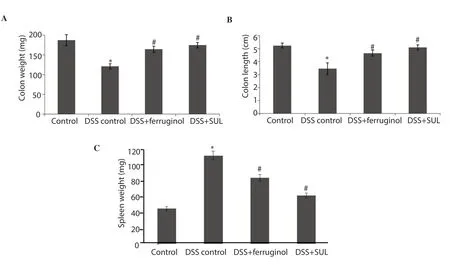
Figure 2.Effect of ferruginol on colon weight (A), colon length (B), and spleen weight (C).Values are given as mean ± SD for groups of 6 mice in each group.*P<0.05 when compared to the normal control, #P<0.05 when compared to the dextran sulfate sodium (DSS)-treated groups.SUL: sulfasalazine.
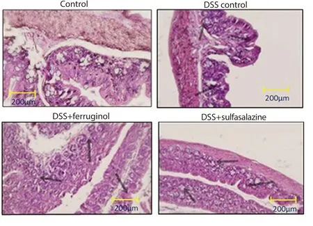
Figure 3.Effect of ferruginol on histological changes.Dextran sulfate sodium (DSS)-induced mice show ulceration and inflammatory cell filtration into the mucosa, which is restored by ferruginol and sulfasalazine.200× magnification.The black arrow marks indicate epithelium damages, ulceration, and leucocyte infiltration.
The significantly elevated levels of IL-6, TNF-α, and IL-1β were detected in DSS-treated mice compared with the normal control mice (P<0.05) (Figure 4B-D).The treatment of FGL significantly decreased the levels of IL-1β, TNF-α, and IL-6 (P<0.05), which was comparable to the effect of sulfasalazine.
3.5.Effect of FGL on the expression of inflammatory cytokines by Western blotting analysis
There were significantly elevated expressions of iNOS, NF-κB p65,COX-2, and MMP-9 proteins in DSS-treated mice compared with normal control mice (P<0.05).The treatment of FGL significantly decreased the levels of iNOS, NF-κB p65, COX-2, and MMP-9(P<0.05), which was comparable to the effect of sulfasalazine(Figure 5).
3.6.Effect of FGL on immunohistochemical analysis of iNOS and COX-2
The protein expressions of iNOS and COX-2 were notably increased in DSS-treated mice compared with the normal control mice; while the expressions of iNOS and COX-2 were decreased in FGL administrated animals (Figure 6).
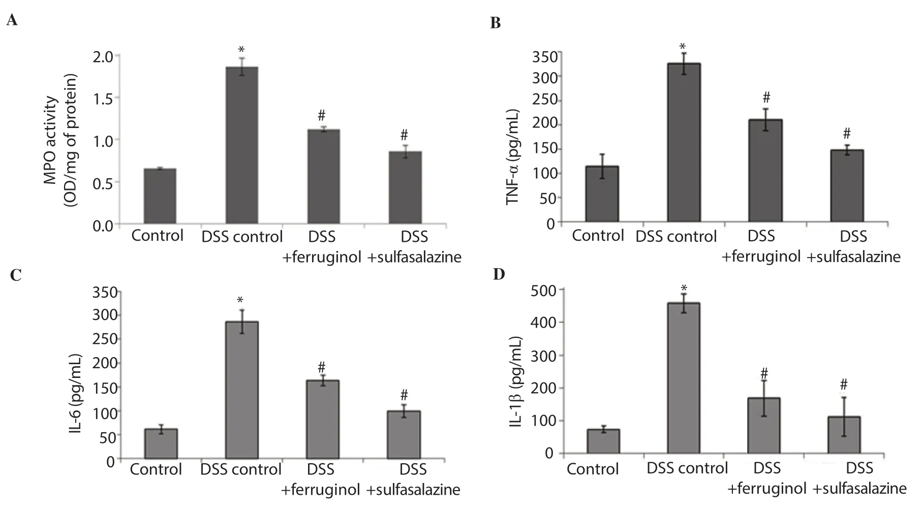
Figure 4.Effect of ferruginol on the myeloperoxidase (MPO) (A), tumor necrosis factor-α (TNF-α) (B), interleukin (IL)-6 (C), and IL-1β (D).Values are given as mean ± SD for groups of 6 mice in each group.*P<0.05 when compared to the normal control, #P<0.01 when compared to the dextran sulfate sodium(DSS)-treated groups.
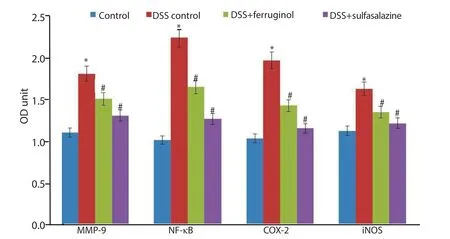
Figure 5.Effect of ferruginol on nuclear factor-κB (NF-κB) p65, inducible nitric oxide synthase (iNOS), cyclooxygenase (COX)-2 and matrix metalloproteinases (MMP)-9.Values are given as mean ± SD for groups of 6 mice in each group.*P<0.05 versus the normal control, #P<0.05 versus the dextran sulfate sodium (DSS)-treated groups.
4.Discussion
DSS results in mucosal ulceration, diarrhea, bodyweight loss, bloody stool and shortened colonic length[19].The decreased colon length and colon weight are the pathological and histological changes of ulcerative colitis, indicating the sternness of inflammatory bowel disease[20].These symptoms including diarrhea and bloody stool are attributed to the penetration of abdominal cells to the lumen caused by DSS[21].In our study, DSS-treated colitis mice displayed significant body weight loss, decreased colon length and colon weight, and higher DAI scores.Rajendiranet al.[22], Arecheet al.[23] and Rodriguezet al.[24]all described the link between DAI scores and pathophysiological alterations of DSS-induced chronic and acute ulcerative colitis.Our findings showed that FGL administration could significantly increase the body weight, colon weight, colon length, and decrease DAI score.MPO is a typical indicator of inflammatory condition, injury of tissues and penetration of neutrophils.The level of MPO in colonic tissues depends on the production of superoxide anion, which contributes to the mucosal dysfunction and tissue necrosis[25].In this study, we observed the level of MPO was significantly elevated in DSS-treated animals, while it was significantly reduced after the FGL treatment,indicting the anti-inflammatory potential of FGL.
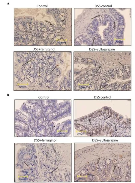
Figure 6.Immunohistochemical analysis of inducible nitric oxide synthase (iNOS) and cyclooxygenase-2 (COX-2).The images at 40× magnification revealed that the ferruginol effectively diminished the expressions of iNOS (A) and COX-2 (B) in dextran sulfate sodium (DSS)-treated mice.Note: The expressions of iNOS and COX-2 expressions are indicated by arrows.
NF-κB is considered as an essential mediator of inflammatory cytokines.It is involved in the pathologic process of ulcerative colitis,colon cancer, and inflammatory bowel disease[26,27], and can lead to the losing of colonic epithelial cells[28].The research of Kasinathanet al.[26] noted the elevated expression of NF-κB in DSS administered animals, which proved the induction of inflammation.Our study also displayed increased NF-κB p65 expression in DSS-treated mice and significantly decreased expressions after FGL treatment.The levels of pro-inflammatory cytokines namely TNF-α, IL-1β, and IL-6 are usually increased during the pathological process of colitis, and these inflammatory cytokines also regulate the initiation of colorectal cancer[29].The previous study reported that the expression of IL-6, IL-1β, and TNF-α was associated with NF-κB activation that regulates the inflammatory response and the duration of continual inflammation[30].In this view, the suppression of these inflammatory cytokines like IL-6,TNF-α, and IL-1β would be a possible and efficient treatment of colon cancer[31].Our study showed no NF-κB translocation to the nucleus in FGL treated colitis mice, which directly supported the finding of Rodríguezet al.[32] and proved the anti-inflammatory effect of FGL.
The COX-2 and iNOS are good indicators for colon cancer diagnosis in bothin vitroandin vivoexperiments[33].In our work,we also observed elevated expression of COX-2 and iNOS in DSS administered colitis mice, which is similar to the findings of Umesalma and Sudhandiran[34].The FGL treatment effectively attenuated the increased expression of iNOS and COX-2 as a gastroprotective agent[32].Some studies described the overexpression of MMP-9 in the lesions of the healthy mucosa, which was considered as an important indicator for the early diagnosis of colorectal cancer[35].It also described that the MMP-9 regulated the metastatic progression in colon carcinoma, expressed by macrophages, mast cells and neutrophils[36].Our study showed that the treatment with FGL significantly prevented the increased MMP-9 expression in DSS-treated colitis mice.In addition, the histopathological assessment of colon tissues exhibited severe ulceration and infiltration of leucocytes in DSS-induced colitis mice.But, the FGL treatment effectively reversed and ameliorated the mucosal sores, reduced the penetration of the inflammatory cells and histological alterations in mucosa induced by DSS.
In conclusion, the findings of this work proved that the administration of FGL efficiently inhibited the DSS-induced ulcerative colitis in mice and also ameliorated the severe inflammation by preventing the NF-κB pathway in colon cells.Besides, the effect of FGL is comparable with sulfasalazine.FGL could be an effective herbal-based compound for treating ulcerative colitis.
Conflict of interest statement
We declare that there is no conflict of interest.
Authors’ contributions
The original ideas, formulation of this research goals and aims were performed by MYD and CLZ.The verification of research data and the overall experimental results were performed by XYZ and YL.The manuscript was originally written by the XYZ, and review,editing and proof reading were performed by MYD, CLZ, and XYZ.
 Asian Pacific Journal of Tropical Biomedicine2020年7期
- Asian Pacific Journal of Tropical Biomedicine的其它文章
- Anticancer effect of Psidium guajava (Guava) leaf extracts against colorectal cancer through inhibition of angiogenesis
- Sesamum indicum (sesame) enhances NK anti-cancer activity, modulates Th1/Th2 balance, and suppresses macrophage inflammatory response
- Antibiofilm activity of alpha-mangostin loaded nanoparticles against Streptococcus mutans
- Anti-proliferative potential of sodium thiosulfate against HT 29 human colon cancer cells with augmented effect in the presence of mitochondrial electron transport chain inhibitors
