Correlation between interleukin 32 and familial clustering of hepatocellular carcinoma in an HCC-epidemic region of Guangxi, China
PENG Jinlin, WU Jizhou*, LI Guojian, WU Jianlin, XI Yumei, LI Xiaoqing, WANG Lei
(1. Department of Infectious Diseases, the First Affiliated Hospital,Guangxi Medical University, Nanning 530021, China; 2. College of Health and Rehabilitation, Chengdu University of Chinese Medicine, Chengdu 610075, China)
[Abstract]Objective: To investigate the association between the human serum level of interleukin-32 (IL-32) and the familial aggregation of hepatocellular carcinoma (HCC) in a HCC-epidemic region of Guangxi, China. Methods: A total of 230 individuals without liver cancer were enrolled, including 115 from families with high HCC risk (HCC family group) and 115 from cancer-free families (non-cancer family group). Enzyme-linked immunosorbent assay was used to determine serum IL-32 level in the peripheral blood of all subjects. IL-32 expression difference was compared between two groups from general serum IL-32 level and aspects of different demographic characteristics, namely, ethnicity, HBsAg status, gender and age. Thereafter, the relationship between IL-32 expression and familial aggregation of HCC in Guangxi was analyzed. Results: The overall IL-32 expression in the HCC family group was higher than that in the non-cancer family group (Z=-2.702, P=0.007). The serum IL-32 expression in males, HBsAg-negative carriers, family members below 25 years old, and the Yao population in the HCC family group was significantly higher than that in the non-cancer family group (P<0.05). In the HCC family group, no statistical differences were observed in IL-32 level among members with different blood kinship to the probands (P=0.429) and among members of families with different numbers of probands (P=0.298). The prevalence rate of HCC in males significantly increased compared with that in females. Conclusion: The increased IL-32 expression might be correlated with the familial aggregation of HCC in Guangxi, and its overexpression might be a susceptibility factor that contributes to the familial clustering of HCC in Guangxi, China.
[Key words]cytokine; interleukin-32(IL-32); hepatocellular carcinoma; familial aggregation
Human hepatocellular carcinoma (HCC) is one of the major malignancies worldwide[1], and the regions with the highest mortality rate of liver cancer are mostly located in China[2]. HCC incidence and death rate are remarkably high in Guangxi, China[3]. Hepatocarcinogenesis is the final result of a multistage and multifactorial pathological process, including the combination of environmental factors and genetic susceptibility. Epidemiologic studies reported that HCC familial aggregation frequently occurs in Guangxi[4]. The underlying mechanisms of HCC familial aggregation remain unclear, but previous studies have indicated that the inflammatory microenvironment surrounding a tumor plays a crucial role in modulating the carcinogenesis and progression of HCC[5-6].
Interleukin-32(IL-32), a novel type of interleukin, is generated mainly by natural killer cells, T cells, monocytes, and epithelial cells after activation[7-8]. IL-32 is considered as a pro-inflammatory cytokine contributing to HCC[9]. However, the correlation between IL-32 levels and the familial clustering of HCC has not been investigated yet. Consequently, the function of IL-32 in HCC familial aggregation remains unknown. We conducted the present case-control study to investigate the function of IL-32 in the familial clustering of liver cancer in Guangxi, China.
1 Materials and methods
1.1 Subjects
All participants enrolled in this study were rural residents in the regions of Guangxi province with a high prevalence of HCC. The study was conducted from January 2012 to December 2014. A total of 115 fourth-generation individuals associated with probands from 18 families with a high incidence of liver cancer were chosen as the HCC family group. Another 115 individuals were recruited from normal families without any history of malignancy and accordingly referred to be the non-cancer family group. All individuals of the non-cancer family group were paired with those from the HCC family group in terms of age, socio-economic background, gender, ethnicity and HBV infection. The present research was performed in accordance with the guideline of Ethics Committee of the First Affiliated Hospital of Guangxi Medical University (Nanning, China) with the reference number 2018 (KY-E-061).
1.2 Inclusion and exclusion criteria
Inclusion criteria: All liver cancer patients met the diagnostic criteria of liver cancer revised by the 4thNational Academic Conference on liver cancer. Pathological diagnosis was based on intrahepatic or extrahepatic pathological examination that confirmed primary liver cancer[10]. The probands were patients pathologically diagnosed with HCC in the First Affiliated Hospital of Guangxi Medical University.
Exclusion Criteria: All subjects were excluded from HAV, HCV, HDV, HEV, and other types of hepatitis; participants were further excluded when they presented thyroid disease, diabetes, kidney disease, cardiovascular disease, or diseases of other systems.
1.3 Blood sample collection and storage
The peripheral venous blood (3 mL) of each subject was collected in the early morning. All samples were required to congeal for 1 h at room temperature and then centrifuged at 3 000 r/min for 5 min to separate the upper serum (2 mL) with a pipette. Subsequently, the samples were placed in frozen pipes for dispensing, numbering, capping, and sealing, and then stored in a refrigerator at -80 ℃ for testing. Subject recruitment and serum specimen collection were performed only after written informed consent was obtained from all subjects.
1.4 Detection of IL-32 level by ELISA
The existence of IL-32 in each blood sample was detected using an enzyme-linked immunosorbent assay (ELISA) kit (Shanghai Enzyme-linked Biotechnology Co., Ltd., Shanghai, China) according to the manufacturer’s instructions.
Absorbance was measured using an iMark automatic microplate absorbance reader (Bio-Rad, CA, USA).
1.5 Statistical analysis
The skewed distribution of the data was represented by a median range [M(P25,P75)]. Wilcoxon rank sum test was conducted to examine the overall differences between the groups. Single factor analysis was performed to identify the statistical correlation between the expression of IL-32 and the familial clustering of liver cancer.Pvalues were two tailed, and differences in data were considered significant atP<0.05. Statistical data were analyzed using SPSS (v. 17.0).
2 Results
2.1 Characteristic information of the participants
The HCC family group included 62 males and 53 females, 50 cases were younger than or equal to 25 years old, whereas 65 cases were older than 25 years old, 26 and 89 cases were from the Yao and Zhuang populations, respectively, and 37 cases were HBsAg positive whereas 78 cases were HBsAg negative, respectively. The mean±SD age of the HCC family group was (31.548±17.270) years (range of 2~78 years). In terms of the genetic relationship with probands, the population in the HCC family group was further classified into first-degree (n=45), second-degree (n=22), third-degree (n=11), and fourth-degree (n=37) relatives of the probands. In terms of the number of probands, family members with a high incidence of liver cancer were further divided into 2~3 cases and 4-case subgroups. The (mean±SD) age of the non-cancer family group was (32.157±17.176) years (range of 4~80 years) (Table 1).

Table 1 Characteristic information of the participants
2.2 Comparison of serum IL-32 level between two groups
Significant differences were observed in the total IL-32 data between the HCC and non-cancer family groups (Z=-2.702,P=0.007), and the general presence of IL-32 was significantly higher in the HCC family group than that in the non-cancer family group (Table 2).

Table 2 Comparison of IL-32 data between two groups M(P25, P75)
2.3 Analysis of the general demographic characteristics of serum IL-32 level between two groups
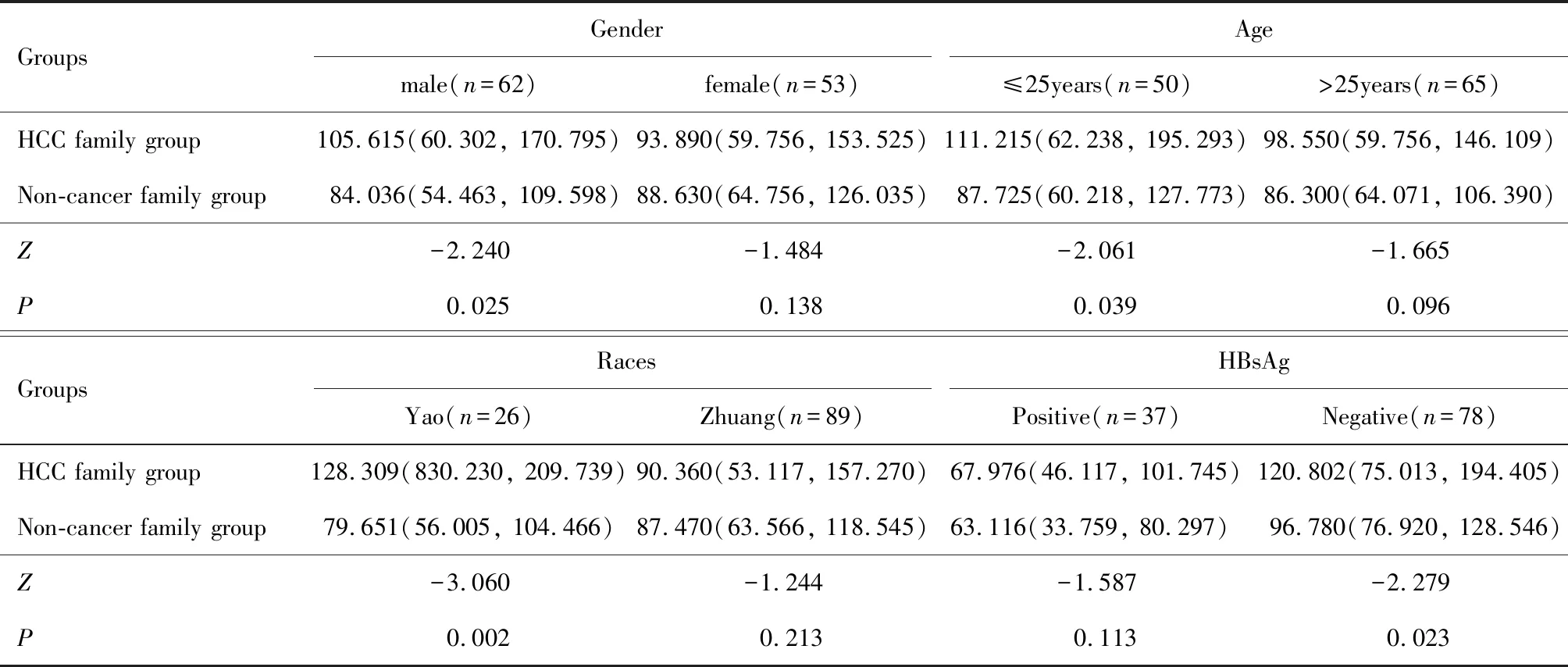
Further analysis revealed that the serum IL-32 expression in males in the HCC family group was significantly higher than that in the non-cancer family group (Z=-2.240,P=0.025). Moreover, the cases aged ≤ 25 years in the HCC family group showed remarkably higher IL-32 level than those in the non-cancer family group (Z=-2.061,P=0.039). The IL-32 expression of HBsAg-negative members of families was significantly different between the two groups (Z=-2.279,P=0.023). Meanwhile, the serum IL-32 levels of the Yao ethnic group were significantly different between the two groups (Z=-3.060,P=0.002)(Table 3).

Table 3 Analysis of the general demographic characteristics of serum IL-32 level between two groups M(P25, P75)/(ng·L-1)
2.4 Distribution of IL-32 data among the relatives of probands in HCC family group
Stratified analysis of the kinship study found no significant difference in the overall distribution of IL-32 data among the relatives of probands in the HCC family group (Z=2.769,P=0.429, Table 4).
2.5 Distribution of IL-32 level in members with different numbers of probands in HCC family group
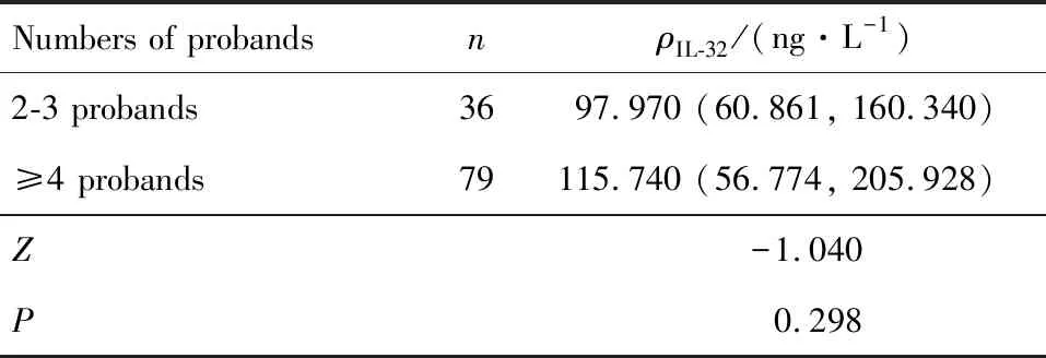
Statistical analysis of the serum IL-32 level in the HCC family members with different numbers of probands showed no marked difference (Z=-1.040,P=0.298, Table 5).
Table 4 Distribution of IL-32 data among blood relatives of probands in HCC family groupM(P25,P75)

GradesnρIL-32/(ng·L-1)First-degree relatives45105.710 (68.328, 169.540)Second-degree relatives2286.575 (49.344, 140.473)Third-degree relatives11119.413 (61.826, 262.105)Fourth-degree relatives3783.789 (51.892, 164.705)Z2.769P0.429
Table 5 Analysis of IL-32 levels in members with different numbers of probands in HCC family groupM(P25,P75)

Numbers of probandsnρIL-32/(ng·L-1)2-3 probands3697.970 (60.861, 160.340)≥4 probands79115.740 (56.774, 205.928)Z-1.040P0.298
2.6 Comparison of gender distribution of the probands in HCC family group
A total of 60 probands comprised 46 males and 14 females. The gender distribution of the probands was significantly different, and males were frequently associated with higher incidence rate of HCC than females (76.667% vs. 23.333%).
3 Discussion
Human HCC is a classic paradigm of inflammation-associated cancer. The inflammatory microenvironment serves as a crucial part in liver carcinogenesis, thereby modulating sustained initiation, promotion, and tumor metastasis by activating proliferation, dampening the acquired immunity, and evading the inherent immune surveillance and elimination. IL-32 is an inflammation-related cytokine in tumor microenvironment that contributes to the occurrence and progression of liver cancer[11]However, the association between IL-32 expression and familial aggregation of HCC has not been studied. In this study, the effect of IL-32 on the familial aggregation of HCC in a high-risk population of HCC were evaluated.
IL-32 is a multifunctional cytokine with a wide range of effects, such as controlling the balance between inflammation and anticancer immune response in cancer microenvironments[12]. This cytokine is also involved in the innate and adaptive immune systems of the host[13]. IL-32 is an important pro-inflammatory factor in human and implicated in cancer growth and invasion. Moreover, it is associated with the occurrence and development of various cancer types and has been discovered in liver[11], colon[14], lung[15], and thyroid cancers[16], demonstrating that it can accelerate tumor cell proliferation, angiogenesis, and tumor metastasis by locally inhibiting antitumor immune reactions[17].
Our preliminary data showed that the average IL-32 level in the HCC family group was significantly higher than that in the non-cancer family group. Thus, an increase in IL-32 expression may increase susceptibility of members in the HCC family group to liver cancer. Stratification analysis indicated that the serum IL-32 expression in HBsAg-negative carriers, family members aged ≤ 25 years, and the Yao population was upregulated in the HCC family group compared with those in the non-cancer family group. The results were consistent with previous findings that the rate of HCC in Yao population from HCC family group was relatively higher than that from the non-cancer family group in Guangxi[18]. These findings may be due to racial differences and genetic effects. IL-32 induces liver cancer[19], and its regulatory mechanism on tumor-promoting effects is complex. IL-32 can also increase the secretion of various pro-inflammatory cytokines and chemokines in stromal cells after the immune cells are stimulated[9, 20-21], a phenomenon that has a strong connection with tumor invasion and metastasis[22-23]. Previous studies also showed that IL-32α inhibits apoptosis by activating NFκB[24-25]and p38 MAPK[26]in HCC. IL-32α knockdown inhibits cell growth and induces intrinsic apoptosis by decreasing p38MAPK, NF-κB, and Bcl-2[9, 27], suggesting that the overexpression of IL-32 in family members with a high incidence of HCC may directly or indirectly interfere with the antitumor immune response of human body and may be explained as a depressed immune response.
Human HBV infection is strongly associated with the development of HCC worldwide[28]. In the present study, stratified analysis of the two groups demonstrated that the overall serum IL-32 level of HBsAg-negative members in the HCC family group significantly increased compared with that in the non-cancer family group (P<0.05). The IL-32 expression of HBsAg-positive members in the HCC family group was not significantly different from that in the non-cancer family group. Homeostasis of the immune microenvironment in the body is disrupted before the onset of carcinogenesis. This event suggests that genetic susceptibility may be related to liver cancer rather than to HBV infection. The increased IL-32 expression leading to the familial clustering of liver cancer does not directly correlate with the presence of HBV infection. The present study found no significant correlation between IL-32 level and genetic relationship with the probands or numbers of probands in the HCC family group or numbers of probands. We speculate that the occurrence of the familial aggregation of HCC in Guangxi may be unrelated to the genetic relationship with the probands and the number of probands in the family. However, our research should be repeated in a population with a larger sample size or subjected to an in-depth research on gene expression. A total of 60 cases of probands were randomly assigned in this study. HCC was more prevalent among males than females, and this observation was consistent with previous findings on gender distribution worldwide[29]. Thus, changes in cytokine levels in the immune microenvironment of males may be more remarkable than those of females.
In conclusion, our findings provided further evidence that increased IL-32 expression may be closely correlated with a high incidence of familial clustering of liver cancer in a Chinese population with a high risk of HCC in Guangxi province. The detailed molecular mechanism will be explored in our subsequent study.
Overall, this study has two limitations. First, this research is an observational analysis, which does not allow the generation of causal inferences. Second, the limited sample size of HBV-positive participants may result in selective bias. Therefore, further research with a large sample size should be performed to firmly establish the conclusions.

