In vitro anti-inflammatory, anti-oxidant and in vivo anti-arthritic properties of stem bark extracts from Nauclea pobeguinii (Rubiaceae) in rats
Tsafack Eric Gonzal, Djuichou Nguemnang Stephanie Flore, Atsamo Albert Donatien, Nana Yousseu William,Tadjoua Tchoumbou Herve, Matah Marthe Mba Vanessa, Mbiantcha Marius✉, Ateufack Gilbert✉
1Laboratory of Animal Physiology and Phytopharmacology, Department of Animal Biology, Faculty of Science, University of Dschang, P.O. Box 67,Dschang, Cameroon
2Laboratory of Animal Physiology, Faculty of Science, University of Yaounde I, PO Box 812, Yaoundé, Cameroon
ABSTRACT Objective: To explore the immunomodulatory, anti-inflammatory,anti-oxidant and anti-arthritic activity of aqueous and methanolic extracts of Nauclea pobeguinii stem bark.Methods: For in vitro assays, the production of reactive oxygen species (chemiluminescence technique), the proliferation of T cells (liquid scintillation counter method), as well as the inhibition of cyclooxygenase, lipoxygenase, protein denaturation, and free radicals [DPPH, ABTS and nitric oxide (NO) inhibition methods]were evaluated. For in vivo assays, a polyarthritis model was induced by complete Freund's adjuvant in rats. The aqueous and methanolic extracts of Nauclea pobeguinii stem bark were administered orally at 150 and 300 mg/kg. After 28 days of treatment, the total blood was taken to quantify the hematological parameters and the serum was used to evaluate the biochemical parameters(alanine aminotransferase, aspartate transaminase, phenylalnine ammonialyase, and proteins) and oxidative stress parameters(malondialdehyde, catalase, superoxide dismutase, glutathione and NO), and then the knee joint was removed for histological analysis.Results: The extracts of Nauclea pobeguinii significantly reduced the production of intra- and extracellular reactive oxygen species and decreased T cell proliferation. They had an inhibitory effect on cyclooxygenase, lipoxygenase, and protein denaturation, and both extracts had antioxidant capacity on DPPH, ABTS and NO.Both extracts alleviated joint inflammation and pain sensitivity after complete Freund's adjuvant injection, reduced alanine aminotransferase, aspartate transaminase, alkaline phosphatase,NO and malondialdehyde levels, increased protein concentration,superoxide dismutase, catalase and glutathione activity, and restored the cytoarchitecture of the joint after complete Freund's adjuvant injection.Conclusions: The aqueous and methanolic extracts of Nauclea pobeguinii have immunomodulatory, anti-inflammatory, anti-oxidant and anti-arthritic properties.
KEYWORDS: Nauclea pobeguinii; Immunomodulation; Antioxidant;
1. Introduction
As a physiological process to protect the body against external or internal aggression, inflammation could limit and repair damage,reduce the size of the lesions, and restore the homeostasis of damaged tissues. Nevertheless, when the inflammatory process is prolonged, it is very harmful to the body and can lead to the development of many inflammatory diseases including rheumatoid arthritis[1]. Inflammatory, systemic, autoimmune and multifactorial rheumatism involves endocrine, psychological, genetic and environmental factors that are particularly responsible for joint destruction, pain, destruction of bone and cartilage with consequent functional disability[2]. In the rheumatoid synovium, the activation of polymorphonuclear cells, macrophages, and synoviocytes leads to the excessive production of T cells and then triggers the inflammatory process, during which leukocytes and many proinflammatory cytokines (IL-1 beta and/or TNF alpha) are produced.These could lead to the production of proteolytic enzymes that are responsible for the damage to the joint such as destruction of cartilage tissue, chronic cell proliferation and bone resorption[2].
During the development of rheumatoid arthritis, the proteins in the articular cartilage can lose their structures (secondary and/or tertiary), thus cause the denaturation and the aggravation of the pathology[3]. Similarly, the phospholipids in the cartilage can be denatured by phospholipase A2 to produce arachidonic acid.This process takes place in a double way: the cyclooxygenase pathway with a production of thromboxane A2, prostacyclin, and prostaglandins E2(PGE2) and F2, as well as the lipooxygenase pathway with leukotriene production[4].
In the physiopathology mechanism of rheumatoid arthritis, reactive oxygen species (ROS) play a key role in chronic inflammation.Indeed, the onset of the phagocytosis process by polymorphonuclear neutrophils, which represent more than 90% of the cells in the synovial fluid during rheumatoid arthritis, requires an increase in oxygen consumption with the direct consequence of the production of several potentially toxic free radicals (oxygenated water,superoxide, radical hydroxyl), which are capable of destabilizing cell membranes promoting cytolysis[5]. This process can either lead to a significant decrease in anti-oxidant defense systems including the reduced glutathione (GSH) (non-enzymatic antioxidant), catalase(CAT) and superoxide dismutase (SOD) (enzymatic antioxidants)or a significant increase in malondialdehyde (MDA) (for lipid peroxidation rate)[6]. In addition, many liver disorders may occur during the development of rheumatoid arthritis, such as changes in some liver enzymes (transaminases, alkaline phosphatase, and gamma transferases)[7].
The debilitating and uncomfortable pathological status results in the cessation of work in more than 50% of affected people and this disease affects nearly 1% of the world’s population[8]. The knowledge of rheumatoid arthritis, its mediators as well as the molecular and cellular mechanisms involved in its pathophysiology has made it possible to develop multitude of therapeutic targets for its control and relief. Corticoids and nonsteroidal anti-inflammatory drugs are used for symptomatic treatments, while biotherapies and immunosuppressants are used in combination with diseasemodifying anti-rheumatic drugs. But these drugs have numerous undesirable effects such as heartburn, digestive bleeding, renal insufficiency, hypertension and even increased risk of microbial infection[9]. Besides, the costs of these treatments are high, so many scientists are turning to alternative treatments that are accessible and have no side effects.
For both developing countries and low-income populations in developed countries, plants are important sources of herbal treatment for many diseases. Traditional medicine remains the main recourse for approximately 85% of the African population[10]. The efficacy and safety of these plant species have become the subject of many pharmacological and clinical investigations. Belonging to the family Rubiaceae, Nauclea pobeguinii (N. pobeguinii) is a widespread plant in Cameroon, where it is widely used in traditional medicine to relieve stomach pains, fevers, joint pain, and even inflammation.Several studies have already been carried out on this plant and have shown that this plant has anti-diabetic[11], antibacterial[12],analgesics and anti-inflammatories activities[13]. In addition, Xu et al[14]showed the presence of flavonoids, tannins, terpenoids,alkaloids, and steroids in this plant in a phytochemical study. The effects of N. pobeguinii on polyarthritis, immunological changes and oxidative stress parameters related to polyarthritis have not yet been demonstrated. Thus, this study aims to evaluate anti-arthritic properties in vivo in polyarthritis rats induced by complete Freund’s adjuvant (CFA); and to evaluate in vitro immunomodulatory, antiinflammatory and antioxidant potential.
2. Materials and methods
2.1. Plant material and extraction
N. pobeguinii (fresh barks, leaves and flowers) was harvested in August 2016 in Mbalmayo, Department of Nyon and So’o,Central Cameroon region, and was then identified at the National Herbarium of Cameroon (Youndé) by simple comparison with an already existing sample [R. Letouzey sample No. 11,367 (28,335/SRFcam)]. After identification, the barks were dried in the shade(shelter from the sun) and ground (roping) to give a fine powder.Part of this powder (500 g) was soaked in distilled water (5 L), and the mixture was boiled for 30 min. The another part (500 g) was quenched in methanol (2.5 L), then the mixture was macerated for 72 h. At the end of each preparation, the two mixtures were filtered via filter paper. The aqueous filtration was evaporated in an oven at 40 ℃ while the methanol filtration was concentrated using a rotary evaporator at 65 ℃ (reduced pressure). This operation yielded 22 g of aqueous extract (4.4% yield) and 42 g of methanolic extract (8.4%yield).
2.2. Reagents, chemicals, and equipments
1,1-Diphenyl-2-picrylhydrazyl (DPPH) was procured from Sigma Aldrich (St. Louis, MO, USA). Sodium nitroprusside was purchased from Merck Ltd. India, Mumbai. Phosphoric acid, sodium hydroxide,sodium linoleate, sulfanilamide, sulfuric acid, linoleic acid, ascorbic acid, and arachidonic acid were purchased from S D Fine Chem.Ltd, Mumbai. Thiobarbituric acid, lipopolysaccharide, hemoglobin,ethylenediaminetetraacetic acid and bovine albumin were procured from Central Drug House Pvt. Ltd, New Delhi. Casein and trypsin were procured from Hi-Media Lab. Ltd, Mumbai. N-(1-Naphthyl)ethylenediamine dihydrochloride, potassium persulfate, potassium phosphate buffer, glutathione, and benzene were obtained from LOBA CHEME Pvt. Ltd. Mumbai. Phytohemagglutinin and RPMI 1640 medium were from HIMEDIA. CFA was purchased from Sigma Chemical Co. (St Louis, MO, USA), while diclofenac (Olfen-100 SR) was bought in a locally certified pharmacy. All the other chemicals and reagents were of pure analytical grade and obtained from the local suppliers.
2.3. Phytochemical assay of N. pobeguinii extracts
To determine the chemical compounds in the aqueous and methanolic extracts of N. pobeguinii, several tests were used based on visual observation of a formed precipitate and/or changes in the coloration of the solution after addition of one or more characteristic reagents. The presence of chemical compounds such as flavonoids,saponins, tannins, anthraquinones, terpenoids, steroids, and alkaloids was verified using the method described by Matos[15].
2.3.1. Tannins
A solution of FeCl3(a few drops) was mixed with 2 mL of each extract dissolved in 2 mL of distilled water, and the appearance of a green precipitate indicated the presence of tannins.
2.3.2. Saponins
The presence of saponins is generally demonstrated by the formation of a stable foam after stirring a solution. A solution of 5 mL of distilled water and 5 mL of each extract was vigorously shaken to check the presence of saponins.
2.3.3. Flavonoids
By mixing 1 mL of lead acetate solution (10%) with 1 mL of each extract, the appearance of a yellow precipitate indicated the presence of flavonoids.
2.3.4. Anthraquinone
The presence of free anthraquinones and/or anthraquinone derivatives in a mixture is indicated by a pink, violet or red coloration in the lower phase (ammoniacal phase) of the mixture after stirring. Thus, two methods were used:
(i) stirring a solution prepared in the following manner: extract (3 mL) and benzene (3 mL), then filtrating and adding 5 mL ammonia(10%) in the filtrate.
(ii) Bring a boiling mixture of extract (3 mL) and sulfuric acid (3 mL), filtering while hot and adding benzene (3 mL) to the filtrate followed by stirring. After separating the benzene layer, adding ammonia prepared at 10% (3 mL).
2.3.5. Terpenoids
A solution of chloroform (2 mL) and each extract (2 mL) was prepared and evaporated to dryness, then the concentrated sulfuric acid (2 mL) was added to the solution and the mixture was heated for 2 min. The appearance of a greyish color was characteristic of the presence of terpenoid.
2.3.6. Steroids
The presence of steroids was characteristic of a greenish color. A total of 2 mL of each extract was dissolved in 2 mL of chloroform,then acetic and sulfuric acids were added.
2.3.7. Alkaloids
A solution of 3 mL of each extract, 3 mL of HCl with the addition of Wagner’s reagents and Layers was stirred and the appearance of turbidity of the precipitate indicated the presence of alkaloids.
2.4. Anti-inflammatory effects of aqueous and methanolic extracts of N. pobeguinii
2.4.1. Inhibition of protein denaturation
To evaluate the anti-inflammatory properties of N. pobeguinii extract, the method previously described by Padmanabhan and Jangle[16]with small modifications was used. Briefly, 1 mL of different extracts or diclofenac sodium of different concentrations(100, 200, 500 and 1 000 µg/mL) was mixed with 5% bovine serum albumin solution (1 mL), then the mixture was incubated 27 ℃ for 1 h. The control tube consisted of a mixture of distilled water and bull serum albumin. Denaturation of the proteins was initiated by placing the mixture in water for 10 min at 70 ℃. After cooling the solution to room temperature, the activity of each mixture was measured at 660 nm. Each test was performed in three repetitions. The formula below was used to calculate the percent inhibition.

2.4.2. Assay of cyclooxygenase and 5-lipoxygenase inhibition
To grow human peripheral lymphocytes, RPMI 1640 medium(HIMEDIA) supplemented with heat-inactivated fetal calf serum(20%) and antibiotics (penicillin and streptomycin) were used. Cell proliferation was stimulated by phytohemagglutinin (HIMEDIA).Under aseptic conditions, after filtration of the culture (cellulose acetate filter, 0.2 µm of the pore), it was incubated for 72 h and activated after 24 h by addition of lipopolysaccharide (1 µL), then incubated again for 24 h. Then, aqueous and methanolic extracts, as well as ibuprofen at 100, 200, 500 and 1 000 µg/mL, were added and incubated for 24 h. After centrifugation at 6 000 rpm for 10 min, the supernatant was isolated, then 50 µL of cell lysis buffer was added followed by further centrifugation at 6 000 rpm for 10 min. The supernatant was isolated and the inflammation test was performed as a pellet suspended in a small amount of supernatant[17].
Tris-HCl, glutathione, hemoglobin and an enzyme were used to prepare the mixing solution. After the addition of the arachidonic acid in this mixture followed by incubation at 37 ℃ for 20 min,0.2 mL of 10% trichloroacetic acid in 1 N HCl and 0.2 mL of thiobarbituric acid were added to the solution, and heated in a boiling water bath for 20 min, and then centrifuged at 1 000 rpm for 3 min. The supernatant was used to evaluate cyclooxygenase activity at 632 nm[17].
Linoleic acid (70 mg) and an equal weight of interpolation were dissolved in 4 mL of oxygen-free water and pipetted. Sodium hydroxide (0.5 N) and water without oxygen (25 mL) were added, then the solution was divided into 0.5 mL portions, rinsed with nitrogen and stored in ice until further use. A quartz cuvette at 25 ℃ was used for the reaction. Tris buffer (2.75 mL, pH 7.4), sodium linoleate (0.2 mL) and enzyme (50 mL) constituted the assay mixture. Optical density was measured at 234 nm[17]. To calculate the percent inhibition, the following formula was used:

2.5. Antioxidant activity
2.5.1. DPPH radical scavenging activity
To determine the DPPH radical scavenging activity of aqueous and methanolic extracts of N. pobeguinii, the method described by Brand-Williams et al.[18]was used with some modifications. DPPH (24 mg)dissolved in 100 mL of methanol was used as the stock solution.The working solution was obtained by diluting the stock solution with methanol until an absorbance of (0.98 ± 0.02) at 517 nm was obtained. The working solution of DPPH (3 mL) was mixed with 100 µL of each extract (1 mg/mL) or standard in a test tube, while the control contained only 100 µL of methanol. The absorbance was measured for 30 min at 517 nm. The following formula was used to calculate the antioxidant activity:

Where, Asand Acare the absorbance of the sample and the control,respectively.
2.5.2. ABTS·+discoloration test
The ABTS·+bleaching test was performed using the procedure described by Re et al[19]with some modifications. For this, 9.5 mL of ABTS reagent (7 mmol/L) was prepared with 245 µL of potassium persulfate (100 mmol/L), then the volume was supplemented to 10 mL with distilled water. The solution was diluted with potassium phosphate buffer (0.1 mol/L, pH 7.4) to an absorbance of (0.70 ±0.02) at 734 nm after being kept in the dark at room temperature for 18 h, then samples of each extract were prepared in methanol with dilutions at 0.1, 0.25, 0.5 and 1 mg/mL. A sample (10 µL) was placed in a test tube and thoroughly mixed with 2.99 mL of ABTS radical working solution. The absorbance of the resulting clear mixture was recorded at 734 nm. The percentage of antioxidant activity was determined using the following formula:% Antioxidant activity = [(Ac-As)/Ac] ×100
Where Asand Acare the absorbance of the sample and the control,respectively.
2.5.3. Nitric oxide (NO) trapping test
Sodium nitroprusside (0.5 mL, 5 mmol/L, pH 7.4) was combined with the test compounds (0.1, 0.25, 0.5 and 1 mg/mL), ascorbic acid or an equivalent amount of methanol and incubated for 180 min at 25 ℃. The Griess reagent (1.5 mL) consisting of sulfanilamide(1%), phosphoric acid (2%) and N-(1-naphthyl)ethylenediamine dihydrochloride (0.1%) was added and the mixture was again incubated for 30 min. The absorbance was measured at 546 nm and the percentage of activity was determined relative to the standard[20].The activity was expressed as percent inhibition using the following equation:
2.6. Immunomodulatory effects of aqueous and methanolic extracts of N. pobeguinii
2.6.1. Isolation of polymorphonuclear neutrophil
2.6.2. Isolation of macrophages on mice
To isolate macrophages in Naval Medical Research Institute mice,the technique previously described by Mbiantcha et al[21]was used. Three adult mice (20-25 g, 4 months) were immunized by intraperitoneal injection of fetal calf serum (1 mL). These animals were observed for 72 h and killed by cervical dislocation. The body of each mouse was soaked in ethanol (70%) for decontamination,then RPMI (Roswell Park Memorial Institute Institute) (10 mL,10%) was injected into the peritoneal cavity, followed by a massage of the peritoneal cavity for 2 min. The abdominal skin was cut to expose the peritoneal cavity. A sterile syringe was introduced into the peritoneum of each mouse and RPMI-containing macrophages were collected and introduced into a dry sterile tube and then centrifuged at 400 g for 20 min at 4 ℃. Then, the supernatant was removed,5 mL of RPMI was reintroduced into the tube and the tube was centrifuged at 300 g for 10 min at 4 ℃. The supernatant was again decanted, 1 mL of RPMI/HBSS (1 mL) was added to the tube and then the tube was kept in ice. The viability of the cells was verified using the trypan blue exclusion method and the cells were counted using a hemocytometer. For the different tests, a cell concentration of 1×10⁶cells/mL was used.

2.6.3. Determination of production of ROS
The production of ROS was evaluated according to the method previously described by Mbiantcha et al[21]with some adjustments.Plates (96 wells) were used for this experiment. A total of 25µL of each extract (3.1 to 100 µg/mL) or ibuprofen were mixed(same volume) with whole blood, polymorphonuclear neutrophils(1×10⁶cells/mL) or macrophages (2×10⁶cells/mL) diluted in HBSS++(1:50), while control wells with only HBSS++, blood or polymorphonuclear neutrophils or macrophages. The plate was incubated at 37 ℃ for 30 min in the thermostatic chamber of the luminometer. Then, 25 µL of zymosan/phorbol myristate acetate and/or 25 µL of luminol/lucigenin (7×10⁵mol/L) were added. In this test, the results were obtained in relative light units (RLU). The following formula was used to calculate the percent inhibition (%) of each extract[21]:

2.6.4. T cell proliferation assay
A volume of 50 µL of each extract (2, 10 and 50 µg/mL) or prednisolone diluted in RPMI (5%) were introduced into plates(96 wells), then the isolated T cells (50 µL, 2×10⁶cells/mL) and phytohemagglutin (PHA)-L (PHAL) (50 µL, 7.5 µg/mL) were added. Negative control wells received only certain cells (50 µL) and RPMI (5%, 150 µL), while positive control wells received cells (50µL), PHA (50 µL) and RPMI (5%, 100 µL). After incubating for 72 h at 37 ℃ in a CO2incubator (5%), the culture was stimulated with 25 µL of 0.5 µCi/well (methyl 7 3H) thymidine solution. Then the plates were incubated for 18 h and the cells were harvested using a filter (fiberglass). The level of thymidine integrated into the cells was determined using an LS65000 liquid counter. Inhibition percentage was determined using the number per minute (CPM) of each well[21]:

2.6.5. Cytotoxicity assay
26.Ring this bell, and what she wanted would appear: The magical castle, with its invisible servants, appears in Cupid and Psyche as well as Beauty and the Beast. Psyche receives the omnipresent service since she is in the home of a god, Cupid, with the divine powers associated with a mythological81 god. The other heroines live in a home of enchantment where every physical desire is met. They have moved from poverty to complete luxury. Supposedly they should be content and feel no more want, but they also know there is more to life than physical luxury.Return to place in story.
The cytotoxicity of the aqueous and methanolic extracts was evaluated using the in vitro assay method described by Mbiantcha et al[21]. Briefly, 100 µL of cell (Hep G2 cells ) suspension (6×10⁴cells/mL) were incubated for 24 h at 37 ℃, 5% CO2in 96-well plates.The supernatant was removed in each well, then each extract (3.1 to 100 µg/mL) and complete DMEM were added to each well to give a final volume of 200 µL. The standard well contained cells (100µL) and complete DMEM, while 2 µL of triton×100 (0.5%) was added to the negative control well. The supernatant was transferred after incubation for 48 h at 37 ℃ in a CO2incubator and 50 µL of MTT (0.5 mg/mL) diluted in PBS (5 mg/mL) were completed in each well. Then the plates were incubated for 4 h, the MTT was carefully inhaled, 100 µL of DMSO was added as stirring for 10 to 15 min in an orbital shaker. The spectrophotometer was used and the absorbance was increased to 540 nm. The percentage inhibition of cell viability was obtained using the following formula[21]:

2.7. In vivo assay
2.7.1. Animal
Wistar rats that weighed between 160 and 200 g (3-4 months) and were raised under normal conditions (temperature 19-23 ℃, brightness 12 h) at the Animal Physiology and Animal Physiology Research Unit Phytopharmacology of the Department of Biology of the University of Dschang (Cameroon) were used. These animals were housed in cages with six animals per cage.
2.7.2. Induction of rheumatoid arthritis
The animals were selected and grouped into 7 groups with 6 rats in each group according to their weight. The doses were based on our previous work[13]. After acclimation for 3 days, the animals were anesthetized with ether vapor, and the polyarthritis was induced by a double injection of CFA (10 mg/mL, Sigma Chemical Co. St Louis,MO, USA) (100 µL on day 1 and 50 µL on day 2) in the caudal vein except group 1 (normal control). The rats were distributed as follows: group 2: negative control (CFA + NaCl), group 3: positive control (CFA + Diclofenac 5 mg/kg), group 4: treated (CFA +aqueous extract at 150 mg/kg), group 5: treated (CFA + aqueous extract at 300 mg/kg), group 6: treated (CFA + methanol extract at 150 mg/kg), group 7: treated (CFA + methanol extract at 300 mg/kg). The different treatments were administered orally from the 14th day after the first CFA injection, then the treatment continued until the 28th day[22].
2.7.3. Measurement of joint diameter, mechanical hyperalgesia,body weight, and arthritic score
The diameter of the joints was measured with Digital Vernier caliper (Mitutoyo, Japan) on day 0 before the injection of CFA which served as the base value, days 3, 5, 7, 9, 11, 13, 15, 17, 19, 21,23, 25 and 27 after the first injection of CFA[22].
Hyperalgesia was measured with an algesimeter (UGO Basil, Italy)on day 0 before CFA injection and on days 2, 4, 6, 8, 10, 12, 14, 16,18, 20, 22, 24, 26 and 28 after the first CFA injection[22].
The arthritic score was assessed by the number of infected legs,based on leg inflammation, leg distortion and the animal’s inability to use the legs on days 0, 7, 14, 21 and 28, while body weight was measured daily using a scale[21].
2.7.4. Weight of the organs, hematological parameters,biochemical parameters, and oxidative stress
At the end of treatment, each rat was anesthetized by intraperitoneal injection of thiopental (50 mg/kg) and euthanized. The blood was removed by catheterization of the abdominal artery and introduced into two tubes. Blood in tube contained ethylenediaminetetraacetic acid as an anticoagulant was used to evaluate the hematological parameters (red blood cells, globules white, hemoglobin, hematocrit,platelets) by the usual standardized laboratory method[21]. Then blood in a second tube without anticoagulant was centrifuged at 2 500 rpm for 15 min, the serum was taken for the evaluation of biochemical parameters such as transaminases [alanine aminotransferase (ALT), aspartate transaminase (AST)], total proteins, creatinine, alkaline phosphatase[21]and oxidative stress parameters (NO, MDA, CAT, SOD and GSH)[23]. The organs (liver,kidneys, spleen, lungs, and thymus) were removed, cleaned with 0.9% NaCl, dehydrated and weighed with a sensitive balance.
2.7.5. Histological analysis
The joints were gently removed, stripped of all fat, and fixed with PBS-buffered formalin (10%) for histological analysis. This analysis was performed according to the standard histology method using color staining (hematoxylin and eosin). The section was observed using a microscope (Scientico STM-50) equipped with digital camera (Celestron 44421) connected to a computer (Toshiba Tecra A9). The digital microscope software (Suit 2.0) was used for taking microphographs with a magnification (×100).
2.8. Statistical analyses
All data of in vitro tests were expressed as mean ± standard deviation in triplicate, whereas for the in vivo test, the data were presented as mean ± SEM. The differences between the groups were evaluated by ANOVA (unidirectional and bidirectional) followed by the Bonferroni test, the significant differences were considered at P<0.05.
2.9. Ethical statement
Experimental procedures were in accordance with NIH publication Nos.85-23 on “Laboratory Animal Care Principles” and approved by the Laboratory Committee (Laboratory of Animal Physiology and Phytopharmacology, Department of Animal Biology, University of Dschang, Cameroon) (Protocol No. 1209004). The experiments also conformed to the guidelines for the study of pain in awake animals established by the International Association for the Study of Pain,Ministry of Scientific Research and Technology, which has adopted the European Union on Animal Care and Experimentation (CEE Council 86/609) guidelines.
For the donation of human blood samples, all processes were accepted by an independent ethics committee, ICCBS, University of Karachi (N°:ICCBS/IEC-008-BC-2015/Protocol/1.0). The blood donors provided informed approval for the use in this study.
3. Results
3.1. Phytochemical screening
Phytochemical test indicated that both extracts of N. pobeguinii were very rich in secondary metabolites, including flavonoids,tannins, saponins, steroids, and alkaloids, while terpenoids were present only in the methanolic extract.
3.2. Immunomodulatory effects of aqueous and methanolic extracts of N. pobeguinii
For intracellular ROS, the extracts of N. pobeguinii stem bark inhibited the production of intracellular ROS in whole blood(IC50= 6.57 µg/mL for aqueous extract and IC50= 3.73 µg/mL for methanolic extract), polymorphonuclear neutrophils (IC50= 4.27µg/mL for aqueous extract and IC50= 3.27 µg/mL for methanolic extract) and macrophages (IC50= 6.10 µg/mL for aqueous extract and IC50= 4.61 µg/mL for methanolic extract). Concerning the extracellular ROS, the aqueous extract inhibited their production with IC50of 6.42 µg/mL (whole blood), 5.51 µg/mL (polymorphonuclear neutrophils) and 5.11 µg/mL (mouse peritoneal macrophages), while methanol extract with IC50of 3.86 µg/mL (whole blood), 3.15 µg/mL(polymorphonuclear neutrophils) and 3.74 µg/mL (mouse peritoneal macrophages).
Concerning the proliferation of T cells, the methanolic extract, the aqueous extract and prednisolone (reference substance) showed an antiproliferative activity with IC50of 1.15, 4.10 and <3.10 µg/mL,respectively.
The aqueous and methanolic extracts of N. pobeguinii did not show
any toxic effect on the cells as compared to the cyclohexamide (0.1µg/mL). Aqueous extract had IC50greater than 100 µg/mL, while the methanolic extract had IC50of 80.54 µg/mL.
3.3. Antioxidant effects of aqueous and methanolic extracts of N. pobeguinii
Table 1 shows that these extracts had a remarkable antioxidant effect. DPPH assay result revealed that aqueous and methanolic extracts at 1 000 µg/mL had maximum inhibition percentages of 91.01% and 78.80%, respectively. With regard to the ABTS discoloration test, the aqueous extract at 1 000 µg/mL had a maximum percentage inhibition of 82.39%, while the methanolic extract exhibited a maximum inhibition percentage of 79.08%.For the NO inhibition test, the maximum inhibition percentages of 94.66% (aqueous extract) and 88.31% (methanolic extract) were obtained at a concentration of 1 000 µg/mL.
3.4. Anti-inflammatory effects of aqueous and methanolic extracts of N. pobeguinii
Table 2 shows the effect of aqueous and methanolic extracts of N.pobeguinii on protein denaturation, inhibition of cyclooxygenase and inhibition of 5-lipoxygenase. The extracts of N. pobeguinii, as well as diclofenac, inhibited the denaturation of proteins in a concentrationdependent manner. Diclofenac, aqueous and methanolic extracts at 1 000 µg/mL had maximum inhibition of 89.19%, 80.13%,and 60.39%, respectively. For cyclooxygenase, the aqueous and methanolic extracts as well as ibuprofen at 1 000 µg/mL exhibited a maximum percentage inhibition of 91.14%, 70.23%, and 97.88%,respectively. With respect to 5-lipoxygenase, the aqueous and methanolic extracts of N. pobeguinii as well as ibuprofen showed a maximum inhibition of 94.56%, 78.17% and 95.31%, respectively.
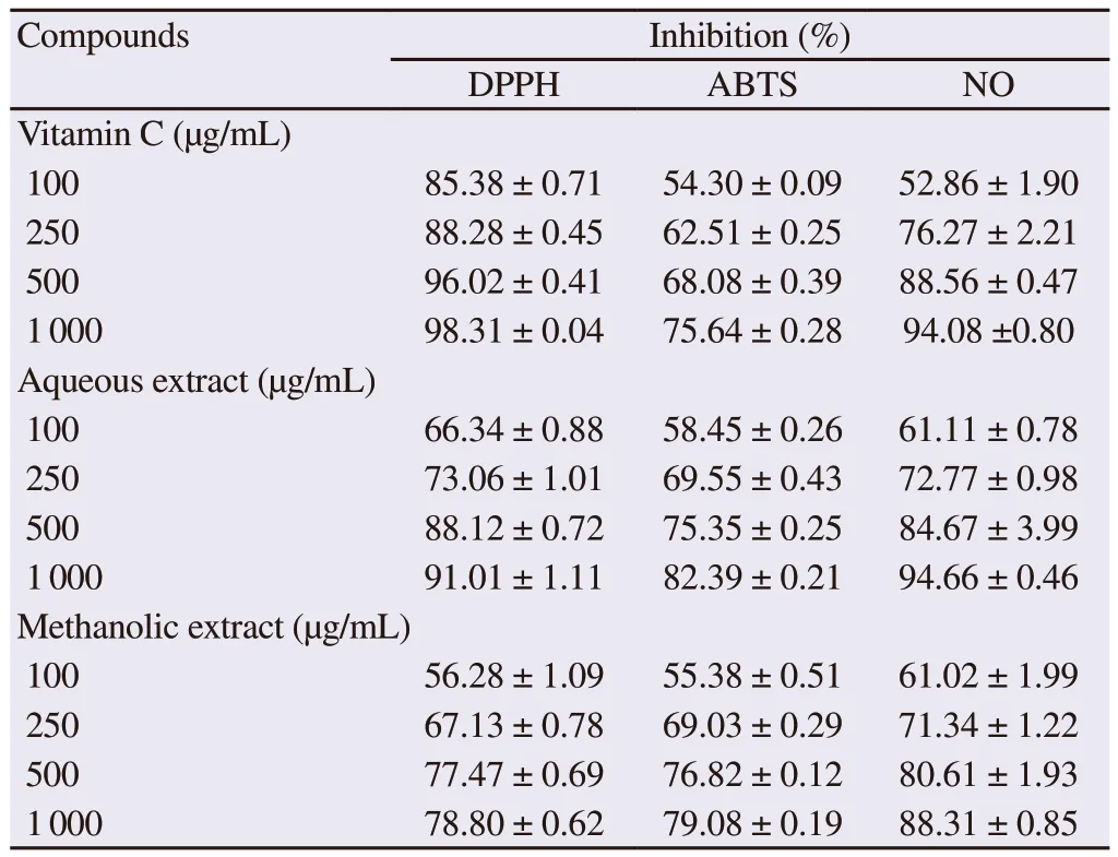
Table 1. In vitro antioxidant property of aqueous and methanolic extracts of Nauclea pobeguinii.
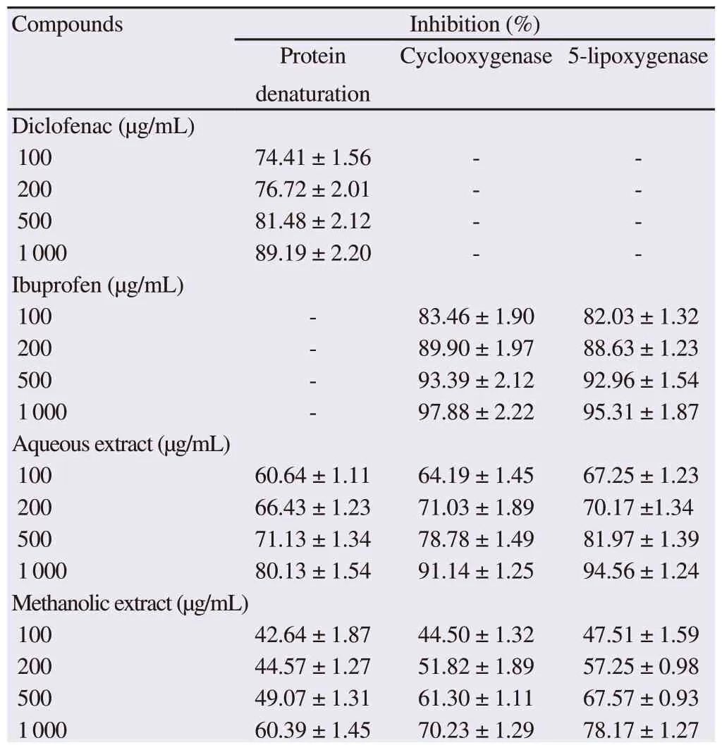
In vitro antiinflammatory property of aqueous and methanolic extracts of Nauclea pobeguinii.
The percentage values were obtained using various concentrations of test compounds and readings are presented as mean ± SD of triplicates.
3.5. In vivo test results
3.5.1. Effect of aqueous and methanolic extracts of N.pobeguinii on the joint diameter
Figure 1A shows the effect of stem bark extracts of N. pobeguinii on the change in joint diameter. The injection of CFA significantly increased (P<0.01) the diameter of the joint from the 9th day after the administration compared to the normal control. Oral administration of the aqueous and methanolic extracts of N. pobeguinii as well as diclofenac decreased significantly the diameter of the joint from the first administration until the 28th day. Aqueous extract at 300 mg/kg on the 27th day showed the maximum inhibition as 68.39%,while the methanolic extract at 300 mg/kg on the 19th day showed 48.92%. Moreover, the maximum effect of diclofenac (40.20%) was observed on the 17th day.
3.5.2. Effect of aqueous and methanolic extracts of N.pobeguinii on mechanical hyperalgesia
The CFA injection significantly reduced (P<0.001) the pain threshold assessed with the analgesimeter in all groups from the 6th day after CFA injection. Treatment with N. pobeguinii extracts at different doses significantly increased (P<0.05) the pain threshold(Figure 1B). This activity reached a maximal level on the 28th day by aqueous extract at 300 mg/kg (75.00%) and the 24th day by the methanolic extract at 300 mg/kg (58.91%). Diclofenac used at a dose of 5 mg/kg induced a maximum inhibition on day 26 (38.80%).
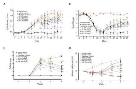
Figure 1. Effect of aqueous (AE) and methanolic (ME) extracts of Nauclea pobeguinii on joint diameter (A), mechanical hyperalgesia (B), arthritis score (C)and body weight (D) in complete Freund's adjuvant-induced arthritis. Values are expressed as mean± SEM for six animals and analyses by two-way ANOVA(One-way analysis of variance) followed by Bonferroni post-hoc test, αP<0.05; βP<0.01, λP<0.001 when compared to the normal control, aP<0.05, bP<0.01,cP<0.001 when compared to the arthritis control group.

Table 3. Effect of aqueous and methanolic extracts of Nauclea pobeguinii on organ weight.
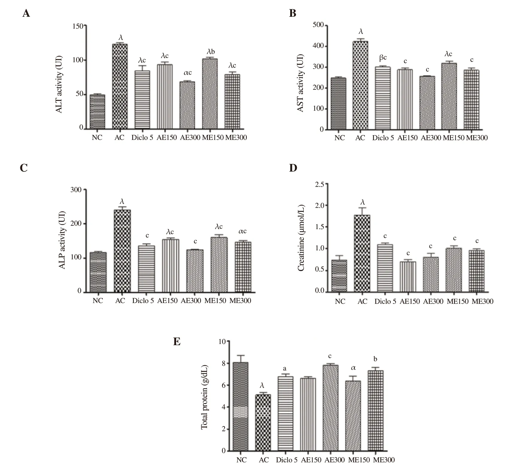
Figure 2. Effect of aqueous and methanolic extracts of Nauclea pobeguinii on serum parameters. Values are expressed as mean ± SEM for six animals and analyses by one-way ANOVA followed by Bonferroni post-hoc test, αP<0.05, βP<0.01, λP<0.001 when compared to the normal control, aP<0.05, bP<0.01,cP<0.001 when compared to the arthritis control. ALT: Alanine aminotransferase; AST: Aspartate aminotransferase; ALP: Alkaline phosphatase; NC: Normal control; AC: Arthritis control; Diclo: Diclofenac; AE 150: Aqueous extract (150 mg/kg); AE 300: Aqueous extract (300 mg/kg); ME 150: Methanolic extract (150 mg/kg); ME 300: Methanolic extract (300 mg/kg).

Table 4. Effect of aqueous and methanolic extracts of Nauclea pobeguinii on hematological parameters.
3.5.3. Effect of aqueous and methanolic extracts of N.pobeguinii on arthritis score
The arthritic score represented by the number of affected limbs increased significantly (P<0.001) in all groups compared to the normal group two weeks after CFA injection. Both extracts of N.pobeguinii significantly decreased (P<0.05) the arthritic score in treated animals one week after treatment. Both extracts at 300 mg/kg showed greater effects than that of diclofenac (5 mg/kg) (Figure 1C).
3.5.4. Effect of aqueous and methanolic extracts of N.pobeguinii on the body weight and organ weight
Figure 1D shows the effect of different treatments on the body weight 4 weeks after CFA injection. In arthritis animals, body weight decreased gradually and the decrease became significant (P<0.01)from the 1st week compared to the normal control. Aqueous and methanolic extracts (300 and 150 mg/kg) and diclofenac (5 mg/kg)had an increase in body weight from week 2, and this increase was significant the in the 4th week.
Table 3 shows the effect of the extracts on the weight of the organs.CFA administration significantly increased the weights of liver(P<0.01), kidney (P<0.001), spleen (P<0.001) and significantly reduced the weights of thymus (P<0.01) and lungs (P<0.01).Oral administration of the aqueous and methanolic extracts of N.pobeguinii significantly improved the weights of these organs.
3.5.5. Effect of aqueous and methanolic extracts of N.pobeguinii on hematological parameters
Table 4 shows the effect of different treatments on hematological parameters 28 days after the CFA administration. CFA significantly elevated levels of white blood cells (P<0.001), platelets (P<0.001),and significantly reduced levels of red blood cells (P<0.05) and hematocrit (P<0.001) compared with animals of the normal control group. Oral administration of the extracts significantly reduced the number of leukocytes and platelets and significantly increased red blood cell and hematocrit levels. The effect of the extracts at 300 mg/kg was more significant than that of diclofenac (5 mg/kg).
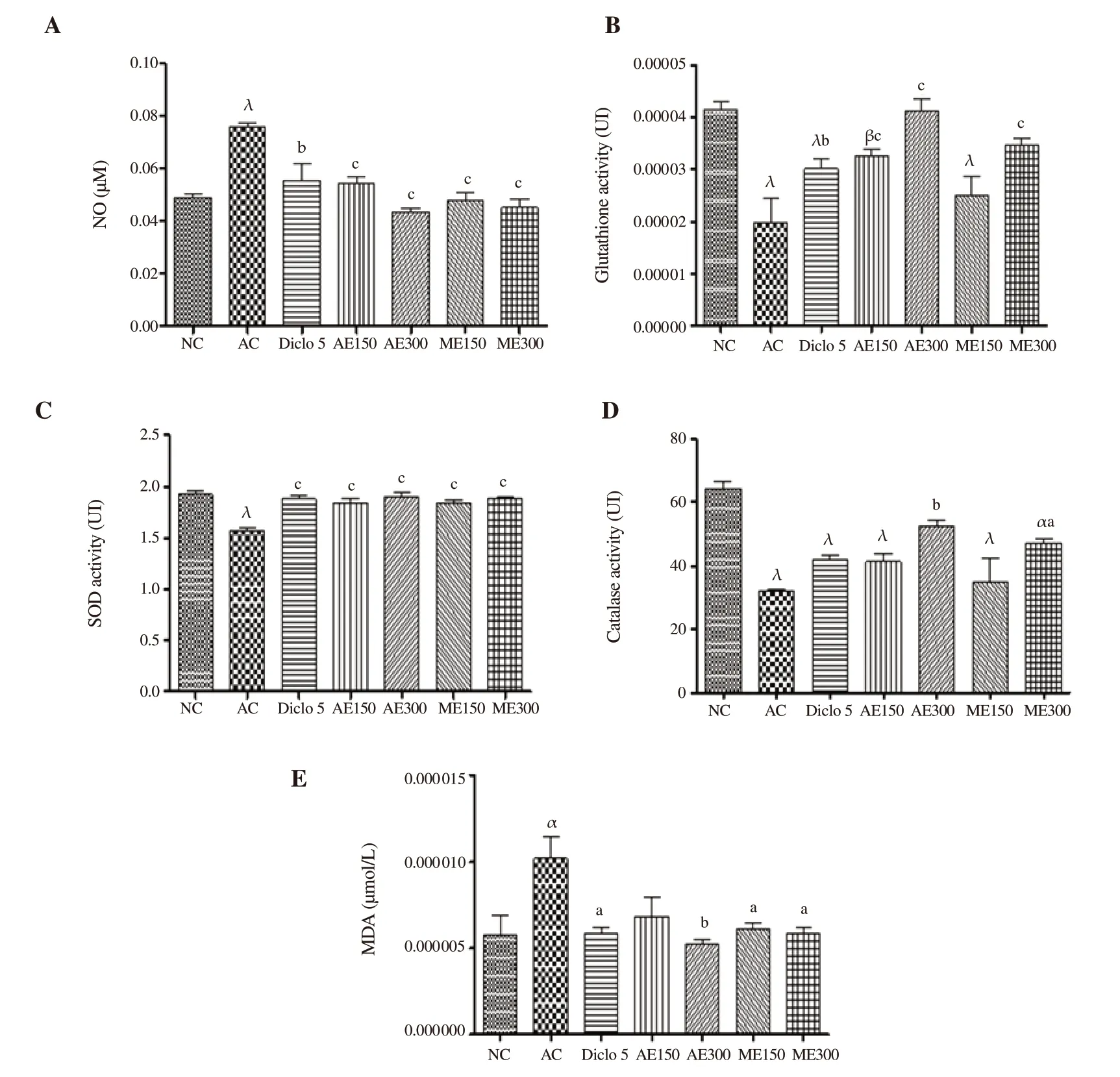
Figure 3. Effect of aqueous and methanolic extracts of Nauclea pobeguinii on some parameters of oxidative stress. Values are expressed as mean ± SEM for six animals and analyses by one-way ANOVA followed by Bonferroni post-hoc test, αP<0.05, βP<0.01, λP<0.001 when compared to normal control, aP<0.05,bP<0.01, cP<0.001 when compared to arthritis control. NO: nitric oxide; SOD: superoxide dismutase; MDA: malondialdehyde; GSH: glutathione; NC: Normal control; AC: Arthritis control; Diclo: Diclofenac; AE 150: Aqueous extract (150 mg/kg); AE 300: Aqueous extract (300 mg/kg); ME 150: Methanolic extract (150 mg/kg); ME 300: Methanolic extract (300 mg/kg).
3.5.6. Effect of aqueous and methanolic extracts of N.pobeguinii on biochemical parameters
Arthritic group significantly elevated serum ALT (P<0.001) (Figure 2A), AST (P<0.001) (Figure 2B), ALP (P<0.001) (Figure 2C)and creatinine (P<0.001) (Figure 2D), and significantly decreased the level of proteins (P<0.001) (Figure 2E). Both extracts of N.pobeguinii significantly reduced ALT, AST, ALP, and creatinine levels and significantly increased total protein level. Extracts at 300 mg/kg showed greater effects than diclofenac on all evaluated parameters.
3.5.7. Effect of aqueous and methanolic extracts of N.pobeguinii on oxidative stress parameters
The effect of aqueous and methanolic extracts of N. pobeguinii on the oxidative stress is illustrated by Figure 3. Th extracts and diclofenac markedly reduced (P<0.05) NO and MDA activities,and significantly increased (P<0.05) CAT, SOD and GSH activities compared to arthritic control group.
3.5.8. Effect of aqueous and methanolic extracts on histopathology
The histopathology of the joints in the normal control (Figure 4A)showed cartilage and intact bones without infiltration of leucocytes;while arthritic control (Figure 4B) showed infiltration of leucocytes,bone destruction, and cartilage. Animals treated with diclofenac (5 mg/kg) (Figure 4C), aqueous extract (150 and 300 mg/kg) (Figures 4D, 4E) and methanolic extract (150 and 300 mg/kg) (Figures 4F,4G) showed a significant reduction in bone erosion, leukocyte infiltration and destruction of articular cartilage.
4. Discussion
This study aims to evaluate the effects of aqueous and methanolic extracts of N. pobeguinii via in vitro and in vivo studies. The in vitro results show that the extracts of N. pobeguinii possess antiinflammatory (inhibition of the denaturation of proteins, the activity of cyclooxygenase, the activity of 5-lipoxygenase, the proliferation of T lymphocytes, production of intracellular and extracellular ROS) and antioxidants properties (DPPH, ABTS and NO), while in vivo results show that the extracts have anti-arthritic activity using arthritis models induced by CFA.
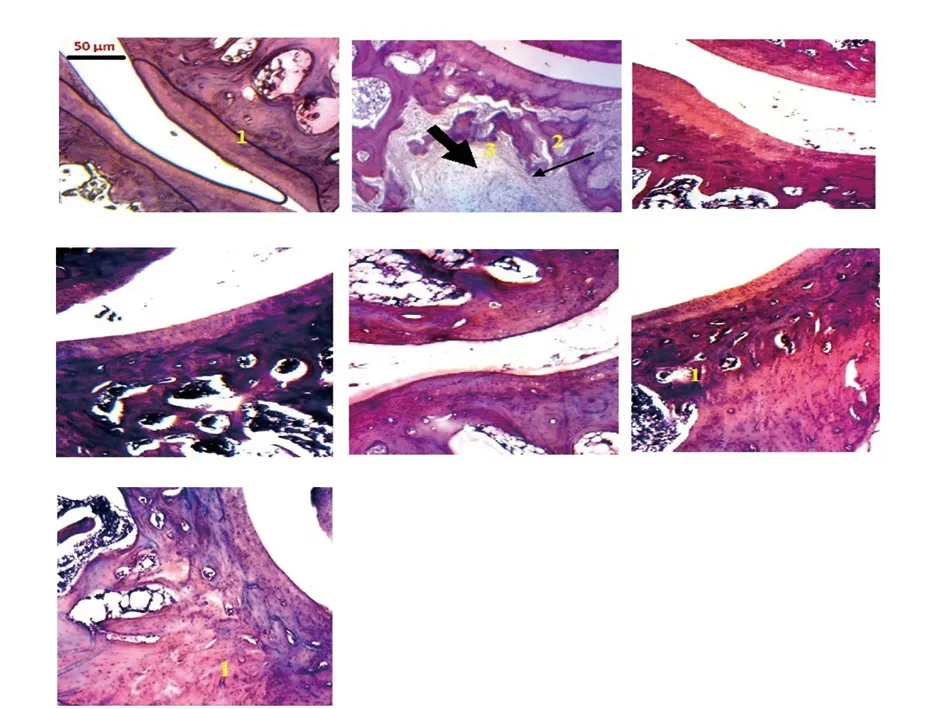
Figure 4. Histopathology of the joints (×100). A: Normal control with normal bone and cartilage (1), no leukocyte infiltration; B: Arthritis control with leukocyte infiltration (2) and bone destruction (3); C: Diclofenac (5 mg/kg), D and E: aqueous extract (150 and 300 mg/kg), F and G: Methanolic extract (150 and 300 mg/kg): reduction of bone erosion (1) and leukocyte infiltration (1).
Rheumatoid arthritis is systemic inflammatory rheumatism characterized by joint pain, inflammation of the synovial membrane,bone erosion and destruction of articular cartilage. Articular cartilage is made up of proteins, whose denaturation can cause rheumatoid arthritis and can cause the aggravation of the pathology[24]. These proteins can lose their secondary and tertiary structures, which can cause a constraint generated in the cell and cause the modification of the hydrogen bonds[6,24]. The aqueous and methanolic extracts of N. pobeguinii inhibited protein denaturation in this study.These effects would be related to the ability of these extracts to protect the structure of the protein and prevent the change of the aforementioned bonds. Actually, flavonoids, tannins, and alkaloids are able to interact with the aliphatic region around the lysine residue on albumin protein and prevent protein denaturation[25].During the inflammatory process, activation of protein kinase C promotes the activity of phospholipase A2 which causes the denaturation of membrane phospholipids to form arachidonic acid[25]. This arachidonic acid is responsible for the formation of eicosanoids which will stimulate enzymes (cyclooxygenase and 5-lipoxygenase) causing the release of inflammatory mediators(PGE2, thromboxanes, and leukotrienes). Our results show that these extracts have an inhibitory effect on the activities of cyclooxygenase and 5-lipoxygenase, and these properties could be attributed to the presence of secondary metabolites belonging to the flavonoids,terpenoids and/or saponins classes. Some of these compounds are able to affect the metabolism of arachidonic acid in several ways,then to specifically inhibit the activities of cyclooxygenase and/or lipoxygenase[26]. Polymorphonuclear neutrophils, macrophages and T cells induce activation of the immune response in patients with rheumatoid arthritis. These agents provoke an immune response by activation of the toll-like receptor, and triggering an inflammatory reaction with cascade release of many proinflammatory cytokines,particularly TNF-α and interleukin 1β. These mediators released by the immune system play an important role in the initiation and progression of rheumatoid arthritis. They are likely to cause phosphorylation and the deterioration of proteins following activation of energy nuclear factor kappa B which is likely to influence the activity of immune cells[27]. Thus, to evaluate the effects of aqueous and methanolic extracts of N. pobeguinii on the modulation of the immune system, the T cell proliferation assay was used. The results show that these extracts significantly inhibit the proliferation of T lymphocytes. It is known that some flavonoids and/or saponins are able to inhibit or block PI3k/Akt/mTOR and NF-κB pathways,thus significantly reduce cell proliferation[28]. Moreover, during the progression of polyarthritis, proinflammatory cytokines (particularly TNF-α and IL-1β) are capable of activating the nicotinamide adenine dinucleotide phosphate-oxidase which through the respiratory chain could cause the release of the superoxide and then induce oxidative stress. Increased production of free radicals and proinflammatory cytokines is responsible for accelerated joint cartilage degradation,bone destruction and osteoclast stimulation[29]. Thus, the antioxidant potential of the extracts was tested in vitro by DPPH, ABTS and NO assays. It shows that both extracts significantly reduce DPPH,ABTS discoloration and NO production. These results suggest that N. pobeguinii has antioxidant properties in vitro. Compounds such as flavonoids and saponins in the two extracts have already shown in vitro antioxidant activities by reducing DPPH, ABTS discoloration and NO production[30].
ROS are involved in the pathophysiology of inflammatory diseases such as rheumatoid arthritis. They lead to the release of cytokines and the activation of proinflammatory enzymes such as cyclooxygenase, lipoxygenase and inducible nitric oxide[7]. Lack of control of these ROS is responsible for cartilage destruction, bone erosion, and nuclear factor expression. The results of our study show a significant inhibitory effect of the extracts on the production of intracellular and extracellular ROS. On the one hand, these extracts had the inhibitory effect on NF-κB phosphorylation, preventing it from releasing proinflammatory cytokines. This effect could be attributed to some terpenoids, flavonoids and/saponins, which are able to inhibit NF-κB/AP1 axes and reduce TNF-α, IL-1β, NO and PGE2production[31,32]. On the other hand, these extracts could inhibit the denaturation of proteins. The activities of cyclooxygenase and 5-lipoxygenase are potent mediators of inflammation, since these enzymes are capable of forming the hydroperoxides precursors of leukotrienes.
Cyclooxygenase is an enzyme that catalyzes the biosynthesis of prostaglandins, an anti-pain, and anti-inflammatory agent, while free radicals oxidize the lipids of the cell membrane, resulting in the formation of lipid peroxidation[31]. The injection of CFA into the rat causes the activation of these enzymes and the activation of lipid peroxidation. The injection in the paw is associated with the development of a monoarthritis whereas the injection in the tail is associated with the development of a polyarthritis in Wistar rats and Swiss albino mice[32]. CFA-induced arthritis is widely used to test anti-arthritic potential of many substances[23]. This systemic injection is characterized by the migration of leukocytes and lymphocytes into the joint, resulting in the development of joint inflammation on the ninth day after injection[23]. In this study, N. pobeguinii extract significantly reduced joint diameter and arthritis score, which could be associated with the ability of these extracts to inhibit lipoxygenase and ROS mediators[33], and with the antiproliferative power of these extracts[18]. Some flavonoids and/or terpenoids are able to act as a nonsteroidal anti-inflammatory drug (diclofenac) by inhibiting the thromboxane-prostanoid receptor, affecting the arachidonic acid release and uptake, inhibiting lipoxygenase enzymes and inducing antiproliferative effect[28]. Inflammatory stimuli such as CFA result in a cascade of proinflammatory mediators such as cytokines and PGE2. These mediators are responsible for the hypersensitization of nociceptors with subsequent hyperalgesia[33]. The analgesic effect of the extract of N. pobeguinii was evaluated by measuring the pain with an analgesimeter and these extracts showed an antihyperalgesic effect. This activity of N. pobeguinii is attributed to the inhibition of the production and/or activity of proinflammatory mediators released during the development of hyperalgesia, or to the ability to inhibit cyclooxygenase, which is responsible for the production of PGE2.In fact, the nonsteroidal anti-inflammatory drug (diclofenac) and some compounds (flavonoids, saponins and/or triterpenoids) could diminish prostaglandin synthesis by inhibiting cyclooxygenase,which catalyzes the formation of prostaglandin precursors from arachidonic acid[34].
Variation in body weight is an important indicator of health conditions. Rheumatoid arthritis is associated with reduced muscle mass, loss of appetite, or metabolic disorders caused by the inflammatory response[35]. The weight loss observed in untreated animals after CFA injection was alleviated after the administration of the aqueous and methanolic extract of N. pobeguinii as well as diclofenac. All treated animals showed an increase in body weight after treatment. People with rheumatoid arthritis typically develop anemia characterized by elevated white blood cell counts and reduced hemoglobin levels, decreased erythropoietin levels, or bone marrow activity[36]. In addition, an increase in platelet levels, a reduction in red blood cells and hematocrit levels are also observed in these patients. Patients with rheumatoid arthritis also have significant variation in liver enzyme activity such as transaminases(ALT and AST), ALP[37], creatinine and total proteins, with hepatic and renal damage. These enzymes (transaminases and ALP) are usually released into the circulation during bone resorption. They are involved in localized bone loss such as bone erosion and periarticular osteopenia and are also considered as precursors of some proinflammatory mediators such as serotonin, bradykinin,and histamine, which are responsible for the development and maintenance of inflammation[38]. In this study, the results show that extracts of N. pobeguinii reduced the number of white blood cells and increased hemoglobin levels. They also improved the variations in serum parameters. It is known that compounds as alkaloid,flavonoids, tannins, terpenes, and steroids have the ability to protect the erythrocytes from oxidative damage, and possess erythropoietin stimulatory, immune-stimulatory and thrombopoietin stimulatory activities[39].
Free radicals are known to destroy membrane lipids, proteins,DNA and articular cartilage. In patients with rheumatoid arthritis,polymorphonuclear neutrophils and macrophages produce ROS that lead to cartilage damage, lipid peroxidation and increase NOcatalyzed reaction or antioxidant defense[40]. In this study, extracts of N. pobeguinii significantly decreased MDA and NO levels,and increased activity of SOD, GSH, and CAT. The presence of compounds such as flavonoids and saponins could justify the antioxidant activity of the plant because it is known that these compounds are able to significantly increase the activities of SOD,CAT, and glutathione-S-transferase as well as levels of ascorbic acid and GSH, while significantly decrease MDA level[41]. In the present study, histopathology of the joints showed that animals treated with aqueous and methanolic extracts of N. pobeguinii have joints with little cellular infiltration, bones and cartilage are less damaged than the untreated animal, suggesting that these extracts would have restored the normal architecture of the joint. Some flavonoids could inhibit the release of NO, TNF-α, IL-1, IL-6 as well as T-cell proliferation[42], significantly suppress secondary inflammatory paw swelling, pain response, and polyarthritis index in rats by inhibiting production of IL-1, TNF-α, and PGE2in synoviocytes and significantly reduce the joint damage[43]. These effects would be related to the inhibitory activities of these extracts on the proliferation of T cells and the production of ROS, which are considered as the main factors for deterioration and destruction of bones and cartilage[9]. Besides, these effects may also be related to the inhibition of proinflammatory cytokines, which have been shown to be responsible for inflammation.
The in vitro study shows that the aqueous and methanolic extracts of N. pobeguinii have important anti-arthritic properties by inhibition of protein denaturation, cyclooxygenase, 5-lipoxygenase, T cell proliferation and production of free radicals. Moreover, in vivo study also shows that they also could reduce inflammation of the joint, hyperalgesia, improve hematological parameters, reduce liver enzymes, regulate oxidative stress parameters, and restore inflammatory joint. These results confirm the analgesic, antiinflammatory and antiarthritic activities of this plant as a traditional medicine.
Conflict of interest statement
The authors declare that they have no competing interests.
Authors’ contributions
TEG, MM and AG designed the work. DNSF, MM, TEG, AAD and MMMV conducted the work, collected and analysed the data. MM,NYW, AG and TTH drafted the manuscript and revised it critically.All authors agree to be accountable for all aspects of the work.
 Asian Pacific Journal of Tropical Biomedicine2020年2期
- Asian Pacific Journal of Tropical Biomedicine的其它文章
- One-pot synthesis of silver nanocomposites from Achyranthes aspera: An eco-friendly larvicide against Aedes aegypti L.
- Micro RNA deregulation and cancer and medicinal plants as microRNA regulator
- Ethanol extracts of Hizikia fusiforme induce apoptosis in human prostate cancer PC3 cells via modulating a ROS-dependent pathway
- Antioxidant and antibacterial activities and identification of bioactive compounds of various extracts of Caulerpa racemosa from Algerian coast
