Chlamydoconidium-producing Trichophyton tonsurans: Atypical morphological features of strains causing tinea capitis in Ceará, Brazil
Raimunda Sâmia Nogueira Brilhante, Germana Costa Paixão, Jonathas Sales de Oliveira,Vandbergue Santos Pereira, Marcos Fábio Gadelha Rocha,2✉, Reginaldo Gonçalves de Lima-Neto, Débora de Souza Collares Maia Castelo-Branco, Rossana de Aguiar Cordeiro, José Júlio Costa Sidrim
1Specialized Medical Mycology Center, Postgraduate Program in Medical Microbiology, Federal University of Ceará, Rua Nunes de Melo, 1315, Fortaleza,Brazil
2Postgraduate Program in Veterinary Sciences, State University of Ceará; Av. Dr. Silas Munguba, 1700, Fortaleza, Brazil
3Department of Tropical Medicine, Center for Health Sciences, Federal University of Pernambuco, Recife-PE, Brazil
Keywords:Trichophyton tonsurans Chlamydoconidia Dermatophyte Phenotyping
ABSTRACT Objective: To report atypical morphological features of Trichophyton (T.) tonsurans strains associated with tinea capitis.Methods: Eighty-two T. tonsurans strains isolated in Ceará, Brazil, were analyzed regarding macro and micromorphological features and nutritional patterns.Results: Fifty-two samples presented abundant chlamydoconidia, which were produced in chains. Macroscopically, these strains developed small glabrous colonies that were firmly attached to the surface of the culture medium, with few or no aerial mycelia and intense rusty yellow pigmentation. Seven strains did not grow with stimulus from thiamine. Samples were heterogeneous regarding urease production and none presented in vitro hair perforation.Conclusions: The observation of T. tonsurans strains with distinct phenotypic features indicates the need to revise the taxonomic criteria for routine identification of this dermatophyte.
1. Introduction
Dermatophytes are among the oldest microorganisms recognized as human pathogens, and are the main fungi involved in superficial mycoses[1,2]. The incidence of infections caused by species such as Epidermophyton floccosum, Microsporum audouinii and Trichophyton(T.) schoenleinii has markedly decreased in the past 100 years,while T. tonsurans has progressively become more important as an infectious agent, particularly causing tinea capitis[3-10]. Also, T.tonsurans is the most common dermatophyte species isolated from scalp of asymptomatic carriers[11]. In Northeastern Brazil, over the past decades, T. tonsurans has been the main etiological agent of tinea capitis[12-15], being considered an emerging pathogen.
Nowadays, dermatophyte taxonomy is under re-evaluation,because of findings based on molecular phylogenetics[1,16,17]; and several researchers have been focusing on studying the genotypic and protein profile of these fungi[16,18-21]. According to De Hoog et al[1], although the molecular approach has been able to resolve the main issues concerning dermatophyte phylogenetic evolution,it may fail sometimes, and species that are well established and clinically different can be indistinguishable in multilocus analysis.
T. tonsurans is routinely identified by analyzing macromorphological and micromorphological features. This species presents moderate growth, with flat, cottony or powdery colonies,and produces a profusely branched conidial system with numerous microconidia having varied sizes and shapes, sometimes associated with a smaller number of macroconidia[22]. Knowledge on genes,molecular mechanisms of pathogenicity and other biological properties of T. tonsurans is still scarce and inconsistent. This condition, along with its low degree of genetic polymorphism[2,13,23],reinforces the importance of a thorough phenotypic characterization for a more accurate laboratory identification of T. tonsurans.
Thus, the objective of this study is to report the atypical morphological features of strains of T. tonsurans associated with tinea capitis, highlighting the abundant production of chlamydoconidia in chains.
2. Materials and methods
Initially, 82 strains of T. tonsurans obtained from the collection of the Specialized Medical Mycology Center (CEMM) of Federal University of Ceará, isolated from scalp lesions were analyzed.
The strain T. tonsurans ATCC 28942 was used as control. Samples were seeded on Sabouraud dextrose agar (SDA), Sabouraud chloramphenicol agar (SCA) or MycoselTMagar (MYC) and incubated at 28 ℃ for up to 20 d. Soon after visual detection of growth, colony microscopy was evaluated. Chlamydoconidia were quantified by two independent observers, in a double-blind arrangement, and strains were categorized into two groups: 1)isolates with ≤5 chlamydoconidia per field, and 2) isolates with at least one chain of chlamydoconidia formed by >5 chlamydoconidia per field. Then, strains that presented at least one chain of chlamydoconidia/microscopic field were further analyzed for their phenotypical features.
For the analysis of morphological features, strains were inoculated on SDA, SCA or MYC and incubated at 28 ℃ for up to 20 d. Size,margins, texture, relief and pigmentation of colonies were defined according to De Hoog et al[22]. Microscopic structures were daily observed from the 5th to the 15th day and on the 20th day of growth.More detailed analysis of the chlamydoconidia was performed by scanning electron microscopy, with fragments of T. tonsurans colony placed on ThermanoxTM coverslips (Thermo Fisher Scientific, NY,USA) and processed, as described by Brilhante et al[24]. Coverslips were observed with a FEI Inspect S50 scanning electron microscope,in high vacuum mode at 15 kV.

Figure 1. Scanning electron microscopic image showing micromorphological characteristics of Trichophyton tonsurans ATCC 28942 (standard strain) and an atypical Trichophyton tonsurans strain. A and B: Local atypical strain showing chlamydoconidium chains (arrows). C and D: Trichophyton tonsurans ATCC 28942 showing microconidia (arrow) with variable shapes and sizes. Strains were grown on Sabouraud agar at 28 ℃ for 5 days.
Urease production was evaluated using Christensen’s urea agar,incubated at 28 ℃ for 5 d, with daily observations. For in vitro hair perforation test, 1 cm2-fragments of T. tonsurans colonies were inoculated in agar plates containing previously sterilized blond hair from a child. Plates were incubated at 28 ℃, for 40 d, with microscopic observations every 7 d. Nutritional features (vitamin requirements) was determined by inoculating strains on different Trichophyton agar media supplemented with histidine, thiamine,inositol and niacin (T1, T2, T3, T4, T5, T6 and T7 agar) and incubated at 28 ℃, for 14 d. Growth quantification for each strain was compared to the growth of T. tonsurans ATCC 28942, and growth was classified as optimal (+3), moderate (+2), slight (+1) or absent (0), according to Larone[25], with adaptations.
To confirm identification of chlamydoconidia-forming T. tonsurans,strains chosen randomly were identified using matrix-assisted laser desorption-ionization time of flight (MALDI-TOF)[26,27]. The equipment used was MALDI-TOF Autoflex ⅢMass Spectrometer(Bruker Daltonics Inc., Billerica, MA, USA/Germany). The resulting peak lists were exported to the software MALDI BiotyperTM 3.0(Bruker Daltonics, Bremem, Germany), which provided the final identification of the isolates as T. tonsurans.
3. Results
Based on the initial screening, 52/82 strains (63.41%) presented at least one chain of chlamydoconidia formed by >5 chlamydoconidia/microscopic field, of which 18/52 (34.62%) presented up to 1 chain formed by >5 chlamydoconidia/field, 24/52 strains (46.15%)produced between 2 and 5 chains/field and 10/52 strains (19.23%)produced >6 chains/field (Figures 1A and 1B). When strains were grown on SDA, 11/52 (21.15%) produced chlamydoconidia and microconidia and 41/52 strains (78.85%) only produced chlamydoconidia. On SCA, 14/52 strains (26.92%) produced chlamydoconidia and microconidia and 38/52 (73.08%) only produced chlamydoconidia. On MYC, 8/52 strains (15.38%)produced both chlamydoconidia and microconidia, and 44/52 strains(84.62%) only produced chlamydoconidia. Elongated and cylindrical and sometimes fusiform, thin-walled macroconidia, with 3 to 5 septa and no spines, were only shown by 3 strains grown on MYC.T. tonsurans ATCC 28942 presented a branched conidial system with several microconidia with different sizes and shapes, but no chlamydoconidia (Figures 1C and 1D).
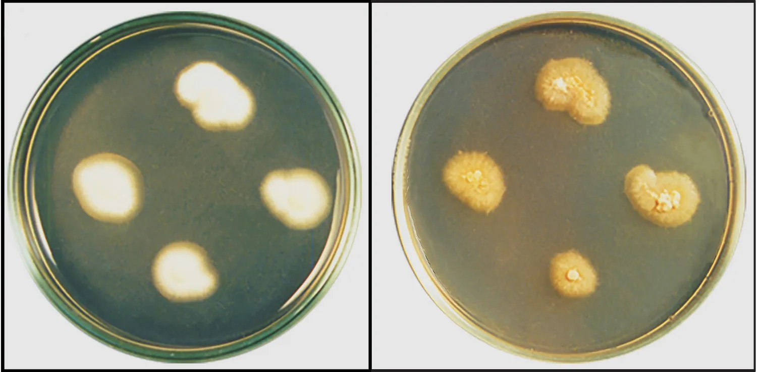
Macroscopically, starting on the 5th day of incubation, all 52 strains presented small colonies (smaller than 1 cm in diameter),with glabrous texture, with slight development of a low cottony aerial mycelium in the central region and an intense rusty yellow color, both on the top and bottom of the colonies, which remained unchanged up to the 20th day of observation (Figure 2A). T.tonsurans ATCC 28942 presented flat white colonies, with an aerial mycelium and no diffusible pigments (Figure 2B). R e g a r d i n g urease production, 27/52 strains (51.92%) were negative and 25/52(48.08%) were positive for the enzyme, 48 h incubation. None of the tested strains perforated the hair under the experimental conditions.Concerning growth on Trichophyton agar, inositol supplementation supported optimal growth for 100% of the strains, while on medium supplemented with both inositol and thiamine, 39/52 (75.00%)strains had optimal growth and 13/52 (25.00%) had slight growth.On thiamine-supplemented medium, 39/52 (75.00%) strains presented optimal growth, 6/52 (11.54%) showed slight growth and 7/52 (13.46%) did not grow. Niacin promoted optimal growth for 41/52 (78.85%) strains and none of the strains grew on agar supplemented with histidine.

Figure 2. Macromorphological characteristics of T. tonsurans ATCC 28942 and an atypical Trichophyton tonsurans strain. A: Local atypical strain showing glabrous texture and central sparse aerial mycelia with an intense rusty pigmentation. B: Trichophyton tonsurans ATCC 28942 (standard strain) showing white cottony colony and aerial mycelia. Colonies grown on Sabouraud dextrose agar and incubated at 28 ℃ for 20 days.
4. Discussion
According to Zhan et al. [16], anthropophilic species of Trichophyton are hard to distinguish by molecular methods and no sexual reproduction structures are known. Additionally, de Hoog et al. [1]stated that sequencing ambiguities and gaps in globally validated genomic databases can obscure the small differences between species, emphasizing the importance of studies that combine molecular, ecological, phenotypic and life-cycle data to establish secure identification of dermatophytes.
T. tonsurans strains analyzed in this study have marked differences when compared to standard strains, in particular regular and abundant production of chlamydoconidia, which result from hyphal modification and cell wall thickening due to the deposition of hydrophobic material. These features are rarely seen in dermatophytes, as they are mainly restricted to the species T.verrucosum and T. violaceum, or to old and pleomorphic cultures[22].Chlamydoconidia are not key structures for the identification of dermatophytes. When present, they are generally associated with mature colonies, reduction of the available nutritional substrate or colony pleomorphism. However, here we show that several chlamydoconidia organized in chains were produced early, after only 5 days of growth. These structures were developed on all three Sabouraud agar media used, which indicates that components of the culture medium, especially cycloheximide, were not determining factors for chlamydoconidium production.
The small number of microconidia and macroconidia produced by the T. tonsurans strains described here is also an uncommon finding,since the classic description of this species is the presence of a branched conidial system, with several microconidia with different shapes and sizes and equally distinct macroconidia.
Mochizuki et al.[7] proposed a presumptive identification of T. tonsurans based on the observation of ovoid structures measuring 7 to 10 µm, similar to intercalary chlamydoconidia. Those authors evaluated the growth of 25 strains of T. tonsurans on Mycosel agar and observed the presence of chlamydoconidia, after 5 days of incubation. The authors described the morphogenesis of these structures in detail and proposed the observation of these structures as a fast and simple method for the presumptive identification of T. tonsurans. However, unlike our findings, the chlamydoconidia described by Mochizukit and colleagues[7] were less numerous and interspersed in the hyphae, while the strains used here presented very numerous chlamydoconidia arranged in chains along the hyphae.
Local atypical T. tonsurans strains also had different characteristics regarding time for growth and macroscopic aspects of the colonies,when compared to standard strains. All strains grew rapidly in 5 to 8 days, producing small glabrous colonies of up to 1 cm in diameter,with a small cottony aerial mycelium in the center of the colony,and intense rusty yellow pigmentation on the top and reverse. These features differ from the flat, cottony colonies with aerial mycelia and no pigment or brownish pigment described for the standard strain of T. tonsurans.
Additionally, the strains of T. tonsurans analyzed here presented a different profile of growth support by thiamine supplementation from that of standard strain, because only 13.46% of our strains did not grow on thiamine-enriched agar. This was unexpected as thiamine is known to promote the growth of T. tonsurans and it is used for the definitive identification of this species. The lack of homogeneity for urease production and the inability to perforate hair in vitro observed for the studied strains are part of the traditional findings for T.tonsurans.
Morphotaxonomic alterations found in the T. tonsurans strains analyzed in this research characterize an atypical presentation of local T. tonsurans and may indicate an unprecedented consistent phenotypic variation, a cryptic species or even a new variety of this dermatophyte. The observation of T. tonsurans strains with these phenotypic features, which had never been described in specialized literature, indicates the need to revise the taxonomic criteria for routine identification of this dermatophyte species and provides precise laboratory data to better understand the epidemiology and configuration or reconfiguration of the global distribution of dermatophytes, especially in Northeastern Brazil.
Conflict of interest statement
We declare that we have no conflict of interest.
Acknowledgments
The authors would like to thank Central Analítica UFC/CT-INFRA/MCTI-SISNANO/Pro-Equipamentos CAPES for technical support in obtaining scanning electron microscopic images.
Foundation project
This work was supported by the Conselho Nacional de Desenvolvimento Cientifico e Tecnológico-CNPq, Brazil(Grant No.:306976/2017-0).
 Asian Pacific Journal of Tropical Medicine2019年8期
- Asian Pacific Journal of Tropical Medicine的其它文章
- Molecular identification of hemoplasmas in free ranging non-human primates in Thailand
- Tioxolone niosomes exert antileishmanial effects on Leishmania tropica by promoting promastigote apoptosis and immunomodulation
- Evaluation of in vitro and in vivo immunostimulatory activities of poly (lactic-co-glycolic acid) nanoparticles loaded with soluble and autoclaved Leishmania infantum antigens: A novel vaccine candidate against visceral leishmaniasis
- The cholera epidemic of 2004 in Douala, Cameroon: A lesson learned
- Pharmacological and analytical aspects of artemisinin for malaria:Advances and challenges
