
Molecular identification of hemoplasmas in free ranging non-human primates in Thailand
2019-08-29 09:44:52ParutSuksaiSupakarnKaewchotPiyaSereerakSirinanBoonnanBongkotchamatPhimsinTaksinaJaruwattananonKacharinRaschasinMutchamonKaewparuehaschaiSorrayaSiriphetBenjapornBhusri
Parut Suksai, Supakarn Kaewchot, Piya Sereerak, Sirinan Boonnan, Bongkotchamat Phimsin,Taksina Jaruwattananon, Kacharin Raschasin, Mutchamon Kaewparuehaschai, Sorraya Siriphet,Benjaporn Bhusri✉
1The Monitoring and Surveillance Center for Zoonotic Diseases in Wildlife and Exotic Animals, Faculty of Veterinary Science, Mahidol University, Salaya,Nakhon Pathom 73170, Thailand
2Department of National Parks, Wildlife and Plant Conservation, Chatuchak, Bangkok 10900, Thailand
Keywords:Hemoplasma Mycoplasma Non-human primate Monkey Thailand
ABSTRACT Objective: To survey hemoplasmas infection in free ranging non-human primates from 8 provinces in Thailand.Methods: DNA from ethylenediaminetetraacetic acid blood of 262 free ranging non-human primates were identified as hemoplasmas using PCR and phylogenetic analysis based on 16S rRNA and rnpB genes.Results: A total of 148 non-human primates (56.49%) were determined positive for Candidatus Mycoplasma haemomacaque, including 125 Macaca fascicularis and 23 Macaca mulatta. Hemoplasmas can cause anemia in monkey but all positive samples were healthy. The positive rates in male and female non-human primates were not significantly different.Conclusions: Candidatus Mycoplasma infection is prevalent in free ranging Macaca fascicularis and Macaca mulatta in Thailand.
1. Introduction
Hemoplasmas or hemotropic mycoplasmas are small pleomorphic and uncultivable bacteria that infect erythrocytes and endothelial cells of several mammalian species[1-4], which can cause anemia[5].Data about the transmission of hemoplasmas is still poor[5].Only one study has experimentally demonstrated transmission of mycoplasmas by Rhipicephalus (R.) sanguineus[6], but the role of this tick in the natural transmission of haemoplasmas is unknown.A recent study investigated a possible trans-stadial route of canine haemoplasma transmission by R. sanguineus in field conditions,but no canine haemoplasma DNA was detected in unfed nymphs or adult stages of ticks[7]. Hemoplasma was reclassified in genus
Mycoplasma from Haemobartonella and Eperythrozoon based on nucleotide sequences of 16S ribosomal (r) RNA (16S rRNA) gene[1].Recently, RNase P RNA (rnpB) gene was recommended to identify and classify hemoplasmas[8]. The hemoplasmas in non-human primates consist of Candidatus Mycoplasma haemomacaque (Ca.M. haemomacaque)[9,10], Candidatus Mycoplasma kahanei (Ca. M.kahanei)[11], and Candidatus Mycoplasma aoti (Ca. M. aoti)[12].
Wildlife has been a major reservoir of infectious diseases and a source of the trouble that threatening humans and domestic animals health[13]. Non-human primates (NHP), as one of the wildlife species, is suggested to be an important reservoir host of several pathogens[13,14]. The infections of hemoplasmas were reported in free ranging NHP[10,15,16]. In Thailand, the large population of free ranging NHP species are long-tailed macaque [Macaca(M.) fascicularis] and rhesus macaque (M. mulatta)[17]. They are closely contact and conflict with humans because of the adaptation of macaque habitats, the human activities and the expansion of human communities[17,18]. Thus, the inter-species transmission of hemoplasmas may be occurred. Hemoplasmas in NHP have been reported in Brazil, French, Japan, the United State of America and the United Kingdom[9,10,12,15,16,19-21]. Unfortunately, there is no information on the hemoplasmas infection in M. fascicularis and M. mulatta in Thailand. Therefore, the present study focused on the genetic screening and preformed the phylogenetic analysis of hemoplasmas species in NHP that were identified from free ranging M. fascicularis and M. mulatta in Thailand.
2. Materials and methods
2.1. Sample collection and DNA extraction
A total of 262 samples of ethylenediaminetetraacetic acid bloods were collected from free ranging NHP including 227 M.fascicularis and 35 M. mulatta from April, 2018 to August, 2018 in 8 provinces of 4 regions in Thailand, including central (Lop Buri and Samut Sakhon), northern (Chiang Rai), southern (Phatthalung and Songkhla), western (Prachuap Khiri Khan), and northeastern(Amnat Charoen and Mukdahan). The NHP were determined healthy by physical examination. All specimens were extracted using DNeasy blood & tissue kit (QIAGEN, Germany) according to the manufacturer’s instructions. Each sample was eluted with 50 µL nuclease free water and stored at -20 ℃.
2.2. Hemoplasmas detection
All DNA were initially screened for hemoplasmas using HBT-F (5’-ATACGGCCCATATTC CTACG-3’) and HBT-R (5’-TGCTCCACCACTTGTTCA-3’) primers of PCR based on 16S rRNA gene[22]. The total volume of PCR mixture was 25 µL that consisted of 1×PCR buffer, 3.5 mM MgCl2, 0.5 µM of each primer,0.25 mM of each deoxynucleotide triphosphate and 1.25 U of i-TaqTM DNA polymerase (iNtRON Biotechnology, Korea), 2 µL of DNA template and made up to 25 µL with nuclease-free water.The PCR conditions included a 3 min initial denaturation at 94 ℃,followed by 30 cycles at 94 ℃ for 30 s, 60 ℃ for 45 s and 72 ℃ for 45 s with a final extension of 72 ℃ for 7 min. The PCR products(595 bp) were electrophoresed in 2% agarose gel and visualized with GelRedTM (Biotium, United States) under UV illumination.The positive 16S rRNA gene was additionally screened by rnpB primers (80F1: 5’-GAGGAAAGTCCRYGCTWGCAC-3’ and 290R1 5’-TCCCYTACCRAAATTTRGGTTTCT-3’)[8]. The final concentration of reaction components and condition were similar to the 16S rRNA screening, except the annealing step was optimized at 44 ℃. The rnpB primers amplified an approximately 220 bp fragment.
2.3. Nucleotide sequence-based analysis
The fragments of 16S rRNA and rnpB genes were purified using QIAquick® Gel Extraction Kit (QIAGEN, Germany) and sent to a private service laboratory (Macrogen Inc., Korea) for nucleotide sequencing. The partial nucleotide sequences of 16S rRNA and rnpB genes were aligned using Clustal W and constructed the phylogenetic relationship of various hemoplasmas using the Maximum Likelihood method in MEGA 7 software[23]. The distances were evaluated by the General Time Reversible model and Tamura 3-parameter model for 16S rRNA and rnpB genes, respectively. The data sets were generated using 1 000 bootstrap resamplings. The reference sequences of hemoplasmas species for comparison were obtained from GenBank NCBI (http://www.ncbi.nlm.nih.gov/genbank).
2.4. Ethnical approval
The present protocol was approved by the Faculty of the Veterinary Science, Mahidol University-Institute Animal Care and Use Committee(Protocol No. MUVS-2018-09-49).
2.5. Statistical analysis
The statistical relationships between positive results of hemoplasmas and sex of free ranging NHP was analyzed by Pearson Chi-Square test using SPSS 18 for Windows (SPSS Inc., United States). Statistical significance was indicated at P value < 0.05.
3. Results
One hundred and forty eight (148) of 262 (56.48%) NHP samples were positive for M. fascicularis (Table 1), which were collected from Lop Buri (n=28), Samut Sakhon (n=24), Mukdahan (n=14),Phatthalung (n=15), Songkhla (n=10), Prachuap Khiri Khan (n=13),Amnat Charoen (n=21) and Chiang Rai (n=23). All positive samples were detected in both host species (125 M. fascicularis and 23 M.mulatta). The positive rate of male NHP was 54.65% (94/172), whereas the positive rate of female NHP was 60.00% (54/90). There is no significant difference in positive rate between male and female NHP(P>0.05).
The partial 16S rRNA and rnpB sequences of hemoplasmas derived from both M. fascicularis (FN55/61-MF) (Accession No. MK192130 and MK192132, respectively) and M. mulatta(FN51/61-MM) (Accession No. MK192131 and MK192133,respectively) were analyzed for sequence homology and phylogeny.The homology between the positive nucleotide sequences and the published nucleotide sequences was analyzed using BLASTn. The obtained sequences of 16S rRNA gene were highly similar to Ca.M. haemomacaque (99%) from M. fuscata (AB820288) and M.fascicularis (KC512401). Conversely, the partial rnpB sequences of positive samples presented low homology (82%) with Ca. M.haemomuris (AB973090) and Ca. M. haemohominis (GU562825)that isolated from Apodemus argenteus and Homo sapiens,respectively. The identity rates of rnpB between the positive and Ca.M. haemomacaque sequences were unknown because rnpB sequence of Ca. M. haemomacaque is still unpublished in GenBank.
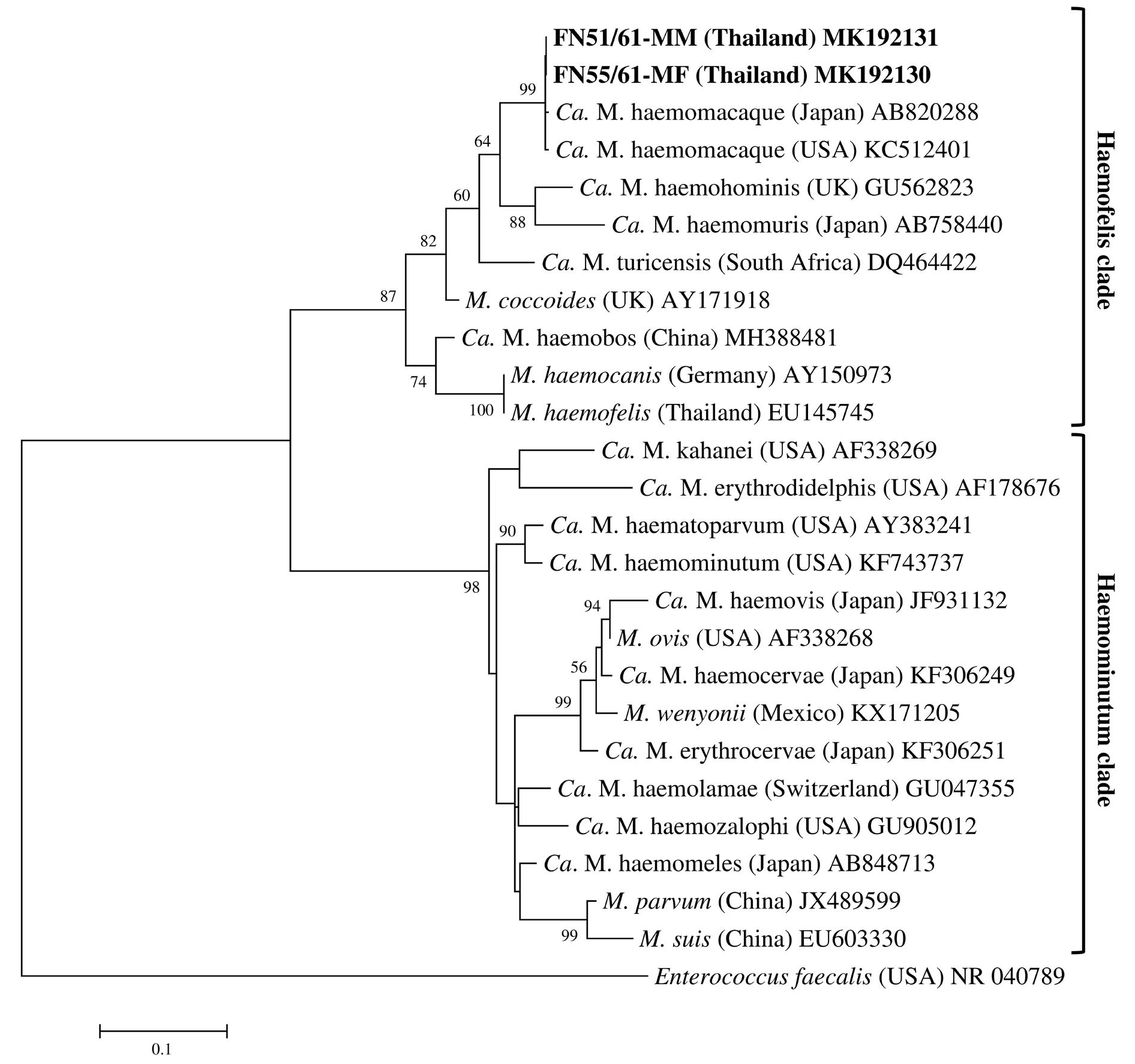
Figure 1. Phylogenetic tree of the partial 16S rRNA sequences of hemoplasma species in free ranging non-human primates and other hemoplasma species from GenBank. ¬
The phylogenetic trees of the partial 16S rRNA and rnpB sequences in M. fascicularis and M. mulatta of the present study were constructed using Enterococcus faecalis as an outgroup. Percentages of bootstrap values were obtained on the nodes of the tree, and values over 50% were displayed. The phylogram of both genes were divided into two clades that were haemofelis and haemominutum.In both phylogeny, the positive sequences showed a number of the haemofelis clade, which were positioned in the same cluster as Ca.M. haemomuris, Ca. M. haemohominis, Ca. M. turicensis, and M.coccoides (Figure 1 and 2). Moreover, the tree of 16S rRNA genes presented the strong relationship between the sequences from the tested NHP and Ca. M. haemomacaque sequence from GenBank(Figure 1).
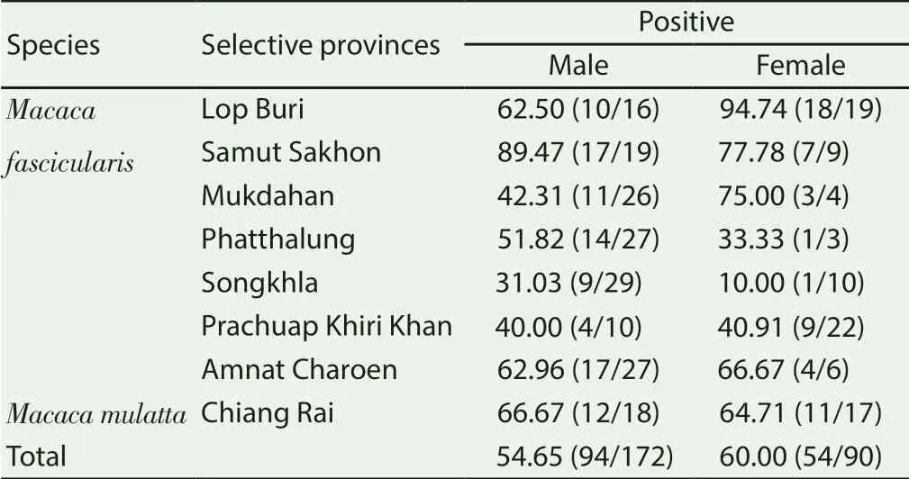
Table 1. Results of hemoplasmas detection in tested non-human primates (%).

Figure 2. Phylogenetic tree of the partial rnpB sequences of hemoplasma species in free ranging non-human primates and other hemoplasma species from GenBank.
4. Discussion
In the present study, the positive rate of hemoplasmas infection among the free ranging NHP was rather high (56.48%). The result was higher than the reports in southern (25%) and northeastern Brazil (35.7%)[16,24] but lower than the study in the United State of America (84.2%)[9] and Japan (100%)[10]. In Thailand, hemoplasma infection in NHP has not been previously reported. Result of this research showed the occurrence of hemoplasmas infection in Thai NHP that including M. fascicularis and M. mulatta. These host species have been found hemoplasmas[9,20,25]. Hemoplasmas infection has presented from no clinical signs to severe anemia in NHP[9,10,19-21,24]. All positive samples in our study did not show clinical signs. Previous reports suggested that anemia may not be associated with hemoplasmas infection in NHP[9,21,24].
The infection rate in male is not significantly different in female,which differ from the previous report[24]. In this study, the greater number of samples was tested, therefore, the results are estimated to be more reliable. Habitats of the tested NHP and most habitats of free ranging NHP in Thailand overlap with communities[17,18],which may cause cross transmission of infection between NHP and humans or domestic animals. Moreover, some species of hemoplasmas were found in humans[26-28], at this point, further studies should be conducted on hemoplasmas infection in humans and other animals in the habitat of NHP, which may effectively monitor the public health of humans and animals.
The positive sequences showed high similarity in 16S rRNA of Ca. M. haemomacaque. Further, the obtained 16S rRNA and rnpB gene sequences belonged to the haemofelis clade and the cluster of Ca. M. turicensis and M. coccoides, which is similar to findings in other studies[9]. Unfortunately, the rnpB sequence of Ca. M. haemomacaque detected in M. fascicularis from previous experiment[9] was not available in GenBank, so the identity rate of the rnpB sequences of hemoplasmas isolated from the positive NHP samples cannot be determined. Although there is no rnpB identity result of Ca. M. haemomacaque, we considered that the rnpB sequences of the positive samples was Ca. M. haemomacaque.This conclusion was supported by the finding that explained above.Previously, Ca. M. haemomacaque had been report in M. fascicularis from the United State of America[9] and M. fuscata from Japan[10]. In Brazil, Mycoplasma spp. closely similar to Ca. M. haemomacaque,were identified in Sapajus apella[16] and Sapajus flavius[21].
Overall, the occurrence showed the widespread of Ca. M.haemomacaque infection in free ranging M. fascicularis and M.mulatta in Thailand, and the infection is associated with male sex.
Conflict of interest statement
We declare that there is no conflict of interest.
Acknowledgements
The present study was supported by Faculty of Veterinary Science,Mahidol University.
 Asian Pacific Journal of Tropical Medicine2019年8期
- Asian Pacific Journal of Tropical Medicine的其它文章
- Chlamydoconidium-producing Trichophyton tonsurans: Atypical morphological features of strains causing tinea capitis in Ceará, Brazil
- Tioxolone niosomes exert antileishmanial effects on Leishmania tropica by promoting promastigote apoptosis and immunomodulation
- Evaluation of in vitro and in vivo immunostimulatory activities of poly (lactic-co-glycolic acid) nanoparticles loaded with soluble and autoclaved Leishmania infantum antigens: A novel vaccine candidate against visceral leishmaniasis
- The cholera epidemic of 2004 in Douala, Cameroon: A lesson learned
- Pharmacological and analytical aspects of artemisinin for malaria:Advances and challenges