Anterior cervical corpectomy decompression and fusion for cervical kyphosis in a girl with Ehlers-Danlos syndrome:A case report
Huang Fang,Peng-Fei Liu,Chang Ge,Wen-Zhi Zhang,Xi-Fu Shang,Cai-Liang Shen,Rui He
Abstract
Key words: Cervical kyphosis;Ehlers-Danlos syndrome;Anterior cervical corpectomy decompression and fusion;Case report
INTRODUCTION
Ehlers-Danlos syndrome(EDS)is an inherited and clinically heterogeneous group of connective tissue disorders[1,2].EDS is characterized by joint laxity and skin fragility[3].Based on the degree of the clinical symptoms,the different biochemical and genetical defects,and the mode of inheritance,six major subtypes of EDS have been recognized since 1998.Among these subtypes,EDS VI(known as the kyphoscoliotic type)is extremely rare.In EDS VI,the kyphoscoliosis is progressive and accompanied with joint hypermobility and abnormal scarring[4].Overall,the incidence rate of EDS is lower than 1 in 100000 live births[5,6].The procollagen-lysine,2-oxoglutatrate 5-dioxygenase(PLOD1)gene encodes lysyl hydroxylase(LH1)to catalyze the formation of hydroxylysine in collagen chains[7,8].The kyphoscoliotic type with a deficiency ofPLOD1/LH1 is of the most interest to a spinal specialist.Among the pediatric population diagnosed with EDS VI,the initial treatment focuses on physical therapy.However,the curve development of scoliosis is rapid.Therefore,once conservative treatment fails or neurologic defects occur because of spinal cord decompression,surgical treatment should be selected[9,10].Many studies have demonstrated surgical treatment for adolescents with thoracolumbar kyphoscoliosis;however,reports involving cervical kyphoscoliosis are few[11].Andrew J.Koberts reported a case where the anterior and posterior approaches were used to treat an infant with EDS type VI with symptomatic cervical kyphoscoliosis[12].Herman and Sonntag reported 20 cases of cervical kyphosis treated by anterior cervical decompression,bone grafting,and anterior cervical plating.The average preoperative kyphosis was 38°,and the postoperative correction was 16°.In addition,10% of patients had a complete remission of preoperative symptoms,and 55% had improved nerve function[13].
As we all know,because of the severe deformity associated with a significant risk of complications,surgery for these patients is extremely difficult.Moroever,it is difficult to develop a surgical standard of treatment for these diseases,so the individual approach may be advisable.This report concerns a 16-year-old girl with EDS VI with symptomatic cervical kyphosis who underwent anterior cervical corpectomy decompression and fusion(ACCF).
CASE PRESENTATION
Chief complaints
The chief complaints of a 16-year-old female with EDS were double upper limb weakness for 7 years and double lower limb walking instability for 2 years.
History of present illness
The patient developed limb weakness without obvious inducement for 7 years,and did not receive enough attention.She did not receive regular treatment else.And she gradually became unable to walk independently before one month.
History of past illness
She denied any history of trauma or surgery and did not have hypertension,diabetes,or cardiovascular and cerebrovascular diseases.
Personal and family history
Her father and brother both had a blue tint to the sclera,joint hypermobility,abnormal scarring,and planovalgus feet.
“一带一路”建设金融支持是近几年重点研究的问题,在“一带一路”建设中,金融占据着十分重要的地位,能够带动我国与沿线国家经济的发展,构建“双赢”局面,凸显出金融的定位服务与引领作用。
Physical examination upon admission
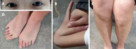
This patient was noted to have a blue tint to the sclera(Figure 1A),joint hypermobility(Figure 1B),abnormal scarring(Figure 1C),and planovalgus feet(Figure 1D).The power of main muscles in four extremities was approximately level III-IV.
Laboratory examinations
Routine blood tests,routine urine tests and urinary sediment examination,routine fecal tests and occult blood test,blood biochemistry,immune indexes,and infection indexes were all normal.
翠姨的妹妹有一张,翠姨有一张,我的所有的同学,几乎每人有一张。就连素不考究的外祖母的肩上也披着一张,只不过披的是蓝色的,没有敢用那最流行的枣红色的就是了。因为她总算年纪大了一点,对年轻人让了一步。
Imaging examinations
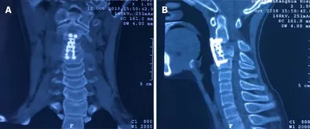
Imaging results(Figure 2)demonstrated that kyphosis at the C3 level was 30° and the cervical spinal cord was compressed from C2 to C4.
TREATMENT
After satisfactory intratracheal anesthesia,the patient was intubated in an appropriate position to avoid excessive activity of the cervical spine.To expose enough field for surgery,a thin pillow was placed behind the shoulder to extend the neck.Through a right-side transverse incision,the cervical spine was exposed carefully.When the gap of C2/3 and C3/4 was certain,a spreader was mounted in the C2 and C4.The intervertebral discs of C3/C4 and C2/C3 were removed to expose the posterior edge of the vertebral body.The operator used rongeur and ultrathin pliers to remove the middle 2/3 of the C3 vertebral body,and the ossified posterior longitudinal ligament was bited.The cartilage surface of C2 and C4 was revealed;the length between C2 and C4 was measured.A titanium mesh was filled with bone from the C3 previously bited and then implanted into the gap between C2 and C4.Because the bone was enough,the iliac crest allograft was not selected.Finally,we used the appropriate titanium nails and plates for the anterior internal fixation.Moreover,during the operation,neuromonitoring was carried out,and no surgical complications were observed.Postoperatively,a satisfactory internal fixation was achieved,and a soft external neck brace was used.
OUTCOME AND FOLLOW-UP
Radiographs 6 wk following anterior cervical corpectomy decompression and fusion showed that the preoperative kyphotic deformity in the cervical spine was significantly corrected(Figure 3).At the 3-mo follow-up,the patient showed significant neurological improvement,as she could walk independly and was able to perform fine movements with her double upper limbs.A computed tomography scan at 5 mo after surgery demonstrated a solid fusion of C2-4 with no displacement of internal hardware anteriorly(Figure 4).Moreover,the normal physiological curvature of the cervical spine was restored.
DISCUSSION
Villefranche Nosology demonstrated the major and minor diagnostic standards for the kyphoscoliosis type of EDS in 1998[4].The major standards include joint hypermobility,decrease of muscle tension,and progressive scoliosis.The secondary standards include abnormal scarring and tissue fragility,osteopenia,miceocorneae,and arterial burst.Hypermobility of the cervical spine may lead to craniocervical unbalance and progressive cervical kyphoscoliosis,usually accompanied by motor disfunction,radiculopathy,myelopathy,and sensory disorders[5].Some studies demonstrated that congenital severe hypotonia was always accompanied by a decrease in muscle power.Patients diagnosed with EDS VI may suffer from myopathy[14].Yişet al[15]revealed that the patients diagnosed with EDS VI were homozygous forPLOD1mutations,leading to either loss of function of LH1 or to its deficit.Surgery may be selected when neurological changes or deformity are progressive.However,there is no article describing only anterior surgical management for patients with cervical kyphosis diagnosed with EDS VI.

Figure1 Clinical features of the girl with Ehlers-Danlos syndrome VI.
The purpose of surgery is to relieve the compression of the cervical spinal cord,restore the normal curvature of the cervical spine,and stabilize the cervical vertebrae.However,internal fixation in underage girls is challenging due to the anatomy and lack of devices designed specifically for this patient.Moreover,in such limited space,when adequate decompression of the cervical spinal cord and restoration of the physiological curvature are performed,the risk of iatrogenic injury to the spinal cord is high.To prevent screw backout,anterior cervical plates need to be used individually.The autograft and allograft have been found to lead to clinical success,but the first is usually recognized as the gold standard[16].Specifically,the bited bone from C3 is enough for grafting in this case,so we did not use any other materials.The external immobilization is an important process after surgery.Halo fixation may be a good choice,but due to the complications of pin loosening and infection,its use is sometimes limited[17].Due to the reliable internal fixation,in this case,we only used soft fixation for 4-6 wk.Lacking of convincing materials,the long-term effect of internal fixation in pediatric patients is not fully clear.There is still much work to be done.However,many studies have found that few children achieve continued growth after fusion of the subaxial spine[18].
CONCLUSION
We report the first case of cervical kyphosis in a pediatric patient with EDS VI,who achieved a satisfactory clinical consequence only following ACCF.There is still some work needed to reveal the details about this case.In addition to some biomolecular and genetic experiments,we may need more time to follow her.

Figure2 Preoperative imaging of a 16-year-old-female with Ehlers-Danlos syndrome VI.

Figure4 Follow-up computed tomography images.
——基于对口述史料的文献分析
 World Journal of Clinical Cases2019年4期
- World Journal of Clinical Cases的其它文章
- Rhombencephalitis caused by Listeria monocytogenes with hydrocephalus and intracranial hemorrhage:A case report and review of the literature
- Primary hepatic leiomyosarcoma successfully treated by transcatheter arterial chemoembolization:A case report
- Long-term follow-up of a patient with venlafaxine-induced diurnal bruxism treated with an occlusal splint:A case report
- Hydrochloric acid enhanced radiofrequency ablation for treatment of large hepatocellular carcinoma in the caudate lobe:Report of three cases
- Therapeutic plasma exchange and continuous renal replacement therapy for severe hyperthyroidism and multi-organ failure:A case report
- Recurrent acute liver failure associated with novel SCYL1 mutation:A case report
