Advances in Evaluation of Facial Asymmetry
Jia-meng HUANG,Juan LI,Jun LIN
Department of Stomatology,the FirstAffiliated Hospital of Zhejiang University,Zhejiang Province,310003,China.
ABSTRACT Facial asymmetry is commonly observed sub clinically in overall population.However,clinically significant facial asymmetry can lead to both functional and aesthetic problems.For patients with facial asymmetry,in order to make appropriate treatment plan,investigation of the underlying etiology and precise clinical examination are essential.The principal aim of this article is to present an invaluable insight into pathogenesis,myriad classifications and various systematic diagnostic approaches indispensable for formulation of treatment plan and appropriate management of facial asymmetry.
KEY WORDS Facial asymmetry;Pathogenesis;Diagnosis
Normally,the human maxillofacial region develops symmetrically on both sides.Theoretically,mirror symmetry can be formed on both sides.However,due to the inherent biological and environmental factors in human development process,there is almost no absolute bilateral symmetry[1,2].Facial symmetry is one of the key determinants of facial attractiveness in psychology and anthropology [3].Human face usually shows minor asymmetry,and minor non-pathological asymmetry is usually difficult to detect and normal.When patients or their parents notice obvious differences on two sides of the face,and clinicians can quantitatively evaluate such differences,this is defined as facial asymmetry[4,5].However,due to the subjectivity of facial aesthetics,the critical value of its clinical significance is not easy to determine,which may depend on the asymmetric region,the clinician’s sense of balance and the patient’s perception of imbalance[4,6,7].
Significant facial asymmetry can cause aesthetic and functional problems,which may adversely affect the patient’s appearance,chewing,breathing and sleeping functions and psychosocial development.Any treatment that fails to achieve the desired results will lead to patients’dissatisfaction [4,5].Therefore,clinicians need more accurate diagnostic assessment and etiological analysis for patients with facial asymmetry,so as to formulate a more reasonable treatment plan.The purpose of this paper is to summarize the pathogenesis,classification and diagnosis of facial asymmetry deformity,which is conducive to accurate diagnosis and provide a basis for the formulation of appropriate treatment plans.
CAUSES OF FACIAL ASYMMETRY
Understanding the causes of facial asymmetry is essential for making treatment plans,facial management and maintaining long-term stability after treatment.
Causes of facial asymmetry can be divided into three different origins [8-9]:i)congenital,which originates from prenatal;ii)developmental,which gradually shows up during development,the etiology of which is not clear;iii)acquired,which is caused by functional mandibular displacement,trauma or disease.
Congenital factors include hereditary and prenatal factors,such as cleft lip and palate,hemifacial micromalformation,neurofibromatosis,congenital muscular torticollis,premature closure of unilateral coronal cranial suture,postural flat skull,etc.[9-11].
Acquired factors included craniofacial trauma,fracture,TMJ infection and arthritis,ankylosis,craniofacial lesions and tumors,Parry-Romberg syndrome and unilateral condylar hyperplasia or dysplasia[8,9,12].
Developmental facial asymmetry is asymptomatic and idiopathic,which is not uncommon in the population.Unlike the other two causes,developmental asymmetry does not exist in infancy.However,as age grows,it gradually appears in the whole process of craniofacial development.It can be obviously observed in puberty without related history of facial trauma or disease[9,13].The etiology is not clear.The possible causes reported in the literature are unilateral chewing,fixed unilateral sleep,bad oral habits or unilateral crossbite.These factors will lead to stimulation of unilateral skeletal development.However,these hypotheses are still controversial and cannot be scientifically validated due to the lack of comparative longitudinal studies[14-16].
CLASSIFICATIONS OF FACIAL ASYMMETRY
There are many classifications for facial asymmetry deformity[2,16-20],two of which are commonly used in clinic.First,Bishara et al.[2]assessed the structures involved in asymmetry and classified facial asymmetry into four categories:dental,osseous,muscular and functional,as shown in Table 2.Second,Wolford[17]proposed that facial asymmetry should be classified into four categories:pseudo-asymmetry,non-pathological asymmetry,unilateral overgrowth,unilateral undergrowth or degeneration,as shown in Table 3.
Various classifications of facial asymmetry deformity reflect the diversity and complexity of clinical manifestations,and provide a reference for clinicians in etiological analysis and diagnosis.
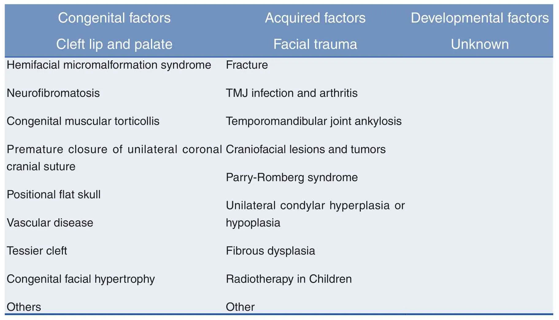
Table 1:Main Causes of Facial Asymmetry
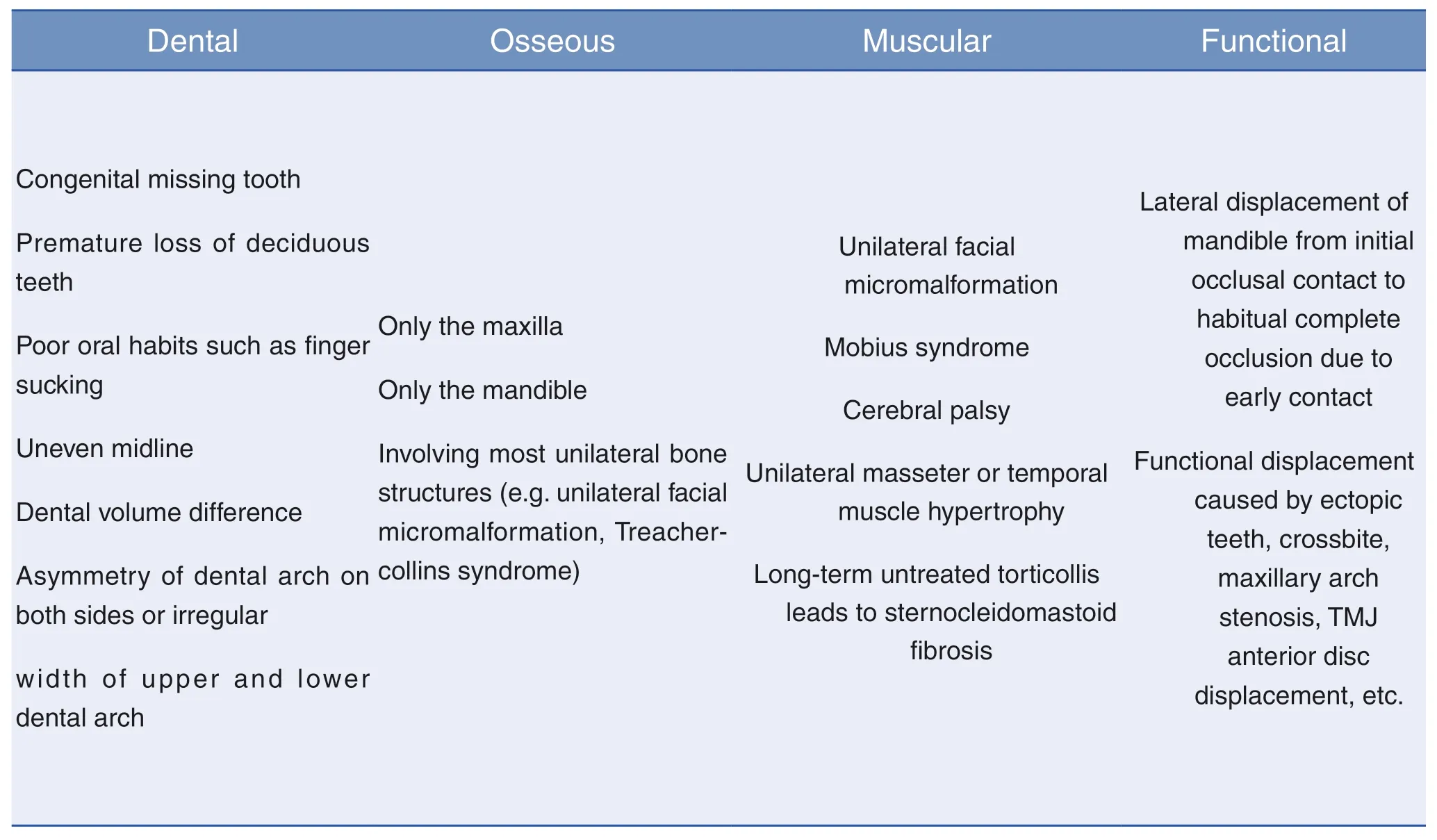
Table 2:Bishara Classification of Facial Asymmetry
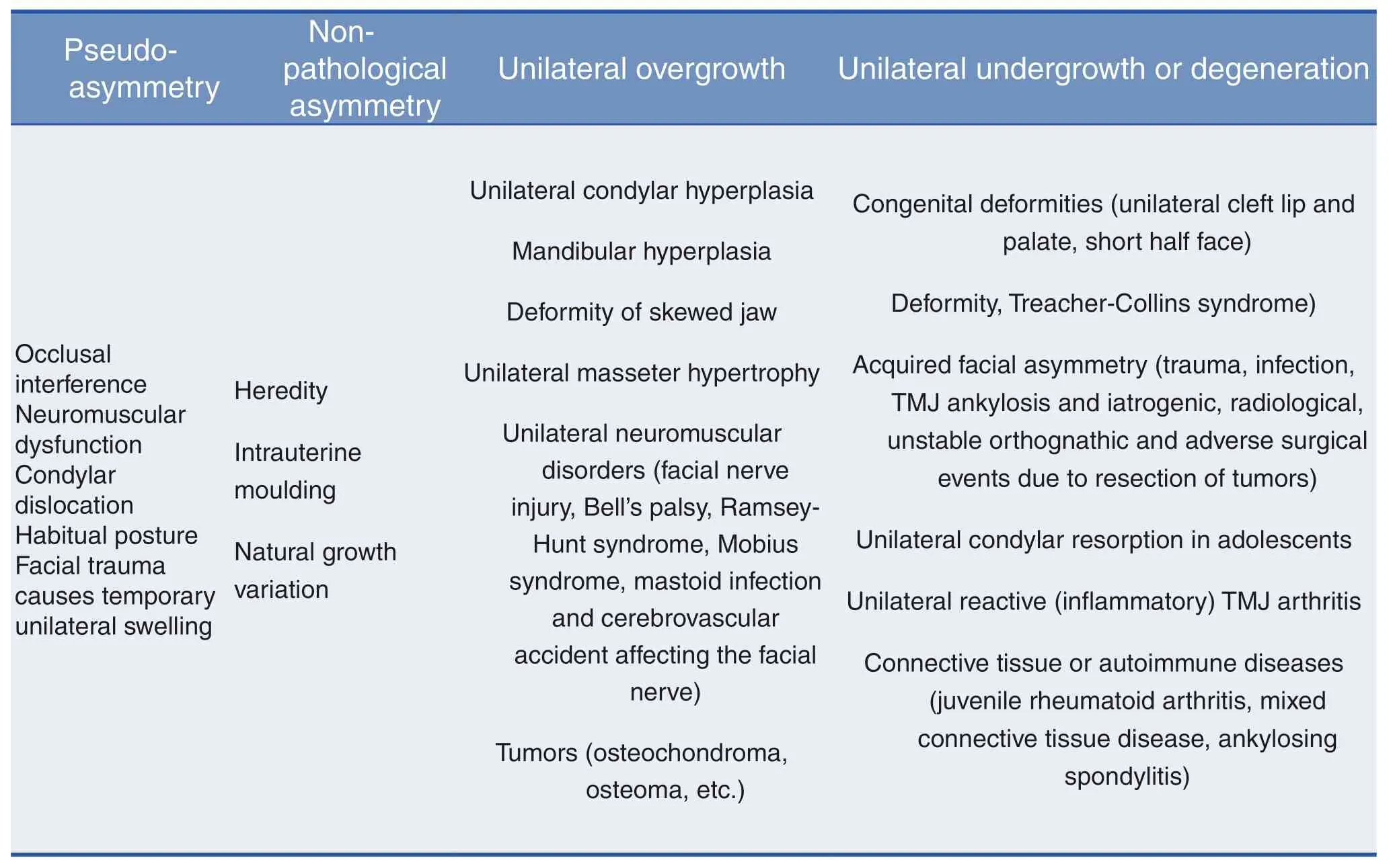
Table 3:Wolford Classification of Facial Asymmetr
EPIDEMIOLOGY AND RELATED FACTORS
Epidemiological studies assess the prevalence of facial asymmetry in orthodontic patients at about 10% [22-23].If the asymmetry is assessed by X-ray,the morbidity could be as high as 50% [24].
Facial asymmetry often occurs in the left mandible,and there is a certain correlation with skeletal growth,temporomandibular joint and other factors.
Most studies have shown that facial asymmetry is more common in the lower 1/3 of the mandible [25].This may be due to the longer growth cycle of the mandible,and the mandible is the skeletal support of the lower 1/3 soft tissues of the face [26-27].The upper jaw has a stable joint area at the base of the skull,and it provides a smaller soft tissue support,which has only a secondary influence on facial asymmetry [28].In addition,facial lateral guidance is mainly on the left side,i.e.mandibular deviation is mostly on the left side [16,29,30].There is no significant difference between men and women [24,31,32].This may be due to the larger size of the right skull and brain and the greater growth potential of the right cranial surface [8].Another potential congenital mechanism may be related to the unbalanced distribution and development of neural crest cells.According to research reports,the migration of neural crest cells occurs earlier on the right side and often delays on the left side [20,28].
Facial asymmetry is associated with skeletal growth.In sagittal direction,facial asymmetry is associated with skeletal type III malocclusion more closely than skeletal type II malocclusion [25,33.As the incidence of type III malocclusion is higher in Asians than in Caucasians,facial asymmetry is more common in normal Asians than in Western countries [9].Some scholars have reported that the prevalence of facial asymmetry in patients with skeletal type I,type II and type III is the same [16].In vertical direction,facial asymmetry is the most common in patients with vertical growth type [25,33].
Facial asymmetry is associated with structural and functional disorders of temporomandibular joint [4,21,34-36].The displacement of articular disc and the change of condylar asymmetry are more serious in patients with facial asymmetry:the skewed condyle moves backward and the contralateral condyle moves forward and downward;the incidence of irreversible anterior disc displacement of articular disc is significantly higher;the mandible rotates and displaces relative to the skull base,and the mouth-opening abnormality [34-36].The changes of condylar position and abnormal mandibular movement aggravate the burden of joint,and the adaptive and compensatory ability of joint itself is gradually weakened,which is very easy to cause temporomandibular joint disorders [34,35].
DIAGNOSIS
For the diagnosis of facial asymmetry,it is necessary to assess the patient’s complaints and expectations,and obtain a detailed and comprehensive medical history,including the history of tooth eruption and treatment,craniofacial lesions,trauma,infection,arthritis,and progressive developmental history [8,37].Then,through the perfect clinical examination,comprehensive imaging examination (including X-ray,CBCT,TMJ imaging,etc.),model analysis,occlusal plate,laboratory examination(electromyogram and ultrasound evaluation),the structure and function of patients are comprehensively evaluated,and the asymmetry in sagittal,coronal and horizontal directions is revealed [1,4,38].These are essential for accurate diagnosis.
CLINICAL EXAMINATION
Extraoral examination involves visual examination of facial morphology,palpation of facial structure and contour to distinguish soft tissue and bone defects,symmetry between bilateral mandibular angle and mandibular contour,mandibular deviation,inclination of lower chin,inclination of occlusal plane,TMJ evaluation,etc.1,4,9,17];when patients are smiling,midline of teeth and midface are examined.Line comparison is used to measure gingival exposure on both sides.Smile symmetry is assessed by the parallelism between the corners of mouth and the pupil lines [4,17,39.Intraoral examination includes crowded dentition,narrow arch,inclination of anterior and posterior teeth,open occlusion,cross occlusion,midline deviation,functional mandibular displacement and maximum opening [9,19,20,37].
EVALUATION OF SOFT TISSUES
The subjective perception evaluation of facial asymmetry depends on soft tissues [1,40,41].Sella-nasion (SN)and Frankfort (FH)planes have been used as horizontal reference planes in various head measurements and clinical assessments.However,for many patients,these reference planes do not match the natural head position and cannot accurately represent the aesthetic perception in daily life.In addition,even through surgery,it is difficult to correct the asymmetry caused by the position of the neck and head.Therefore,clinical evaluation should be carried out in the natural head position [1,42].
2D and 3D facial photographs are helpful in assessing general facial features,asymmetry,and the relationship between upper,middle and lower facial parts,nose and lip.3D facial images have obvious advantages in predicting soft tissue changes [32,40,41]].The commonly used markers,reference lines,linear and angular measurements for soft tissue assessment are shown in Table 4 [43-45].
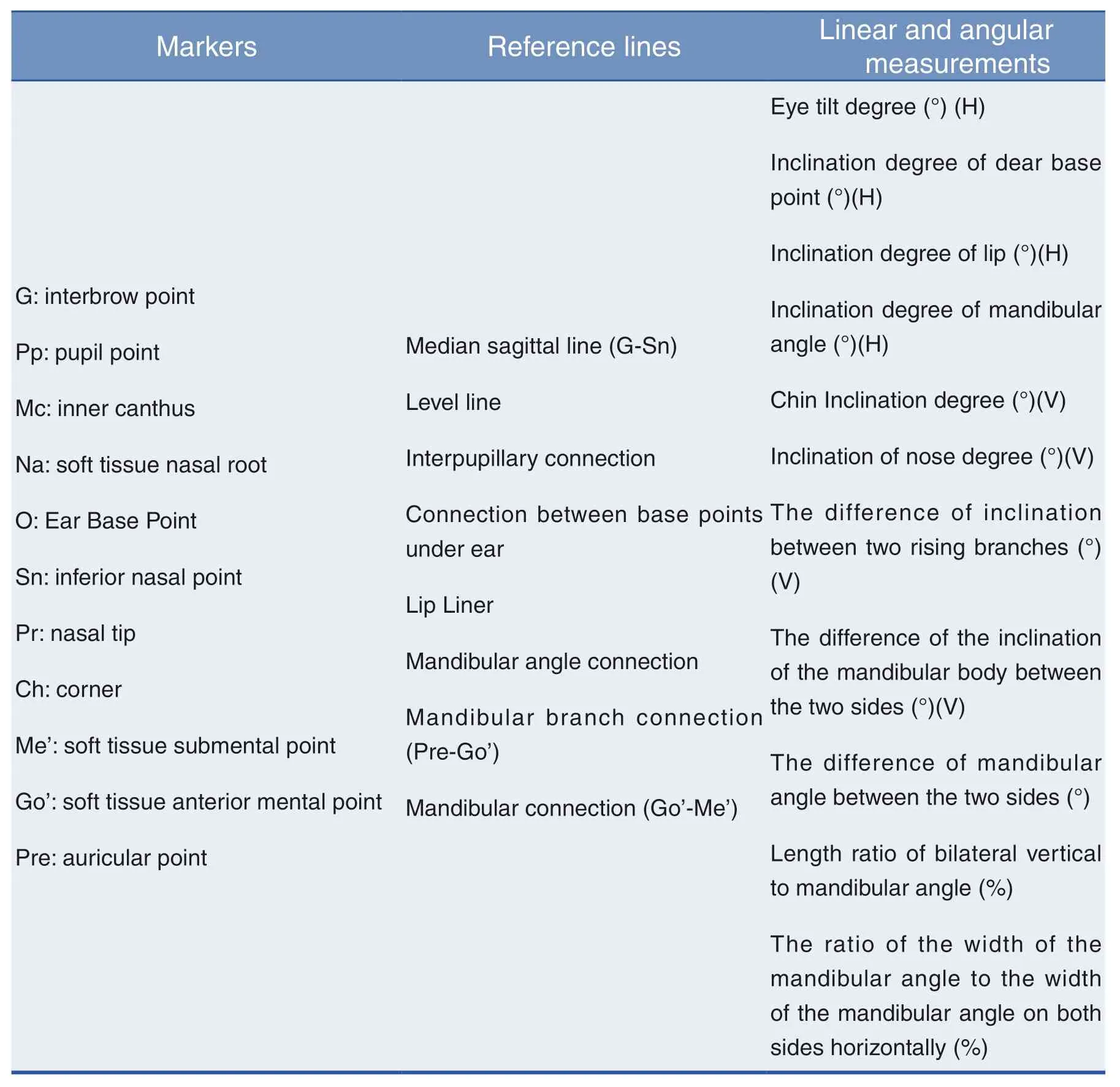
Table 4:Soft Tissue Markers,Reference Lines,Linear and Angular Measurements
ANALYSIS OF OCCLUSION AND TEMPOROMANDIBULAR JOINT
The midline of the teeth should be evaluated in four states:open mouth position,centric relation position (CR),initial contact position and centric occlusion position (CO)[1,4,21].Pure skeletal asymmetry and dental asymmetry have similar midline dislocation in CO and CR positions,but when the asymmetry is caused by occlusal interference,functional mandibular displacement occurs from the initial contact position to CO position.This inclination can occur in the same or opposite direction,and the asymmetry can become more prominent or less obvious[1,4,9].
The lateral inclination of occlusal plane may be caused by the asymmetric development of both sides of mandibular condyle or ascending ramus.The examination can easily determine the slope of occlusal plane[8,17,37]by requiring the patient to bite the tongue depressor,which represents the horizontal occlusal plane,compared with the line between pupils.Inclination of occlusal plane more than 4 degrees often leads to obvious asymmetry of the patient’s face[46].
For unilateral posterior crossbite or open bite,careful examination is needed to determine whether the asymmetry is caused by skeletal,dental or functional occlusion.For example,in the case of facial asymmetry with unilateral posterior progressive open bite,it may be due to the growth of the vertical height of unilateral condyle or mandibular ramus[9,15,18].
The occlusal model can provide indispensable information about the position and arrangement of teeth,the shape and symmetry of dental arch,the occlusal relationship and coordination between upper and lower archs[4,21,47].The occlusal model is transferred to the occlusal framework by facial arch transfer for analysis.The skew and rotation angles of maxilla and mandible relative to skull could be evaluated,and the skew angles of maxilla and mandible relative to occlusal plane and facial midline could be determined[1,9,21].In addition,doctors should assess the presence of asymmetry caused by temporomandibular joint (TMJ)disorders [34-36].For patients with facial asymmetry accompanied by TMJ symptoms,when CO and CR are found to be imbalanced and functional deviation is suspected,the occlusal plate should be worn for a period of time.The occlusal plate can remove the influence of occlusal interference,make the neuromuscular system de-programmed,and guide the mandible to its normal position,so as to present the true facial misalignment of patients.On this basis,further examination and analysis are carried out [15,36,48].
IMAGING EXAMINATION
The panoramic photo shows the same proportion of the upper and lower parts,which is more accurate in vertical analysis.It is mostly used to compare the symmetry of condyle,mandibular ramus and other parts,and treatment plan can be designed on such basis[21,51].
Posterior-anterior radiographs (positive radiographs)are of great significance in the comparison of left and right structures.In addition,the degree of functional skewness can be determined by comparing the two positive radiographs in the open mouth position and the closed mouth position[6].Continuous orthographic charts in years can assess whether asymmetry is progressive[49].In the analysis of positive radiographs,the first step is to establish accurate reference lines:one is to use the line passing through the median structure (such as ethmoid crest,nasal septum,anterior nasal ridge and chin)as the vertical reference line;the other is to use the line connecting the inferior orbital rim as the horizontal reference line,and its vertical line as the vertical reference line.The main analysis methods include Ricketts analysis and Grummons analysis[49-50].
However,the traditional 2D images have some problems,such as image change with head position changing,image overlap,and inconsistent magnification,which have different degrees of impact on facial symmetry research,and the accuracy is not high[26,30,51].
3D images,including CBCT,CT and so on,can analyze the craniofacial complex in three-dimensional,which is conducive to the accurate diagnosis of facial asymmetry[26,38,50].In recent years,CBCT has been widely used in the field of Stomatology[26,52-53].It has the advantages of fast scanning speed,low radiation dose,high resolution of reconstructed image,short reconstruction time,good imaging effect on skull,maxillofacial bone and soft tissue,and can be directly measured and analyzed by threedimensional measurement.By comparing the difference between left and right sides,it can be evaluated.Assessing the degree of asymmetry is of great help to diagnosis,treatment design,treatment simulation and evaluation.The makers and measurements of CBCT analysis are shown in Table 5.
In addition,the application of digital 3D diagnostic and therapeutic design imaging software (Dolphin Imaging,etc.)can integrate CBCT,dental scanning data and facial three-dimensional photographs quickly and accurately,and create patient-specific three-dimensional computer model,which is helpful for three-dimensional measurement and analysis,accurate diagnosis and design,and more intuitive evaluation and prediction of skeletal and soft tissue changes before and after orthognathic surgery,better communication between clinicians and patients[1,4,38,50].
There are also some special imaging methods,such as skeletal scintillation scanning[55](radionuclide scanning),which is a dynamic growth analysis method based on the uptake of Tc-99m by metabolically active bone.It is helpful for the diagnosis and treatment of unilateral skeletal hyperplasia.
According to the order of macro-aesthetics and microaesthetics,the steps of diagnostic evaluation of facial asymmetry are summarized as follows:
(1)Asymmetry of overall appearance is assessed by upper body images and frontal photographs.
(2)Use front and side facial photographs to evaluate facial shape,vertical ratio and width,and obtain 3D photographsif necessary.
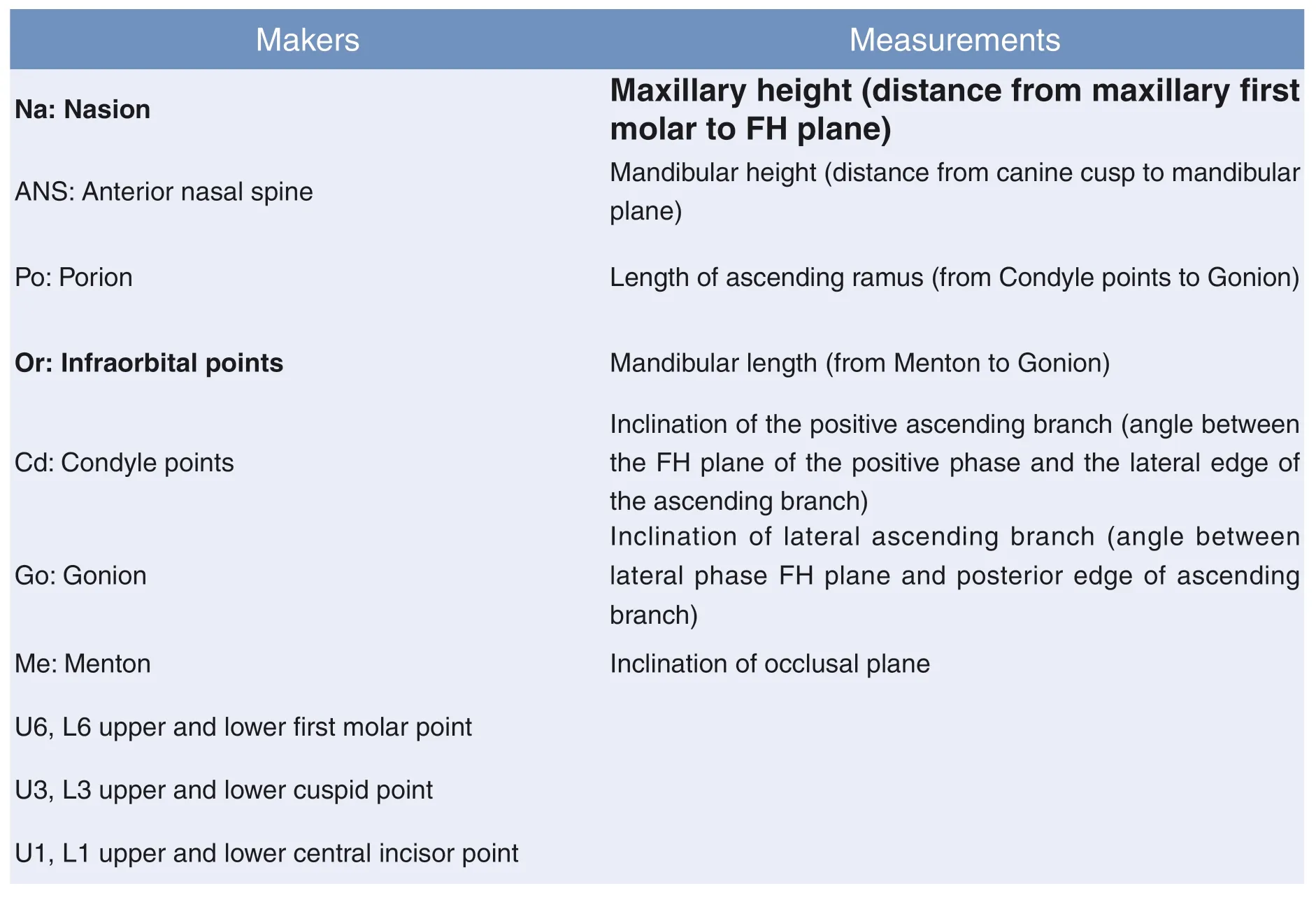
Table 4:Soft Tissue Markers,Reference Lines,Linear and Angular Measurements
(3)The occlusal plane and the midline of the teeth are evaluated directly and indirectly.
(4)Occlusal analysis and TMJ evaluation are performed by intraoral examination and TMJ examination.
(5)Take 2-D and 3-D images,such as orthographic,lateral,panoramic,CBCT,and MRI.Combine with model analysis,make accurate measurement and evaluation,and verify whether they are consistent with the previous information.
(6)Comprehend all data,measure and verify the direction and degree of asymmetry,and analyze the causes of asymmetry.
CONCLUSIONS
This paper summarizes the causes,classifications,epidemic and related factors of facial asymmetry,and diagnostic methods.A deep understanding of facial asymmetry will help to analyze clinical features more rigorously and quantify the degree of asymmetry accurately,formulate the best treatment plan,optimize aesthetics and function,and improve patient satisfaction.
Fund program
Foundation project:Zhejiang Natural Science
Foundation Project (No:LQ18H140004);Zhejiang
Medical and Health Science and Technology Project
(No:2018256920)
 Chinese Journal of Plastic and Reconstructive Surgery2019年2期
- Chinese Journal of Plastic and Reconstructive Surgery的其它文章
- Experience of VSD in the treatment of foot tophi rupture with severe infection wounds
- Clinical Observation of Anesthesia Effect of 2% Lidocaine by Air Pressure Injection in Facial Skin Aesthetic Treatment
- Implantation Instrument on the Expression of Collagen-I and Cell Proliferation Markers in Pathological Scar
- Inhibitory Effect of Photodynamic Therapy on Keloids
- Experimental Effect of Umbilical Cord Mesenchymal Stem Cells on Stress Urinary Incontinence Rats Model and Efficient Tracking in vivo with MRI
- The common complications associated with osteotomy genioplasty:A literature review and meta-analysis
