Experience of VSD in the treatment of foot tophi rupture with severe infection wounds
Peng WANG,Yong-jing HE,Ji-hua WANG,Wei-qi YANG,Li-kun ZHU
Department of Plastic Surgery,Second Affiliated Hospital,Kunming Medical University,Yunnan Province,650101,China
ABSTRACT Objective Investigate the clinical effects of Vacuum Sealing Drainage (VSD)in the treatment of 11 cases of foot tophi rupture with severely infected wounds.Methods From January 2017 to January 2019,11 patients with foot tophi rupture and severe infection were enrolled in our department.There were 9 males and 2 females,aged from 27 to 68 years old.All patients were treated with VSD after debridement.The treatment time was 7d-42d,with an average of 17d.Results All patients were followed up for 6 months after VSD treatment.All the wounds healed well without complications.Conclusion VSD is used to treat foot tophus rupture with severe infection of wounds.It is easy to operate and satisfactory in clinical results.
KEY WORDS vacuum sealing drainage;foot tophi;rupture;severe infection;wound healing
INTRODUCTION
Tophi are characteristic clinical manifestations of chronic gout.They are caused by the continuous deposition of monosodium urate,resulting in deformity with limited joint movement and combined with skin ulcers and infections,which seriously affects the physical and mental health of patients[].Such infected wounds with multi-system diseases,if not treated promptly and effectively,may greatly aggravate the disease and even amputation.Therefore,it is valuable to explore new methods for the treatment of such infectious wounds.
MATERIALS AND METHODS
General Information
From January 2017 to January 2019,11 patients with foot tophi rupture and severe infections were enrolled in our department,including 9 males and 2 females,aged 27-68 years.A total of 13 wounds,the wound formation time was about 3 weeks to 2 months,the shape of the wound was different,the smallest area was 2cm×1cm,the maximum was 7cm×5cm,and the depth was different.All wounds were exudated with foul odor secretions and creating redness and swelling.
Materials
The built-in straw closed negative pressure material(Wuhan Deqiabaier Surgical Implant Co.,Ltd.)contains:(1)The polyethylene ethanol hydrated seaweed foam,models have 15cm×10cm×1cm,15cm×5cm×1cm,5cm×5cm×1cm three kinds;(2)The foam distributes two silicone drainage tubes;(3)The three-way joints and drainage tube clamps;(4)The medical transparent film.
TREATMENT METHODS
Basic Treatment
At the time of admission,blood routine,blood uric acid and biochemical tests were conducted to understand uric acid.X-rays of the foot were used to understand the bone destruction.Colchicine and anti-inflammatory analgesics were choosed to control the acute episode of gout.After the acute episode of gout control,benzbromarone tablets were used to reduce blood uric acid.During the period,the clinician should pay more attention to patients’s pain and notice to review blood uric acid.At the same time,the patients were given symptomatic and supportive treatment such as promoting blood circulation and removing blood stasis,correcting anemia,malnutrition and electrolyte disorder.After admission,the secretion culture and drug sensitivity test were taken.According to the culture results,sensitive antibiotics were used to resist infection.If there is multiple drug resistance,they should be isolated.
Topical Treatment
Debridement:After satisfactory anesthesia,the patients were repeatedly rinsed with hydrogen peroxide and normal saline,and then rinsed with 5%iodophor and normal saline.All open sinus passages were explored,necrotic tissues and non-functional tendon and bone tissues were thoroughly removed,and clotting was thoroughly conducted with electric knife.Installation of VSD:Carefully wipe the dead skin cells at the edge of the wound with sterile alcohol gauze before applying the film.Select different types of foam material to cover the wound surface according to the size of the wound surface,and then attach the medical transparent film.The film coverage covers more than 3cm around the wound surface.If there is a sinus or tunnel,the foam with the pipe should be trimmed to fill the cavity.
Connection negative pressure:connect the tee pipe,connect the extension drainage tube,and then connect the negative pressure source of the bed center.The negative pressure value depends on the size of the wound surface and the liquid leakage condition.Adjust the negative pressure at 100-450mm Hg (1mm Hg=Within 0.133kpa),the foam dressing can be quickly shrunk,the liquid flow in the drainage tube is smooth,and there is no air leakage sound at the listening film,which indicates the sealing is good and the drainage is smooth.
Postoperative management:the situation of negative pressure drainage should be closely monitored after the operation.such as poor drainage can be retrogradely pushed into 15ml-20ml saline,after soaking for 5 minutes,it was taken out and repeated several times until it was smooth.Some patients have pain in the affected area after surgery, which is likely to be caused by excessive negative pressure,the clinician can fine-tune the low negative pressure.Continuously attract 5d-10d,remove the VSD device,observe the wound condition:If the wound was clean,it could be closed and stitched.After suture,VSD treatment was continued.If there are still more necrotic secretions on the wound surface,VSD can be installed after debridement until the wound surface is clean,the granulation is“strawberry-like”,and the wound is reduced and then sutured.
RESULTS
11 patients were followed up for 6 months after treating with VSD for 7d-42d,and all wounds healead well.One of them was located on the left first metatarsophalangeal joint,and the infected wound was at least 2cm×1cm×1cm.After 7 days of VSD treatment,the wound secretion was reduced,the granulation grew freshly,and the wound became smaller and then crawled and healed.The remaining wounds were healed after 3-5 VSD treatments.
TYPICAL CASE
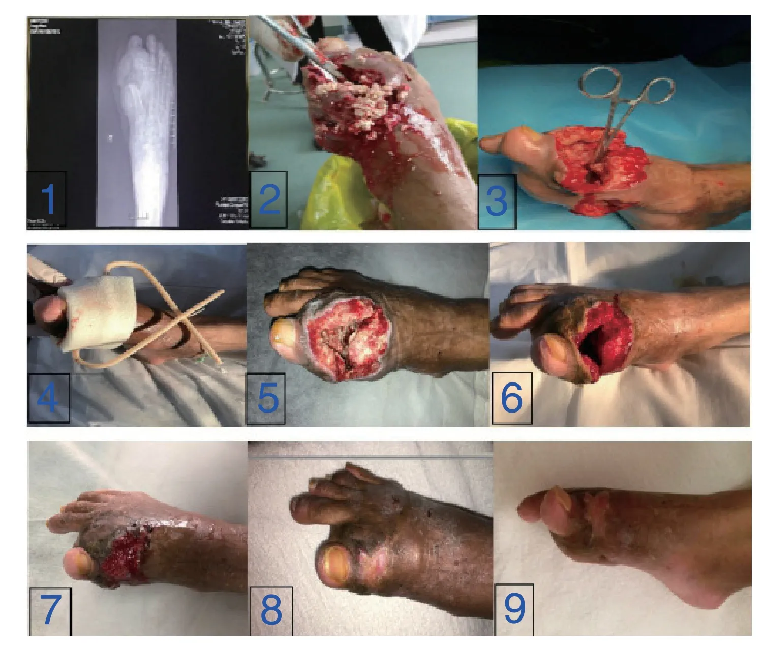
The patient,male,54 years old,was admitted to the hospital at 16:02,September 6,2018,due to“formation of right foot tophi for more than 20 years,ulceration and pus for 3 weeks”.Physical examination:The first metatarsophalangeal joint on the right side was red and swollen,and with a bulge of about 3cm×3cm×3cm in size.The pus was spread around the wound with stench,and a little yellow-white bean curd was exuded.The skin around the wound was black.Auxiliary examination:foot positive position (right side):multiple bone destruction of the right foot bone (Fig.1);test results:blood uric acid 571 umol/L;white blood cells 18.65×10⁹/L.After admission,wound secretion culture was detected:infection with Proteus mirabilis,Staphylococcus aureus,and Klebsiella acidophilus (multi-drug resistant strain).At the same time,according to the results of drug sensitivity test,levofloxacin lactate sodium chloride injection was used to prevent infection,and benzbromarone tablets were used to lower blood uric acid and etoricoxib tablets to relieve pain.The tophi in wounded toe and surface necrotic tissue were removed,and the deep ulcer tissue and part of the bone were removed under the general anesthesia of On September 8,2018.The first VSD was installed for 5 days after the electric knife was completely hemostasis(Fig.3,Fig.4).Postoperative pathological diagnosis:consistent with (right foot)tophi with ulcer formation.On the 5th day after surgery,the VSD was opened,and the granulation of the right foot toe was found.The base was rosy and the cavity was reduced (Fig.5),which proved that VSD treatment was effective.The second time after debridement,VSD was installed for 7 days.During the period,the blood biochemistry was reviewed:uric acid 330 umol/L;blood routine:white blood cells 6.17x10⁹/L.On the 7th day,the closed negative pressure material was opened,and the wound surface was healed better and the cavity was further reduced (Fig.6).After debridement,the wound was sutured in the local anesthesia,and the third time VSD was installed for 9 days.On the 9th day,the wound cavity had been filled with fresh granulation tissue(Fig.7).The combined rheumatoid-associated antibody test results were negative.After discharge from hospital,the patient was combined with diet and oral medication to control blood uric acid,and regular review.The patient was followed up for 6 months,the wound was healed well(Fig.8,Fig.9).

Fig.1.foot positive position (right side);Fig.2.Preoperative;Fig.3.Intraoperative debridement;Fig.4.VSD was installed postoperatively;Fig.5.Immediately after the first VSD 5d;Fig.6.Immediately after the second VSD 7d;Fig.7.Immediately after the third VSD 9d;Fig.8.The frontal photos was followed up after 6 months of discharge;Fig.9.The profile photos was followed up after 6 months of discharge.
DISCUSSION
Tophi Diagnosis
There are many methods can be used in the diagnosis and assessment of the tophus,including X-ray、ultrasound (US)、magnetic resonance (MRI)、computed tomography (CT)and dual energy CT (DECT)[].In addition to assessing the location and number of deposition of monal-sodium urate crystals,the damage degree of joints and inflammatory reactions,the above examinations can also evaluate the therapeutic effect of lowering uric acid[].The detection of monosodium urate crystals in puncture contents,which remains the gold standard for the diagnosis of gout[].Furthermore,CT has a good differentiation effect on tophi,which is superior to MRI and ultrasound in the evaluation of internal joint tophi.Tophi present a high-density mass on CT,and the CT value of tophi is different from other space-occupying lesions3.DECT is an imaging examination method developed in recent years.It uses X-ray with different energy and CT value changes corresponding to tissues to obtain color coding reflecting histochemical composition through relevant software processing,which can find more deposits of gouout than physical examination,bringing hope for non-invasive diagnosis of gouout4.However,it should be recognized that DECT has many limitations.For example,it is not consistent with the results measured by physical methods,and it can only detect the highdensity tophi (the uric acid content in the tophi is at least 15%~20%).When the uric acid content is lower than this level,no matter the size of the tophi,DECT cannot detect it[].
How to Treat Tophi
Foot tophi rupture is a common complication of gouty arthritis[],clinical manifestations include swollen pain in the affected area,localized ulceration,exudation of yellowwhite bean dregs-like secretions,and even inclusion of dead bones,pus and blood,some patients with deeper wounds form multiple sinuses,fistulas or concurrent infections.The wounds are difficult to heal in the short term.In severe cases,the wounds lead to walking obstacles and may even face the risk of puncture or amputation,causing pain to the patient and increasing the burden on the family and society[].Therefore,the treatment of patients with foot tophi rupture and severe infection should be paid attention to by clinicians.Clinically,there are many treatments for foot tophi,including surgical therapy[],extracorporeal shock wave[],vacuum sealing drainage technique[],urate oxidase in vitro dissolution of tophi[],Chinese medicine non-drug therapy[1],etc.,all of which can achieve certain effects.
The Advantage of The VSD
VSD was first proposed by Fleisehmann[],Department of Trauma Surgery,ULM University,Germany,and applied to the treatment of soft tissue injury of limbs,and achieved good results.VSD is a wound formed by hydrating seaweed salt foam with polyethylene glycol or covering skin or soft tissue defects,infection and necrosis.It promotes wound healing by providing a moist,negative pressure,hypoxia and slightly acid environment for wounds,and improving microcirculation in the wound[].It is widely used in trauma,orthopedics,burns,general surgery and other departments,suitable for the repair of various types of infected wounds and dead space,except active bleeding,anaerobic infection and cancerous wounds.
The main advantages are:(1)VSD negative pressure foam material achieves drainage through strong capillary siphon action;Multi-side hole drainage tube facilitates timely elimination of necrotic tissue and exudate from wounds;(2)VSD negative pressure foam material is a hydrophilic material,and granulation tissue does not grow into the interior of the material (a unique hydration membrane protects granulation tissue,demolition of materials will not pull bleeding),which promotes wound healing;(3)Simultaneously fill the VSD negative pressure foam material in the wound surface and wound cavity,which can be multi-directional drainage,drainage is more sufficient;(4)Reduce the number of dressing changes,greatly reducing the suffering of patients,to some extent,also reduce the workload of medical staff;(5)Avoid the opportunity of cross-infection in the hospital during opening dressing;(6)Shorten the wound healing time of patients and improv the clinical turnover rate.
Treatment Experience
Currently,benzbromarone and probenecidare commonly used in clinic to promote uric acid excretion and allopurinol that inhibits the synthesis of uric acid[7].The author selected colchicine and anti-inflammatory painkiller in the first few days after the patients were admitted to hospital.After the acute gout outbreak was controlled,the benzbromarone was used to decrease serum uric acid.According to international recommended indicators,serum uric acid level should be kept below 360 mmol/L for a long time[].During this period,the author paid more attention to the pain relief of the patients and paid attention to the review of serum uric acid.At the same time,the wound secretions were retained for pathogen culture and drug sensitivity test during the first debridement and dressing change,and the antibiotic sufficient course of treatment was selected according to the drug sensitivity test results.This group of patients with foot tophi rupture with severe infection,if not timely and effective treatment,it is likely to face truncated toe or amputation.Therefore,with the help of scholars at home and abroad,the author applies VSD to such wound treatment and has satisfactory clinical results.Zhao kun et al.[]have used surgical resection combined with VSD to treat patients with tophi combined with infection.For some urates that are difficult to remove,during the operation,repeating washing with sodium bicarbonate solution or hydrogen peroxide solution and placing VSD after the operation can promote the discharge of urate,and obtained certain therapy.Tejera et al.[]believe that combined operation with VSD for the treatment of patients with gout complicated with infection can significantly inhibit the reproduction of wound bacteria and prevent the occurrence of cross infection.Meanwhile,VSD can create moist and protective base wounds for patients with gout,which is conducive to the recovery of postoperative joint function and improvement of pain score.Fei zhijun et al.[]reported that 16 cases of giant tophi combined with bone destruction and soft tissue necrosis treated with surgical excision combined with vacuum sealing drainage,and the clinical effect was satisfactory.Zhang zhanlei et al.[]reported that for patients with giant tophi,the clinical effect of surgical excision of stones combined with drug therapy was significantly better than that of drug therapy alone.For asymptomatic patients with gout whose tophi are larger than 1 cm,surgical resection of tophi is also recommended to better control the acute onset of gout.For patients with tophus smaller than 1cm,active conservative treatment,diet control,close observation and surgical resection are recommended.
During the treatment,the author found that it is difficult to completely remove the tophi from the debridement under local anesthesia.If the VSD negative pressure treatment is used at this time,the VSD drainage tube is easily blocked and the treatment effect is not good.Therefore,the author recommends:First,in general anesthesia or in the case of spinal anesthesia,according to the X-Xay of positive oblique position of the foot to guide the debridement range,debridement is more thorough and clear the tophi and remove the deep ulcer tissue and partially destroyed bone.Electrocoagulation is able to stop bleeding,after then install a flush tube in the deep part when installing the VSD,which prevents the deep residual stone from blocking the drain tube.Secondly,the first time to install VSD treatment can be advanced to the 4d or 5d,although tophi are very cleaned during the operation.The first time the VSD is installed,outflowing more residual tophi and the VSD is not opened in time,which is easy to cause drainage tube occlusion and delays wound healing.The second and third time of VSD treatment can be extended to the 8d and 9d respectively,and the granulation of the wound is better.
Complications in Treatment
The Pain
All patients have different degrees of pain after installing VSD,and the pain can be tolerated.This may be related to excessive pressure or continuous attraction of negative pressure suction.The medical staff needs to know the location,nature and extent of the patient’s pain and find the cause and symptomatic treatment.
The Obstruction of The Drainage Tube
It may be related to the clogging of the patient’s tophi,the thickening of the drainage fluid,the bulging of the dressing,and the folding of the drainage tube.The 60 ml syringe is used to extract the saline pulse flushing.It can also be pumped when flushing.The pulse flushing has a certain pressure and impact force,which can make blood clots and large secretions adhering to the wall of the VSD drainage tube fall off,decompose and easily be sucked out[].Replace the VSD if necessary.
Negative pressure failure
It may be due to medical transparent film leakage or negative pressure source shutdown.Relabel the membrane in the leaking place,and explain that patients and their families should pay attention to the folding of drainage tube after installing VSD,and timely report any problems.The negative pressure source cannot be turned off by itself.
Anemia
Some patients are afraid of gout attacks and only eat vegetarian food on the diet,leading to chronic anemia and hypoproteinemia.High-quality animal protein and low-salt diet should be promoted under the condition of strict monitoring of serum urea nitrogen and electrolytes.Eggs,milk and fresh water fish should be properly supplemented,and patients should be encouraged to eat more fruits and vegetables rich in vitamins such as cherries and tomatoes to promote uric acid excretion[].
CONCLUSIONS
In summary,for patients with foot tophi rupture and severe infection,antibiotics are selected according to secretion culture and drug sensitivity test,and basic treatments such as uric acid and nutritional symptom support are provided.After thorough debridement,VSD treatment is applied,which is not only easy to operate but also conducive to postoperative wound secretion drainage,reducing infection,improving pain and promoting wound healing.
About the author
Peng WANG,Department of Plastic Surgery,The second affiliated hospital of kunming medical university.Research Fields wound repair and facial contour plasty.Email:1653660076@qq.com.
Li-Kun ZHU,Department of Plastic Surgery,The second affiliated hospital of kunming medical university.Email:623532076@qq.com.
HE Yong-Jing,male,PhD,Working in Department of Plastic Surgery,The second affiliated hospital of kunming medical university.Majoring in microsurgical free flap repairing.Email:30970568@qq.com.
Wei-qi YANG,Department of Plastic Surgery,The second affiliated hospital of kunming medical university.Email:147965925@qq.com.
Corresponding author:Ji-Hua WANG,Department of Plastic Surgery,The second affiliated hospital of kunming medical university.Specialiaed in research of microetaloplasty and reconstruction of whole ear,repairing and reconstruction of nasal defect.Email:wangjihua1966@163.com.
 Chinese Journal of Plastic and Reconstructive Surgery2019年2期
- Chinese Journal of Plastic and Reconstructive Surgery的其它文章
- Clinical Observation of Anesthesia Effect of 2% Lidocaine by Air Pressure Injection in Facial Skin Aesthetic Treatment
- Implantation Instrument on the Expression of Collagen-I and Cell Proliferation Markers in Pathological Scar
- Inhibitory Effect of Photodynamic Therapy on Keloids
- Experimental Effect of Umbilical Cord Mesenchymal Stem Cells on Stress Urinary Incontinence Rats Model and Efficient Tracking in vivo with MRI
- The common complications associated with osteotomy genioplasty:A literature review and meta-analysis
- Advances in Evaluation of Facial Asymmetry
