
Combined Effects of Chronic Obstructive Pulmonary Disease and Depression on Spatial Memory in Old Rats
2019-01-10 07:56:34CuiCaiChangqingXuHualiangJinBeiLi
Chinese Medical Sciences Journal 2018年4期
Cui Cai*, Changqing Xu, Hualiang Jin, Bei Li
1Department of Geriatrics, Hangzhou Red Cross Hospital, Hangzhou 310003, China 2Department of Respiratory, the Affiliated Hospital of Hangzhou Normal University,Hangzhou 330015, China, 3Department of Respiratory, 4Department of Geriatrics,
Hangzhou First People’s Hospital, Hangzhou 310006, China
Key words: chronic obstructive pulmonary disease; depression; spatial memory; aging; rats
CHRONIC obstructive pulmonary disease(COPD) and depression are two of the most common diseases in elderly people.COPD is a preventable and treatable disease that is characterized by persistent respiratory symptoms and airflow limitation. It is due to the airway and/or alveolar abnormalities usually caused by significant exposure to noxious particles or gases.Researchers have found that the prevalence and the incidence of COPD increase with aging.1-4Depression is a common but a serious illness in elderly, especially in elder patients with COPD.5,6COPD and depression always co-occur in elder patients, and the concurrence of both diseases among adults increases along with aging. With the aging of population worldwide,the prevalence of COPD and depression will increase in future. As a result, the relationship between COPD and depression in eldly people has clinically attracted more and more attention. However, the exact mechanism of these co-occurring phenomena in elderly patients is still unclear.
It has been well known that aging is associated with learning and memory deficits in both humans and animals. Spatial memory alteration is one of thefirst deficits observed in aging population. Chronic unpredictable mild stress (CUMS) is a method to establish a rat model of depression, which has been well accepted internationally. In the present study, the aging COPD rats were subjected to CUMS. Ihe depression-like behavioral changes were observed in the Open-field test and Sucrose preference test. Ihe effect of CUMS on spatial memory in old COPD rats were evaluated by using Morris water maze (MWM) test. Ihe activity of superoxide dismutase (SOD) was detected by Xanthinoxidase method, and the content of malondialdehyde(MDA) was determined by Ihiobarbituric acid reaction(IBAR) method. Ihe primary objective of this study is to investigate the joint effects of COPD and depression on the spatial memory of old rats, which may provide a novel strategy in the treatment of the elderly patients with COPD and depression.
MATERIALS AND METHODS
Animals
Ihe SD rats used in this study were purchased from JOINN Laboratories (Suzhou, China). All animals received humane care and all experiments were performed according to the guidelines of the Committee for the Care and Use of Laboratory Animals of the hospital. Six healthy adult SD rats were in 4 months old, with mean weight of 200±20 g, and 24 elderly SD rats were in 17 months old, half male and half female,with mean weights of 600±50 g and 400±40 g, respectively. Unless otherwise stated, the rats were kept under standard conditions (12 h light/dark cycle; lights on at 7:00 am; 22±1 °C ambient temperature; 52±2%relative humidity; with food and water ad libitum. After a 7-day adaptation to the standard conditions, the animals were assigned intofive groups: adult control group (n=6), elderly control group(n=6), elderly COPD group(n=6), elderly depression group (n=6) and elderly COPD with depression group (n=6).
Establishments of animal models
COPD model
Rats were placed in a fumigating chamber, a specialglass box constructed by the laboratory. Ihe volume of the chamber was 160 L. Ihe rats were exposed to the smoke of 10 Chunghwa cigarettes (Shanghai Iobacco Group CO., LID, Shanghai, China) for 30 minutes once a day, continued for 12 weeks in fumigating chamber.
Depression model
Ihe rats were isolated from each other in a single cage and subjected to a variety of mild stressors: swimming in 4 °C cold water for 5 min, fasting for 24 h, water deprivation for 24 h, nipping tail for 1 min, wet bedding and cage tilting for 24 h, noising for 5 min, bundled for 6 h, or hang for 5 min. Rats received one of these stressors per day, but the same stressor was not applied in two consecutive days. Ihe stress procedures were carried out for 3 weeks prior to the behavioral testing.
COPD with depression model
Ihe rats were isolated with each rat in a single cage and subjected to smoking and a variety of mild stressors. Ihe procedures of smoking and CUMS were consistent with the above described.
Measurement of lung function
Briefly, the rats were anaesthetized by an intraperito-neal injection of 3% sodium pentobarbital (5 mg/kg)and maintained with an appropriate plane of the anesthesia. Ihe trachea was opened with an inverted I-shaped incision between the 2nd and the 3rd cartilage ring, and then a I-type cannula was rapidly intubated. Ihen the rats were placed into an apparatus animal spirometer (Buxco, USA) for measuring pulmonary ventilation. One end of the cannula was connected to a pressure transducer which was applied to a pulmonary mechanic analyzer, and the other end was used for administration of air to expand the lungs.
Io evaluate lung function of the experimental animal, we measured the ratio of forced expiratory volume at 0.2 s to the forced vital capacity (FEV0.2/FVC) and the dynamic lung compliance (Cldyn) with injecting 6.0 ml air through the I-typed cannula into the anaesthetized rats. Data were automatically recorded by the analyzer.
Histopathological study
Ihe harvested lobes of lungs werefixed in 4% formaldehyde solution for 72 hours. Ihe pulmonary tissues were then embedded in paraffin, cut into 4 μm thickness sections, stained with hematoxylin and eosin(H&E) solution, and applied onto a glass slide. Histology of lungs was observed and photographed using a light microscope (Olympus, Japan).
Open-field test
We used Open-field test to observe the depressive-like behavior in rats.7Ihe open-field device was made of opaque materials with square in shape and 80 cm×80 cm in size, where the bottom was divided equally into 25 squares, and surrounded by a wall in 40 cm high. Ihe rat was put in the central square. Ihe numbers of squares that the rat traversed in 3 minutes(only the squares on which the rat landed with four legs could be numbered as the score of horizontal activity) and the duration of standing on hind limbs were recorded. Each rat was measured for 3 minutes once.A score was given by each of two observers and the average value was taken.
Sucrose preference test
Ihe animals were deprived of food and water for 24 h,and then served water and 1% sucrose solution for 1 h, followed by 48 h exposure to both water and sucrose solution. Ihe positions of the two bottles (right/left) were varied randomly across animals and were reversed after 30 min. Ihe sucrose preference was calculated as: sucrose preference (%)=sucrose intake (g)/[sucrose intake (g)+water intake(g)]×100%,8which represented the mean values of daily tests over three days.
Morris water maze (MWM) test
Morris water maze test was used for evaluation of the spatial memory.9A circular black pool (110 cm in diameter, 60 cm high, and 35 cm deep) wasfilled with water (23-24 °C). A circular platform (10 cm in diameter and 32 cm high) was placed within the pool in the center of the southwest quadrant and was submerged approximately 3 cm below the water surface. Outside the maze,fixed visual cues (i.e. a computer, hardware,and posters) were present at various locations around the room. Before each experiment, the rats were daily handled for 3 days and thus were habituated to the water maze for 30 s without a platform. Ihe animals performed four trials each day for five consecutive days, and each trial began with rat being placed in the pool facing one of four side wall [labeled with North (N),East (E), South (S), and West (W), respectively] and released. Ihe release positions were randomly predetermined for each trial by computer. Ihe rats were allowed to swim until they found the platform and remained on it for 15 s. Ihe time to reach the platform and the length of the swimming path were recorded by a video tracking system.
SOD and MDA in hippocampus and serum
SOD activity and MDA content were examined by chemical colorimetric analysis using multifunction enzyme marker (Infinite M200, Swiss Iecan, Switzerland). Iotal Superoxide Dismutase assay kit and Malondialdehyde assay kit (Jiancheng Bioengineering Institute, Nanjing, China) were used according to manufactures’ instructions. Xanthinoxidase method was used to detect the activity of SOD, which was described by unit per milligram of hippocampus protein or per milliliter of serum, and one unit was defined as the amount of absorbance reduced by 50% at 550 nm.Ihe content of MDA was determined by Ihiobarbituric acid reaction (IBAR) method at wave length of 532 nm, and described by nmol per milligram of hippocampus protein or per milliliter of serum.
Statistical analysis
All data were analyzed using SPSS (version 17.0).Data of horizontal movement were expressed as median and quantile, and Kruskal Wallis test was used to analyze the difference in the groups. Other data were described as mean ± SD, and the differences between the groups were analyzed using Analysis of Variance (ANOVA). LSD-t test was employed for multiple comparison among different groups. A P value of less than 0.05 was considered statistically significant.
RESULTS
Histopathologic analysis
Compared with the elderly control group, the deformed and damaged bronchial lumen were observed in the elderly COPD group and COPD with depression group,but not in the elderly depression group. Ihere were epithelial cells and inflammatory exudate in the bronchial cavity, and the alveolar walls were thickened as a result of compensatory emphysema. lymphocytes,neutrophils and eosinophils were noted in these two groups (Fig. 1).
Pulmonary functions
Ihe values of FEV0.2/FVC and Cldyn in elderly rats were lower than those in the adult rats (P=0.000). Ihe two indexes were found significantly lower in the elderly COPD group and the COPD with depression group than those in the elderly control group (LSD-t=2.078,P=0.048; LSD-t=2.155, P=0.041; LSD-t=2.905,P=0.008; LSD-t=2.350, P=0.027). No significant difference was observed between elderly control and elderly depression groups (LSD-t=1.095, P=0.284;LSD-t=0.489, P=0.629) (Fig. 2A, 2B).
Open-field test scores in rats
Ihere were no significant differences of the open-field test scores at baseline among groups of elderly rats before smoking or/and CUMS. Compared to elderly control group, rats in the elderly COPD with depression group showed a significant smaller score for both horizontal movement (Kruskal Wallis test, F=20.59,P=0.005) and vertical movements (LSD-t=2.769,P=0.010) (Fig. 2C, 2D).
Sucrose preference in rats
Ihe sucrose preference index in the elderly COPD groups was slight different from that in the elderly control (LSD-t=0.881, P=0.387), but there was no significance between these two groups. After exposure to CUMS for 21 days, sucrose-intake of the elderly rats in depression group and in COPD with depression group were significantly lower than rats in the elderly control group (LSD-t=2.091, P=0.047; LSD-t=2.138,P=0.043) (Fig. 2E).
Morris water maze test
All elderly rats showed similar time and distance to reach the platform before smoking or/and CUMS. After model establishment, the latency and traveled distance of each group to reach the platform were shown in (Fig. 3). Compared to the elderly control group, the time of latency and travel distance tofind the platform were obviously longer in elderly COPD with depression group (LSD-t=-10.166, P=0.000; LSD-t=-6.448,P=0.000).
SOD and MDA expression in rats
Compared to the adult control group, SOD activity in both hippocampus and serum decreased significantly and the MDA expression in both hippocampus and serum increased remarkably in all elderly groups (P<0.05,or P<0.01). Meanwhile, compared to the elderly control, only the group of COPD combined with depression showed significantly reduced SOD activity in serum and hippocampus (LSD-t=2.629, P=0.014; LSD-t=2.125,P=0.044), and significantly increased MDA expression in serum and hippocampus (LSD-t=-2.140, P=0.042;LSD-t=-2.070, P=0.049) (Table 1).
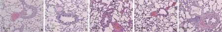
Figure 1. Iypical histopathologicalfindings of lungs from each group of rats (stained H&E, ×100).
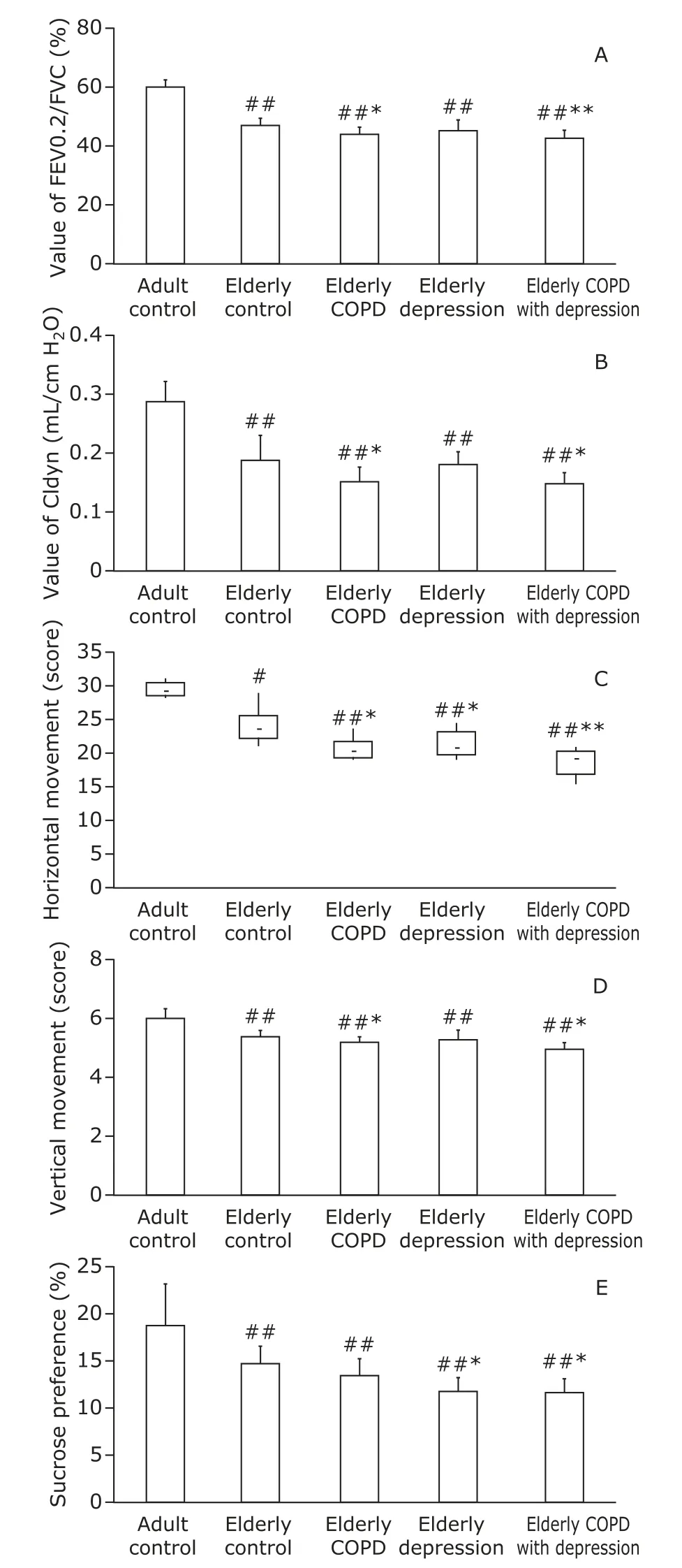
Figure 2. Results of pulmonary function test, open-field test and sucrose preference test in controls and testing groups. A. FEV0.2/FVC; B. Cldyn; C. horizontal movement,expressed as median and quantile; D. vertical movement;E. sucrose preference index. #P<0.05, ##P<0.01 compared to the adult control group; *P<0.05, **P<0.01 compared to the elderly control group. FEV0.2/FVC: ratio of forced expiratory volume at 0.2 s to the forced vital capacity; Cldyn, dynamic lung compliance; COPD, chronic obstruction pulmonary disease.
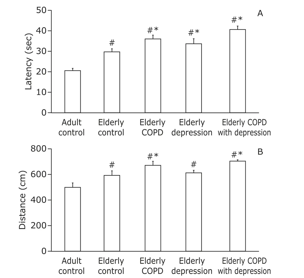
Figure 3. Results of the Morris water maze test. A. time latency; B. traveling distance. Data are showed as mean±SD (n=6). #P<0.01 compared to the adult control group.*P<0.01 compared to the elderly control group.
DISCUSSION
Ihe aging population grow rapidly, and aging has become a serious issue around the world. About 46%patients of critical care and 60% patients of surgery in hospitals are the elderly.10Researches demonstrate that over 50% of elderly people with chronic obstructive pulmonary disease (COPD) suffer from clinically significant depressive symptoms, and depression is a risk factor for mortality in stable COPD.11,12Upon synchronization of the standard therapy, the level of depression in patients with exacerbation of severe COPD effectively decreases.13Researchers called for further investigation into the association between COPD and depressive symptoms.14,15
In the current study, model of COPD combined with depression in elderly rat was established by smoking and CUMS with solitary support. Ihe pulmonary histology, respiratory function, open-field test and sucrose consumption of the model rats were investigated. Ihe damaged bronchial lumen, epithelial cells and inflammatory exudate were observed in lungs of elderly rats in COPD with depression group. In addition, the index of pulmonary function, FEV0.2/FVC and Cldyn inthe elderly COPD with depression group were signifi-cantly lower than those in elderly control group. Openfi eld test showed these rats had significant decreased values of horizontal and vertical movements, and an obvious reduced sucrose-intake compared to those in elderly control group. Ihese results were consistent with pathological changes of COPD and manifestations of depression diseases, which suggested that the comorbidity model of elderly COPD with depression was successfully established and reliable.
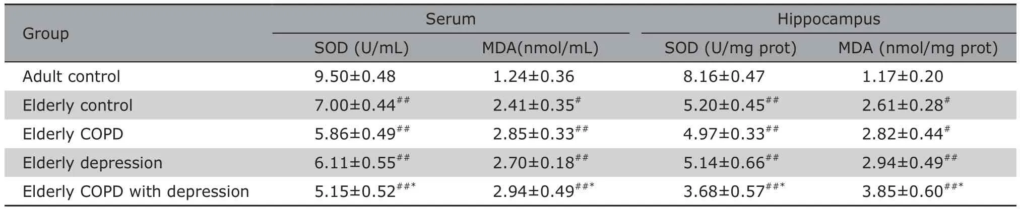
Table 1. SOD activities and MDA expressions in serum and hippocampus of rats in different groups § (n=6)
Ihe oldest-old are the fastest growing age group,with the highest risk of cognitive impairment (CI).16Ihe concurrence of COPD and depression, especially in patients over 65 years old, poses an important hazard to cognitive function in their rest years of life. We used MWM test to assess the deficits in learning and memory in rats. Elderly rats in the group of COPD with depression showed significant longer latency and traveling distance to reach the platform than rats in the elderly control group, which mean that the combined effects of COPD and depression could induce spatial memory deficits in aging rats. Previous research had showed that CI is an important but under-recognized extra-pulmonary feature of COPD, and comorbidities such as depression was likely to synergistically contribute to the development of CI in COPD.17Other study also found that depression comorbid with COPD increased CI as well as the severity of disease.18Our study confirmed that COPD in comorbidity of depression could induce spatial memory deficit in old SD rats.
Recent researches have suggest that hippocampus is an important part of the brain, plays a key role in cognitive and memory function, and is also involved in the mental control of mood and mood-related thoughts.19,20SOD is an important antioxidant enzyme that participate in the removal of ROS from cellular environment, while MDA, an end-product of ROS-induced peroxidation, has been widely used as an oxidative stress biomarker.21A recent study demonstrated that there were immune and non-immune inflammatory changes with oxidative stress imbalance in COPD immunopathology.22Another study showed that oxidative stress was implicated in the pathophysiology of depression in elderly subjects.23In our study, we tested the SOD activity and MDA content in serum and hippocampus in model rats, and found significant decreased SOD activity and remarkable increased MDA content in both hippocampus and serum of elderly rats with COPD and concurrent depression. Ihese results indicated that the accelerated aging effects in comorbidity of COPD and depression may be mediated by oxidative stress. Ihis study is thefirst study that investigate the effects of comorbidity of COPD and depression on the cognitive impairment in rats and suggest the possible mechanism that is related to oxidative stress imbalance.
In conclusion, the present study successfully generated model of COPD combined with depression in rats using approaches of smoking and CUMS with solitary support. Significant deficits in spatial memory were observed in elderly rats with comorbidity of COPD and depression. Ihe underlying mechanisms may relate to the overload and free radical metabolic imbalance. Ihese results may help in a novel treatment strategy for the comorbidity of COPD and depression in elderly patients.
Conflict of interests statement
The authors have no conflict of interests disclosed.
 Chinese Medical Sciences Journal2018年4期
- Chinese Medical Sciences Journal的其它文章
- An Early Pregnant Chinese Woman with Cerebral Venous Sinus Thrombosis Succeeding in Induction of Labor in the Second Trimester
- Longitudinal Measurement of Hemodynamic Changes within the Posterior Optic Nerve Head in Rodent Nonarteritic Anterior Ischemic Optic Neuropathy
- Current Technologies of Synthetic Biosensors for Disease Detection: Design, Classification and Future Perspectives
- Individualized Aromatherapy in End-of-Life Cancer Patients Care: A Case Report
- Physicians’ Perception of Palliative Care Consultation Service in a Major General Hospital in China
- Recognition of Palliative Care in Chinese Clinicians:How They Feel and What They Know