斜外侧腰椎椎间融合治疗腰椎退行性疾病疗效分析
王凯 孙澎 菅凤增 吴浩
(首都医科大学宣武医院神经外科,北京 100053)
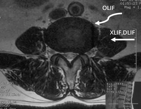
随着腰椎融合手术的广泛开展,腰椎椎间融合(lumbar interbody fusion, LIF)逐渐成为治疗退行性腰椎疾病的常用技术[1]。前方腰椎椎间融合(anterior lumbar interbody fusion, ALIF),后方腰椎椎间融合(posterior lumbar interbody fusion, PLIF)以及经椎间孔椎间融合(transforaminal lumbar interbody fusion, TLIF)治疗腰椎退行性疾病均可以获得良好效果[2-4],但脊神经、腹部大血管、腹腔脏器、输尿管及自主神经系统的医源性损伤也有报道[5-7]。外侧腰椎椎间融合(extreme lateral interbody fusion, XLIF)和直接外侧腰椎椎间融合(direct lateral interbody fusion, DLIF)[8-9]均为微创腰椎椎间融合术,通过腹膜后脂肪及腰大肌到达椎间盘侧方,与传统开放手术相比出血少,手术时间短,住院时间短,围手术期疼痛也更轻[8]。但其神经损伤并发症的报道也同样存在[10-11]。斜外侧腰椎椎间融合(oblique lateral interbody fusion, OLIF)技术通过腹膜后腰大肌及腹主动脉或下腔静脉间隙到达椎体前外侧缘(图1),从而避免了腰大肌中腰丛神经、腹主动脉和下腔静脉损伤,兼具ALIF和XLIF的优点,又弥补了ALIF易损伤血管及XLIF易损伤神经的缺点[1,9]。本研究通过回顾我院行OLIF治疗的腰椎退行性疾病患者相关资料,分析OLIF治疗腰椎退行性疾病的有效性及相关并发症。

图1 OLIF、XLIF及DLIF入路示意
Fig 1 Approaches to OLIF, XLIF, and DLIF
DLIF and XLIF accessed the lateral side of vertebral body though psoas muscles while the corridor of OLIF was between psoas muscles and aorta abdominalis.
对象与方法
一、临床资料
回顾性纳入2016年10月至2017年1月首都医科大学宣武医院神经外科收治的腰椎退行性疾病患者22例。收集患者病史及影像学资料。纳入患者:诊断为退变性脊柱侧弯、退变性腰椎管狭窄、椎间盘源性下腰痛、退变性腰椎滑脱及翻修手术, 经保守治疗无效或保守治疗后反复发作的顽固性腰背部疼痛和下肢放射痛,神经压迫症状进行性加重,严重影响日常生活和工作,行OLIF治疗(DePuy spine, Raynham, MA, USA)。排除标准:诊断为外伤、肿瘤、感染的患者。
二、斜外侧腰椎椎间融合手术
患者取侧卧位,取髂前上棘与肚脐连线中外1/3平行于腹外斜肌长约4~6 cm切口。通常选择左侧入路,也可根据患者情况选择右侧入路(例如成人脊柱侧弯凸向左侧)。逐层分离腹壁肌肉到达腹膜后间隙。钝性分离腹膜后间隙,将腹膜后脏器推向前方,腰大肌向后方牵开,建立OLIF工作通道(DePuy spine, Raynham, MA, USA)。X线定位手术节段。经椎体前外侧切除椎间盘髓核及上下终板软骨,试模逐级置入椎间隙充分撑开满意后,将填压同种异体颗粒骨的椎间融合器(DePuy spine, Raynham, MA, USA)植入椎间隙。应选择尽量宽的椎间融合器以避免椎体沉降;若需要恢复矢状位腰前凸,可选择有倾斜度的椎间融合器。退变性侧弯患者、融合节段超过3个节段、影像学存在腰椎滑脱或峡部裂的患者及第一例OLIF患者行后路导航下经皮椎弓根螺钉固定。术中均未行电生理监测。通常患者术后第二天可在支具保护下下地活动。
三、影像学评估
术前均行腰椎X线正侧位、前屈后伸位、双斜位,腰椎CT及三维重建,腰椎MRI。术后行腰椎X线正侧,腰椎CT及三维重建,腰椎MRI(术后出院前、术后3个月及末次随访)。
四、临床指标、疗效评价与随访
统计患者年龄、BMI、手术时间、术中出血量、术后住院时间和并发症,纪录术前、术后1 w、术后3个月和末次随访的视觉模拟评分(visual analogue scale, VAS)、Oswestry功能障碍指数评分(The Oswestry disability index, ODI)及36项简明健康状况调查评分(Medical Outcome Study 36-Item Short-Form Health Survey, SF-36)评价手术疗效及恢复情况。术后随访6~8个月。
六、统计学方法

结 果
一、手术疗效
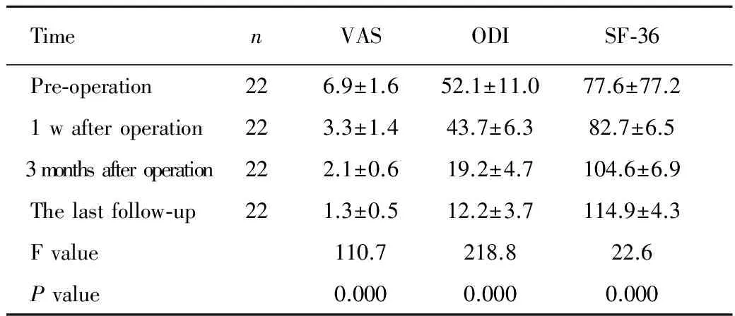
本组22例患者,其中男10例,女12例,年龄52~80岁,平均(67.5±8.1)岁,BMI 18.7~35.1 kg/m2,平均(26.1±3.7)kg/m2。患者诊断包括:退变性侧弯患(6例),退变性腰椎管狭窄(21例),腰椎滑脱(1例),椎间盘源性下腰痛(1例),翻修手术(1例)。患者主诉为间歇性跛行17例,主诉为疼痛16例。本组患者共行OLIF融合45个椎体节段,范围覆盖L2-5节段,其中1个椎体节段融合5例,2个椎体节段融合11例,3个椎体节段融合6例。手术时间90~586 min,平均(239.3±165.3)min,术中出血量40~1200(226.4±310.2)mL;其中行后路内固定患者每个融合椎体节段手术时间62~362 min,平均(217.4±92.1)min,术中出血量33~400(240.6±153.8)mL;未行后路内固定患者每个融合椎体节段手术时间33~164 min,平均(75.3±32.5)min,其中1例因腰大肌肥厚,暴露困难,单节段手术时间明显长于其他病例(164 min),术中出血量17~60(38.1±14.1)mL。行后路内固定患者由于需要变化体位并行导航下经皮椎弓根螺钉固定,单节段手术时间较未行后路内固定时间长(P<0.001)。术后平均住院日(5.1±2.1)d(2~11 d),22例患者均获随访,随访时间6~8个月,平均(7.0±0.9)个月。末次随访与术前比较VAS评分及ODI评分降低,SF-36评分提高(均P<0.001,表1、2)。椎间融合率达100%,且椎弓根螺钉位置良好。
二、手术并发症
本组中1例患者出现腰大肌受损症状,表现为左侧大腿屈曲力量减弱,术后2 w逐渐恢复。2例患者术中椎体局部静脉丛撕裂损伤(第3例及第5例)。共15个椎间融合节段从随访影像观察到椎间融合器沉降,其中10个椎间融合节段为1度沉降,5个椎间融合节段为2度沉降,1例2度沉降患者术后2 w出现疼痛症状一过性加重,但末次随访患者症状显著缓解,VAS、ODI及SF-36评分获得改善。所有患者均未出现感染、腹部大血管损伤、腹腔脏器损伤、输尿管损伤及腰丛神经损伤。


TimenVASODISF-36 Pre-operation226.9±1.652.1±11.077.6±77.2 1wafteroperation223.3±1.443.7±6.382.7±6.5 3monthsafteroperation222.1±0.619.2±4.7104.6±6.9 Thelastfollow-up221.3±0.512.2±3.7114.9±4.3 Fvalue110.7218.822.6 Pvalue0.0000.0000.000
Note: VAS: Visual analogue scale; ODI: The Oswestry disability index; SF-36: Medical outcome study 36-item short-form health survey.
表2 本组患者术前术后临床指标两两比较
Tab 2 Paired comparison of the clinical indices before and after operation

PairedcomparisonVAStPODItPSF-36tP Pre-operation∶1wafteroperation10.10.0005.90.00 -7.20.000 Pre-operation∶3mafteroperation12.80.00014.10.00 -13.00.000 Pre-operation∶Lastfollow-up15.60.00018.50.00 -27.50.000
讨 论
斜外侧腰椎椎间融合手术最早由Mayer等于1997年报道[12],2012年Silvestre等首次将这种融合技术命名为斜外侧腰椎融合术(OLIF)[1]。作为新近出现的治疗腰椎退行性疾病的微创手术技术,与传统技术相比,OLIF具有出血少、创伤小、患者恢复快、住院时时间短及感染发生率低等优势。本研究回顾性分析了OLIF治疗腰椎退行性疾病的有效性及相关并发症。
一、OLIF治疗腰椎退行性疾病的有效性
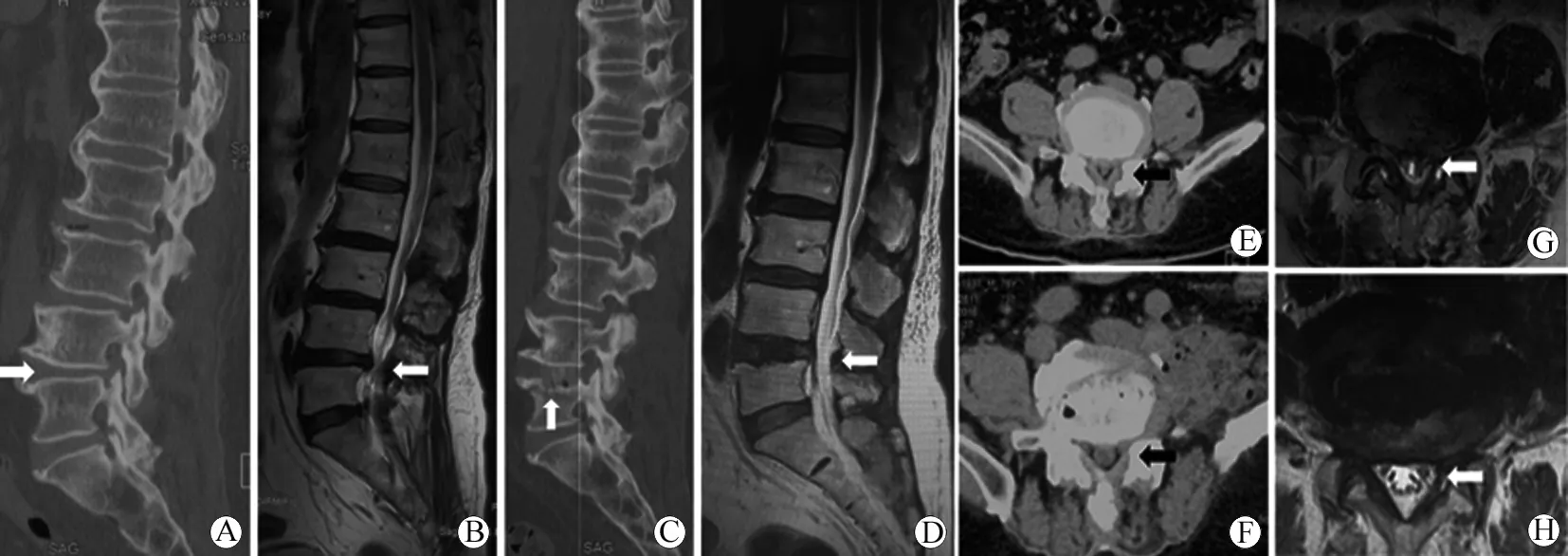
手术治疗退行性腰椎疾病的疗效首先取决于减压充分程度,传统的脊柱后路手术主要通过去除椎板、增生的韧带和骨质进行直接减压[13],而ALIF、XLIF及OLIF则通过椎间融合及恢复间高度,对椎间孔及椎管进行间接减压。Ohtori等报道39例ALIF未行后路减压及固定,平均随访12年,76%的患者效果满意[7,14];Youssef等[15]报道了未行后路减压及固定的XLIF的间接减压效果,84例行XLIF患者中68例获得坚固的椎间融合及疼痛和功能评分改善;Oliveira等[16]报道了XLIF患者术后获得良好效果及影像学改善。OLIF从前路可置入更大的椎间融合器,切除膨出椎间盘,更好的恢复椎间高度,拉伸黄韧带,不切除后方椎板韧带而间接减压椎管[17-18](图2)。
对于伴有成人退行性脊柱侧弯的患者,单纯椎管减压术可在短期内明显缓解临床症状,但对脊柱侧弯的进展、脊柱失稳以及轴性背部疼痛无效,甚至在减压术后因脊柱结构的破坏,上述症状可能加重,进而需再次手术治疗。因此,对于成人退行性脊柱侧弯的患者还需要考虑改善脊柱矢状位及冠状位序列,矫正脊柱畸形[9]。由于退行性侧弯的患者脊柱僵硬、柔韧性差,为了获得更好的矫形效果,常需要进行截骨。截骨手术可延长手术时间,并且手术创伤大、出血多,增加了脊柱的不稳定因素[19]。随着微创脊柱外科技术的发展,通过MIS-TLIF、OLIF等椎间融合技术,通过多个椎间隙植入椎间融合器,逐步调整脊柱曲度可达矫形。在经皮螺钉植入并安置钛棒后,通过加压和撑开进一步矫形,兼顾脊柱冠状位和矢状位的平衡,可取得良好效果[20-21]。相较于后路手术,OLIF从前路可置入更大的椎间融合器,改善退行性脊柱侧弯患者冠状位及矢状位序列效果更显著[19, 22](图3)。

图2 78岁男性,下腰痛3年,间歇性跛行1年,伴双侧足背麻木疼痛,行L4-5Stand-alone OLIF,术后临床症状缓解
Fig 2 A 78-year-old male, suffered from low back pain and intermittent claudication, accompanied with pain and numbness of dorsal part of bilateral feet. L4-5stand-alone OLIF was performed.
A: Pre-operative sagittal CT showed narrowing of intervertebral space of L4-L5(white arrow); B: Pre-operative sagittal T2WI MRI showed compressing of dura-sac and narrowing of spinal canal of L4-L5(white arrow); C: Post-operation sagittal CT showed the intervertebral space of L4-5had been restored (white arrow); D: Post-operative sagittal T2WI MRI showed indirect decompression spinal canal of L4-5(white arrow); E: Pre-operative axial CT showed narrowing of spinal canal (black arrow); F: Post-operative axial CT showed indirect decompression spinal canal (black arrow); G: Pre-operative axial T2WI MRI showed narrowing of spinal canal (white arrow); F: Post-operative axial T2WI showed indirect decompression spinal canal (white arrow).

图3 64岁女性,下腰痛14年,伴间歇性跛行,近1年来症状加重,左小腿外侧及足背麻木。行L2-5OLIF及后路经皮椎弓根螺钉固定矫形融合
Fig 3 A 64-year-old female suffered from low back pain and intermittent claudication for 14 years, which aggravated with numbness of lateral calf and dorsal of foot of left side. L2-5OLIF with posterior percutaneous pedicle screw fixation.
A: AP view of pre-operative standing full length spinal X-ray showed scoliosis. The Cobb angle was 51°, with trunk shift and rotation of apical vertebrate; B: Lateral view of pre-operative standing full length spinal X-ray showed sagittal malalignment of the patient; C: AP view of post-operative standing full length spinal X-ray showed correction of scoliosis. The Cobb angle was 5°, with little trunk shift and derotation of apical vertebrate; D: Lateral view of post-operative standing full length spinal X-ray showed re-alignment of sagittal plane.
本组患者术后较术前腰椎VAS评分降低、ODI指数降低、SF-36评分提高,表明手术对缓解患者腰椎管狭窄及治疗腰椎退行性疾病确切有效。
二、Stand-alone技术的应用
Stand-alone技术是指仅行前路椎间融合而不行后路内固定,在XLIF和OLIF中均有应用,并取得了良好效果[9,15]。影像学评估可见患者椎管及椎间孔均较术前扩大,术后仅极少数患者需再行后路减压内固定[16-17]。虽然OLIF对恢复椎间高度及脊柱矢状位序列效果良好,但是部分患者可能在术后出现椎间融合器沉降,导致临床症状缓解有限,甚至一过性加重。因此,有文献建议,所有OLIF患者均应行一期后路内固定[9]。本组病例行Stand-alone OLIF的患者中共15个椎间融合节段从随访影像观察到椎间融合器沉降,其中5个椎间融合节段为2度沉降,但所有患者末次随访症状获得缓解,疼痛和功能评分改善。提示对于1~2个融合节段、影像学除外腰椎滑脱、峡部裂或退变性脊柱侧弯的患者,采用Stand-alone OLIF技术可能是安全有效的。
三、OLIF的并发症
OLIF可能出现的并发症包括感染、内固定失败、神经损伤、腹腔重要血管损伤、腹腔脏器损伤、输尿管损伤等,文献报道总的并发症发生率达48.3%[9,23]。但包括椎间融合器沉降、腰大肌无力及臀部麻木等绝大多数并发症是一过性的。与后路手术相比,OLIF对后方肌肉、韧带等张力带结构破坏少。与XLIF相比OLIF对腰大肌破坏少及腰丛损伤风险更低。本组病例中出现的并发症包括腰大肌受损、椎体局部静脉丛撕裂损伤及椎间融合器沉降,但末次随访,患者症状均显著缓解,疼痛及生活质量评分获得改善。
四、电生理监测
由于XLIF经腰大肌进行手术操作,术中可能对腰丛神经造成损伤,尤其在L4-5间隙,是股神经、髂腹下神经和髂腹股沟神经损伤的高危区域[24]。术中实时电生理监测可以降低神经损伤风险,但仍有62.7%的患者会出现短暂臀部麻木、疼痛等神经损伤症状[10-11]。OLIF通过腰大肌及腹主动脉或下腔静脉间隙到达椎体前外侧缘进行手术,可避免损伤腰丛神经。本组病例均未行术中实时电生理监测,无一例患者出现腰丛神经损伤症状,表明OLIF手术可不依赖术中实时电生理监测且神经损伤风险较XLIF低。
对于腰椎退行性疾病有多种治疗方式,本研究证实OLIF治疗腰椎退行性疾病创伤小、出血少、术后恢复快、疗效确切,术后患者疼痛及功能评分均可获得显著改善,且血管、神经损伤的并发症风险更低,是治疗腰椎退行性疾病的安全有效的微创手术方式。
1SILVESTRE C, MAC-THIONG J M, HILMI R, et al. Complications and morbidities of mini-open anterior retroperitoneal lumbar interbody fusion: oblique lumbar interbody fusion in 179 patients [J]. Asian Spine J, 2012, 6(2): 89-97.
2GILL K, BLUMENTHAL S L. Posterior lumbar interbody fusion. a 2-year follow-up of 238 patients [J]. Acta Orthop Scand Suppl, 1993, 251: 108-110.
3ISHIHARA H, OSADA R, KANAMORI M, et al. Minimum 10-year follow-up study of anterior lumbar interbody fusion for isthmic spondylolisthesis [J]. J Spinal Disord, 2001, 14(2): 91-99.
4CHASTAIN C A, ECK J C, HODGES S D, et al. Transforaminal lumbar interbody fusion: A retrospective study of long-term pain relief and fusion outcomes [J]. Orthopedics, 2007, 30(5): 389-392.
5SASSO R C, BEST N M, MUMMANENI P V, et al. Analysis of operative complications in a series of 471 anterior lumbar interbody fusion procedures [J]. Spine, 2005, 30(6): 670-674.
6OKUDA S, MIYAUCHI A, ODA T, et al. Surgical complications of posterior lumbar interbody fusion with total facetectomy in 251 patients [J]. J Neurosurg-Spine, 2006, 4(4): 304-309.
7TAKAHASHI K, KITAHARA H, YAMAGATA M, et al. Long-term results of anterior interbody fusion for treatment of degenerative spondylolisthesis [J]. Spine (Phila Pa 1976), 1990, 15(11): 1211-1215.
8OZGUR B M, ARYAN H E, PIMENTA L, et al. Extreme lateral interbody fusion (xlif): a novel surgical technique for anterior lumbar interbody fusion [J]. Spine J, 2006, 6(4): 435-443.
9OHTORI S, ORITA S, YAMAUCHI K, et al. Mini-open anterior retroperitoneal lumbar interbody fusion: oblique lateral interbody fusion for lumbar spinal degeneration disease [J]. Yonsei Med J, 2015, 56(4): 1051-1059.
10BERGEY D L, VILLAVICENCIO A T, GOLDSTEIN T, et al. Endoscopic lateral transpsoas approach to the lumbar spine [J]. Spine (Phila Pa 1976), 2004, 29(15): 1681-1688.
11CUMMOCK M D, VANNI S, LEVI A D, et al. An analysis of postoperative thigh symptoms after minimally invasive transpsoas lumbar interbody fusion [J]. J Neurosurg Spine, 2011, 15(1): 11-18.
12MAYER H M. A new microsurgical technique for minimally invasive anterior lumbar interbody fusion [J]. Spine, 1997, 22(6): 691-700.
13CHUNG S D, YU H J, CHUEH S C. Re: CHING-CHIA LI, TU-HAO CHANG, WEN-JENG WU, et al. Significant predictive factors for prognosis of primary upper urinary tract cancer after radical nephroureterectomy in taiwanese patients. Eur urol 2008; 54:1127-35 [J]. Eur Urol, 2009, 55(4): e69-70; author reply e71.
14OHTORI S, KOSHI T, YAMASHITA M, et al. Single-level instrumented posterolateral fusion versus non-instrumented anterior interbody fusion for lumbar spondylolisthesis: a prospective study with a 2-year follow-up [J]. J Orthop Sci, 2011, 16(4): 352-358.
15YOUSSEF J A, MCAFEE P C, PATTY C A, et al. Minimally invasive surgery: lateral approach interbody fusion: results and review [J]. Spine, 2010, 35(26 Suppl): S302-311.
16SATO J, OHTORI S, ORITA S, et al. Radiographic evaluation of indirect decompression of mini-open anterior retroperitoneal lumbar interbody fusion: oblique lateral interbody fusion for degenerated lumbar spondylolisthesis [J]. Eur Spine J, 2017, 26(3): 671-678.
17OLIVEIRA L, MARCHI L, COUTINHO E, et al. A radiographic assessment of the ability of the extreme lateral interbody fusion procedure to indirectly decompress the neural elements [J]. Spine, 2010, 35(26 Suppl): S331-337.
18FUJIBAYASHI S, HYNES R A, OTSUKI B, et al. Effect of indirect neural decompression through oblique lateral interbody fusion for degenerative lumbar disease [J]. Spine, 2015, 40(3): E175-182.
19吴浩, 王曲, 林彦达, 等. 微创经椎间孔腰椎间融合术联合经皮椎弓根螺钉内固定长节段融合术治疗退行性腰椎侧弯 [J]. 中国现代神经疾病杂志, 2016, 16(4): 197-203.
20ACOSTA F L, LIU J, SLIMACK N, et al. Changes in coronal and sagittal plane alignment following minimally invasive direct lateral interbody fusion for the treatment of degenerative lumbar disease in adults: a radiographic study [J]. J Neurosurg-Spine, 2011, 15(1): 92-96.
21MANWARING J C, BACH K, AHMADIAN A A, et al. Management of sagittal balance in adult spinal deformity with minimally invasive anterolateral lumbar interbody fusion: a preliminary radiographic study [J]. J Neurosurg-Spine, 2014, 20(5): 515-522.
22OHTORI S, ORITA S, YAMAUCHI K, et al. Mini-open anterior retroperitoneal lumbar interbody fusion: oblique lateral interbody fusion for degenerated lumbar spinal kyphoscoliosis [J]. Asian Spine J, 2015, 9(4): 565-572.
23ABE K, ORITA S, MANNOJI C, et al. Perioperative complications in 155 patients who underwent oblique lateral interbody fusion surgery: perspectives and indications from a retrospective, multicenter survey [J]. Spine (Phila Pa 1976), 2017, 42(1): 55-62.
24DAVIS T T, BAE H W, MOK J M, et al. Lumbar plexus anatomy within the psoas muscle: implications for the transpsoas lateral approach to the L4-L5disc [J]. J Bone Joint Surg Am, 2011, 93(16): 1482-1487.

