面向组织工程的多细胞结构三维自组装成形方法的研究进展
朱晓璐,杨逸飞
(河海大学机电工程学院,江苏常州 213022)
面向组织工程的多细胞结构三维自组装成形方法的研究进展
朱晓璐,杨逸飞
(河海大学机电工程学院,江苏常州 213022)
多细胞结构是由细胞群体构成的有机体,其体外构建对于组织工程和再生医学的发展具有重要的基础意义。利用细胞自身的自组织特性构建三维(3D)多细胞结构正成为生物制造和组织再生的一种重要途径,并受到越来越多的关注。对三维多细胞结构的自组装式构建与调控的相关基础研究及关键技术进行了综述及分析,主要涉及凝胶内3D细胞培养、多细胞结构可控形成,及其与图灵反应-扩散机制的联系等方面的研究工作。为进一步研究多细胞3D自组装机理,使得该自组装过程可控,且满足同时调控外部施加和细胞自身分泌的作用因子的浓度梯度分布的需求,提出对内部结构特征梯度化的3D凝胶体内细胞3D自组装模型进行研究,以推进三维多细胞结构及组织前体形成的理性调控技术。
仿生学;生物制造;三维细胞培养; 结构梯度化凝胶; 三维细胞自组装
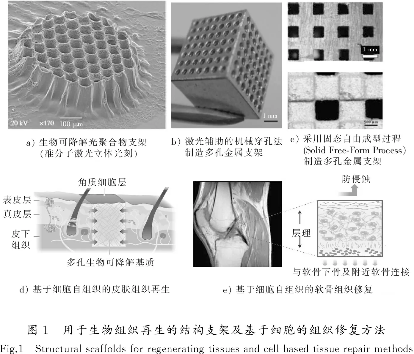
近年来,人体受损组织或器官的再生技术受到越来越多的关注[1-6]。为实现活体组织与器官的结构再造和功能再生(生物制造),研究人员设计并制造各种机械结构支架(Scaffolds)并将其广泛应用于生物组织工程,且支架的制造逐渐向着更为复杂和精细的方向发展[7-12],比如通过基于准分子激光的立体光刻制造生物光聚合物支架[11](图1 a)),通过激光辅助的机械穿孔法制造多孔金属支架[13](图1 b)),以及采用固态自由成型制造多孔金属支架[13](图1 c))等。在重建生物组织在微环境中的结构特征方面,各种各样的支架结构通过各自独特的结构特点和表面涂层及处理,使得相应类型的细胞能够黏附其上并促使细胞按照特定的几何特征排列生长,还可根据设计需求实现相应的降解过程,这为组织工程学的发展作出了一定的贡献。然而,虽然支架的制造更加精细,但支架仍然很有可能由于其自身的降解,诱发的免疫原性反应,以及其他不可预料的并发症而带来应用中的问题[14],而且细胞在生长过程中会发生位置移动,其最终生成的多细胞体系结构有时会偏离最初设计的几何特征。

另外,基于细胞的3D打印技术也是生物制造领域中一项重要的技术手段[15-17],其主要通过将包裹细胞的微单元体(也有文献称之为bio-ink[14])按照逐层堆积打印的方式形成最终的多细胞结构体或器官结构,其能够高效地制造具备特定几何结构的三维多细胞结构体以及组织/器官结构雏形。其主体思想是借助外力(3D打印机)实现三维几何结构的再造,然而组织/器官甚至多细胞体系都非常复杂,主要依靠几何约束(3D逐层堆积),并不一定保证其化学生物方面功能的全面实现[18]。如果能够充分地依靠生物细胞自身所具备的先天性再生能力,使其完全自发的自组装成为组织或器官结构(尽可能模拟自然界中的作用机制),而非主要利用外力将其塑造成一定几何结构,将会为促进生物制造领域的发展进一步拓宽研究思路[18-19]。所以,近年国际上越来越多的相关研究工作正逐渐开始重视细胞和组织的先天固有的自我组织(self-organizing)特性,通常也可以称为细胞自组装。而且,很多种基于细胞的治疗方法被提出并应用于重组或恢复组织功能[6,20-22], 例如,图1 d)所示的皮肤全厚度(含表皮层、真皮层和皮下组织)创伤的修复;图1 e)所示的用于软骨修复的基质与细胞混合植入物,该植入物稳固地附着于软骨下骨及附近软骨,并具有合适的Ⅱ型胶原蛋白密度及定向,以承受关节处的剪切力。这些方法主要依赖于细胞自身的生长和3D自组装过程。因此,研究细胞在三维基质(3D)中的自组装对于组织工程、再生医学和生物制造的发展有着重要意义。
1 面向组织工程的细胞自组装研究进展
研究细胞固有的自组装过程以及调控方法,通常需要构建适合的细胞外基质以达到模拟人体内自然生理环境的目的。目前在国内外相关研究工作中,在凝胶系统中的三维空间(3D)细胞培养[22-24]是一项能够实现上述目标的很有前景的技术,因为3D细胞培养技术相对于传统的2D培养技术提供了更为接近自然生理环境并且充分利用细胞及细胞群体的内在固有属性。
1.1 细胞自组装试验与技术的研究现状及分析
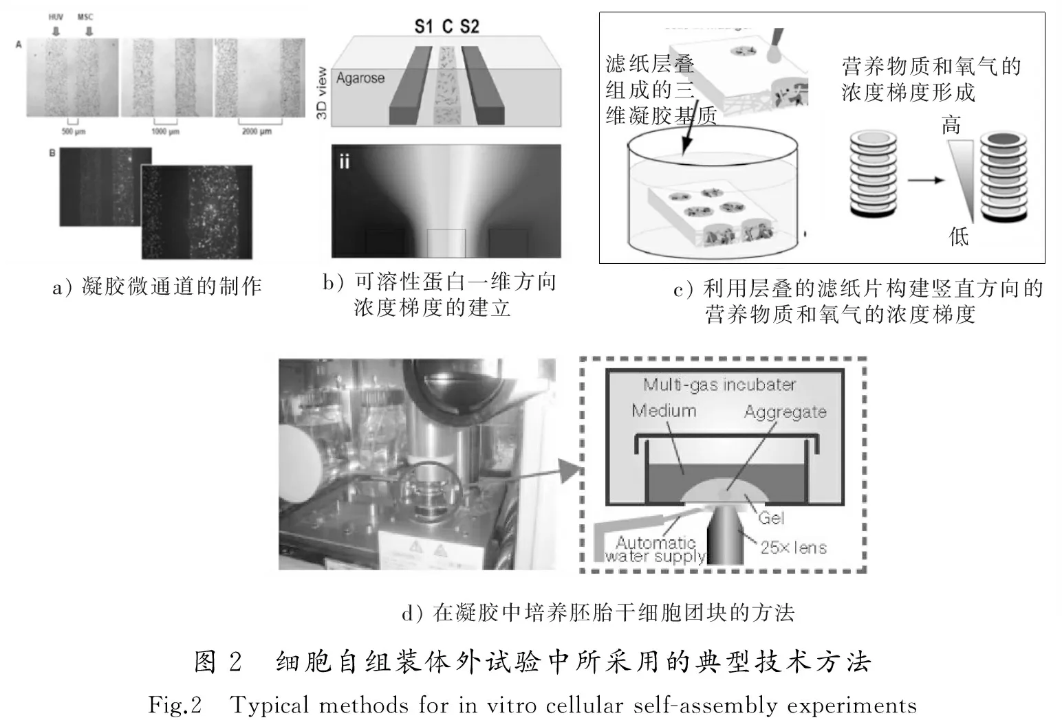
对于细胞的3D自组装研究在试验和技术方法方面,现有的国内外研究工作取得了一些进步和有意义的成果。目前,利用或者结合细胞的自组装特性构建三维的生物组织特征结构的途径主要有:构建并组装含细胞的片层[25-26];直接使细胞沉积或移动到指定位置进而设定细胞的排列特征[16,27-29];以及构建包含细胞的凝胶纤维进而对其组装[30]等。在这些方法中,都避免使用了各类结构支架,而是通过设定初始几何限制或初始细胞分布,然后利用细胞自组装特性以期实现特定多细胞结构或是组织单元结构的构建。此外,还有一部分研究工作通过外部微结构或者外部施加的作用因子梯度[31]以及培养的化学生物条件对细胞生长和自组装的影响,比如凝胶微结构成型 (如图2 a)所示)[32],外基质内的趋化因子浓度梯度的建立(如图2 b)所示)[33],营养物质与氧气浓度梯度的建立(如图2 c)所示)[34]以及在三维凝胶对胚胎干细胞团块的培养(如图2 d)所示)[35-36]等方法被提出并应用于研究细胞对周围微环境的响应乃至器官雏形的形成。上述成果中,实现凝胶微结构的制作,主要是用于研究不同细胞之间的相互作用;外基质内的建立一些作用因子(某种蛋白质)梯度(见图2 b))[33,37]或者氧气及营养物质的浓度梯度(见图2 c))[34],是因为实际生物组织在自然演变过程中所处的微环境内通常大量存在物质浓度的梯度分布,因而需要构建特定的浓度梯度以便更好地模拟自然界中多细胞体系所处的微环境;通过对胚胎干细胞团块在三维(3D)凝胶中的自组装过程能够实现自组装的视网膜原基[35]形态发生,开辟了利用多功能干细胞治疗某些内分泌疾病研究的新途径;此外,通过对胚胎干细胞团块的培养以及细胞群体自组装过程,可在特定条件下实现脑下垂体前叶雏形[36]形态发生,这对于视网膜退化的治疗方法研究极具意义。

1.2 细胞自组装的反应-扩散理论机制的研究现状及分析
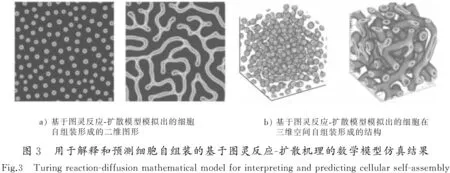
除了上述在生物细胞自组装的试验或支撑技术实现上的研究,为了从理论上研究或从试验上控制细胞在三维空间(3D)的相互作用[32]或自组装[38],建立了若干种基于图灵反应-扩散(reaction-diffusion,RD)机理[39-40]的数学模型[41-44]以探索多细胞结构或组织形成的机理,并预测出可能出现的各种组织结构。这种理论最早由英国科学家图灵(Alan Turing)于1951年提出[39],该理论假设生物图案形成过程中包含2种最基本的作用因子:活化子(activator)和抑制子(inhibitor)。基于简单的分子扩散-反应机理,来解释或预测活化子和抑制子最终的浓度分布,进而得出细胞密度在空间的分布(比如活化子浓度较高的区域,细胞密度也高),这样就有可能近似模拟出一些生物组织的形态图案(见图3)[41]。

这种目前被国内外研究者采纳的图灵反应-扩散模型虽然能够从数学意义上解释自然界中动植物的外部或内部各种图形的形成机理,比如虎、斑马身上的条纹、鱼类身上的图案,此外该模型还能在一定程度上解释人体颅盖骨的形成[45]。其所涉及的基于反应-扩散机理的数学模型包含2个主要参量(作为活化子的蛋白分子BMP2和作为抑制子的蛋白分子Noggin)的扩散反应过程,这2个参量与间充质细胞和成骨细胞之间会发生相互作用,例如促进或抑制细胞生长的作用。从反应-扩散系统的本质机理上讲,如果某些干细胞自身能够分泌出成对的活化子和抑制子,那么便具有了体外自组装成特定多细胞结构的能力,血管间充质干细胞便满足这种条件并能够在二维平面形成特定多细胞结构图案[40,46](见图4 a)),这也在一定程度上进一步论证了反应-扩散模型能够作为预测多细胞结构图案的体外形成。但是,该基本模型目前还不能直接用于指导利用人工方法进行体外3D细胞自组装的机理和定量调控研究(如图4 b)所示的三维乳腺上皮细胞的自组装结构[47]就难以直接采用上述基本模型来预测)。

根据目前国际上该研究领域的发展动态来看,试验研究的发展速度大于理论进展的速度。在最近的3D干细胞自组装成为特定多细胞结构体或器官结构特征的研究中,其试验结果通常很大程度上是依据大量试验探索而得出的经验得到的,在细胞组装的初始时刻并不能事先预测到最后的结果。 这并非根据预测性理论进行演绎推理进而进行有针对性的设计,也不是根据预先设计的3D多细胞结构/器官结构特征进行相应的制造。总体来说,自底向上的基于细胞自组装的生物制造技术的发展仍需要更多科学理论的支持和引导,且系统深入的细胞自组装或细胞团块的3D自组装机理仍是将来研究的重点和热点。
2 细胞自组装研究现状与分析
在试验和相关技术实现方面,虽然通过3D自组装方法研究多细胞结构体系形成的研究工作取得了一定的成果[22-23,32-37],但单个细胞或多细胞结构究竟是以何种机理相互作用最终形成整个器官仍然不完全清楚。 因为平衡稳定状态下的器官形成过程需要多重细胞间相互作用的协调发展,才能实现细胞群体行为的展现并逐渐发展成为器官,而影响细胞活性、增殖与分化的环境因素众多,具体包括细胞间相互作用、细胞与外基质之间的相互作用、生长因子的浓度分布等[38-50],故而细胞(含多潜能干细胞)的自组装过程的机理非常复杂且尚不清晰,包括3D多细胞结构在三维空间形状发生改变的内在机制以及受到均匀施加的作用因子的作用后3D形状发生改变的原因等。另外,上述研究在模拟自然界生物体内普遍存在的作用因子浓度梯度的时候,较多采用外部浓度差方法(比如样品两侧微流道内溶液存在浓度差),但这样一般只能调控外部作用因子(比如蛋白分子)的浓度差,而不能直接调控细胞自身分泌的作用因子,这也对细胞3D自组装机理的研究造成了一定的局限。
在机理和相应的数学模型方面,基于反应-扩散机理的数学模型通常不能直接用于指导这种自底向上的基于细胞3D自组装的生物制造,因为采用人工方法制造出的多细胞微观结构中所涉及的各类试验参数值和材料的参数值还没有与上述基本模型中的物理量建立直接的对应关系。而建立这种直接的定量(或半定量)关系需要尽可能精确地设计相应的试验,并通过对比各类不同的可控试验条件下的3D自组装形成的结构形态和各类不同参数值设置下模型的模拟结果,进而确定数学模型的关键参数值范围以及描述细胞生物学行为的数学表达项(比如考虑到分子和细胞扩散系数的非均匀空间分布等)。本课题组在采用数学模型引导细胞自组装体外试验方面取得了一定的成果[51-53],并部分地解决了上述问题,包括能够通过反应-扩散数学模型引导血管间充质干细胞自组装成为3D管状和网状镂空微结构等。
3 研究展望
在基于凝胶的3D细胞培养的试验和应用方面,研究人员有望设计出更加准确、精细的试验条件,包括凝胶所能提供的基质环境,细胞所受的化学和力学微环境,以及细胞群体的几何分布及生长控制等。更加精细的试验结果将会进一步推进细胞-凝胶复合材料向临床应用的转化。
在理论和相应的数学模型方面,反应-扩散体系学说为从系统整体角度理解3D多细胞结构形成提供了一个思路,其可以避免对细胞自组装过程中各类繁杂细节的试验测量,且相应的部分数学模型可以模拟出与活体组织形态相近的结构形态。这种理论模型的建立和对组织形成过程的仿真预测对于通过3D细胞培养进行组织前体结构的体外支配具有重要的引导作用,因此在该模型的方程解存在的范围内进行大量的参数值试算是探索新的组织结构类型的一种潜在的有效方法,在未来的研究发展中有望引导研究人员设计相应的试验条件并为试验指明方向。
针对前述的国内外研究现状、存在问题及趋势的分析,为进一步研究多细胞3D自组装机理并使自组装过程可控,满足同时调控外部施加和细胞自身分泌的作用因子的浓度梯度分布的需求,提出一种对内部结构特征梯度化的3D凝胶块组合体材料中细胞3D自组装进行研究的思想,建立相应的体外试验模型以及能够描述细胞3D自组装过程的反应-扩散(RD)基本数学模型,结合RD模型研究细胞在这种内部或内外部作用因子(比如蛋白质分子)的浓度梯度作用下的自组装行为的变化规律。这种研究思路通过试验与理论相结合的方式有望论证各种作用因子或者凝胶力学参数值对细胞自组装过程的调控作用,并据此为面向生物制造的3D多细胞结构的可控化制造提供可靠的理论指导和技术基础。
/
[1] KOLESKY D B, TRUBY R L, GLADMAN A S, et al. 3D bioprinting of vascularized, heterogeneous cell-laden tissue constructs[J]. Advanced Materials, 2014, 26(19): 3124-3130.
[2] REISSIS D, TANG Q O, COOPER N C, et al. Current clinical evidence for the use of mesenchymal stem cells in articular cartilage repair[J]. Expert Opinion on Biological Therapy, 2016, 16(4): 535-557.
[3] KEHR N S, RIEHEMANN K. Controlled cell growth and cell migration in periodic mesoporous oganosilica/alginate nanocomposite hydrogels[J]. Advanced Healthcare Materials, 2016, 5(2): 193-197.
[4] JENNEWEIN M, BUBEL M, GUTHORL S, et al. Two- and three-dimensional co-culture models of soft tissue healing: Pericyte-endothelial cell interaction[J]. Cell and Tissue Research, 2016, 365(2): 279-293.
[5] XU T, ZHAO W, ZHU J M, et al. Complex heterogeneous tissue constructs containing multiple cell types prepared by inkjet printing technology[J]. Biomaterials, 2013, 34(1): 130-139.
[6] EIRAKU M, TAKATA N, ISHIBASHI H, et al. Self-organizing optic-cup morphogenesis in three-dimensional culture[J]. Nature, 2011, 472(7341): 51-56.
[7] TSANG V L, BHATIA S N. Three-dimensional tissue fabrication[J]. Adv Drug Deliv Rev, 2004, 56(11): 1635-1647.
[8] HUTMACHER D W, SITTINGER M, RISBUD M V. Scaffold-based tissue engineering: Rationale for computer-aided design and solid free-form fabrication systems[J]. Trends Biotechnol, 2004, 22(7): 354-362.
[9] LI X, FENG Y F, WANG C T, et al. Evaluation of biological properties of electron beam melted Ti6Al4V implant with biomimetic coating in vitro and in vivo[J]. PLoS One, 2012, 7(12): e52049.
[10] BHUMIRATANA S, VUNJAK-NOVAKOVIC G. Concise review: Personalized human bone grafts for reconstructing head and face[J]. Stem Cells Translational Medicine, 2012, 1(1): 64-69.
[11] BEKE S, ANJUM F, TSUSHIMA H, et al. Towards excimer-laser-based stereolithography: A rapid process to fabricate rigid biodegradable photopolymer scaffolds[J]. Journal of the Royal Society Interface, 2012, 9(76): 3017-3026.
[12] 张维杰,连芩,李涤尘,等. 基于 3-D 打印技术的软骨修复及软骨下骨重建[J]. 中国修复重建外科杂志, 2014, 28(3): 318-324.
ZHANG Weijie, LIAN Qin, LI Dichen,et al.Cartilage repair and subchondral bone reconstruction based on three-dimensional printing technique[J]. Chinese Journal of Reparative and Reconstructive Surgery, 2014, 28(3):318-324.
[13] YUSOP A H, BAKIR A A, SHAHAROM N A, et al. Porous biodegradable metals for hard tissue scaffolds: A review[J]. International Journal of Biomaterials, 2012,2012: 641430.
[14] JAKAB K, NOROTTE C, MARGA F, et al. Tissue engineering by self-assembly and bio-printing of living cells[J]. Biofabrication, 2010, 2(2): 022001.
[15] MIRONOV V, KASYANOV V, MARKWALD R R. Organ printing: From bioprinter to organ biofabrication line[J]. Current Opinion in Biotechnology, 2011, 22(5): 667-673.
[16] MIRONOV V, BOLAND T, TRUSK T, et al. Organ printing: Computer-aided jet-based 3D tissue engineering[J]. Trends in Biotechnology, 2003, 21(4): 157-161.
[17] GU G X, SU I, SHARMA S, et al. Three-dimensional-printing of bio-inspired composites[J]. Journal of Biomechanical Engineering, 2016, 138(2): 021006.
[18] ATHANASIOU K A, ESWARAMOORTHY R, HADIDI P, et al. Self-organization and the self-assembling process in tissue engineering[J]. Annual Review of Biomedical Engineering, 2013, 15(1): 115-136.
[19] TAKEBE T, SEKINE K, ENOMURA M, et al. Vascularized and functional human liver from an iPSC-derived organ bud transplant[J]. Nature, 2013, 499(7459): 481-484.
[20] HUH D, MATTHEWS B D, MAMMOTO A, et al. Reconstituting organ-level lung functions on a chip[J]. Science, 2010, 328(5986): 1662-1668.
[21] BERTHIAUME F, MAGUIRE T J, YARMUSH M L. Tissue engineering and regenerative medicine: History, progress, and challenges[J]. Annual Review Chemical & Biomolecular Engineering, 2011, 2(2): 403-430.
[22] HAYCOCK J W. 3D cell culture: A review of current approaches and techniques[J]. Methods in Molecular Biology, 2011, 695: 1-15.
[23] LEE J, CUDDIHY M J, KOTOV N A. Three-dimensional cell culture matrices: State of the art[J]. Tissue Engineering Part B-Reviews, 2008, 14(1): 61-86.
[24] ZHU X L, DING X T. Study on a 3D hydrogel-based culture model for characterizing growth of fibroblasts under viral infection and drug treatment[J]. SLAS Discovery, 2017, 22(5): 626-634.
[25] SHIMIZU T, YAMATO M, KIKUCHI A, et al. Cell sheet engineering for myocardial tissue reconstruction[J]. Biomaterials, 2003, 24(13): 2309-2316.
[26] YANG J, YAMATO M, SHIMIZU T, et al. Reconstruction of functional tissues with cell sheet engineering[J]. Biomaterials, 2007, 28(34): 5033-5043.
[27] ODDE D J, RENN M J. Laser-guided direct writing of living cells[J]. Biotechnology & Bioengineering, 2000, 67(3): 312-318.
[28] HO C T, LIN R Z, CHANG W Y, et al. Rapid heterogeneous liver-cell on-chip patterning via the enhanced field-induced dielectrophoresis trap[J]. Lab on a Chip, 2006, 6(6): 724-734.
[29] MA Z, PIRLO R K, WAN Q, et al. Laser-guidance-based cell deposition microscope for heterotypic single-cell micropatterning[J]. Biofabrication, 2011, 3(3): 034107.
[30] ONOE H, OKITSU T, ITOU A, et al. Metre-long cell-laden microfibres exhibit tissue morphologies and functions[J]. Nature Materials, 2013, 12(6): 584-590.
[31] SOMAWEERA H, IBRAGUIMOV A, PAPPAS D. A review of chemical gradient systems for cell analysis[J]. Analytica Chimica Acta, 2016, 907: 7-17.
[32] TRKOV S, ENG G, DI LIDDO R, et al. Micropatterned three-dimensional hydrogel system to study human endothelial-mesenchymal stem cell interactions[J]. Journal of Tissue Engineering & Regenerative Medicine, 2010, 4(3): 205-215.
[33] HAESSLER U, PISANO M, WU M, et al. Dendritic cell chemotaxis in 3D under defined chemokine gradients reveals differential response to ligands CCL21 and CCL19[J]. Proceedings of the National Academy of Sciences of the United States of America, 2011, 108(14): 5614-5619.
[34] DERDA R, LAROMAINE A, MAMMOTO A, et al. Paper-supported 3D cell culture for tissue-based bioassays[J]. Proceedings of the National Academy of Sciences of the United States of America, 2009, 106(44): 18457-18462.
[35] EIRAKU M, TAKATA N, ISHIBASHI H, et al. Self-organizing optic-cup morphogenesis in three-dimensional culture[J]. Nature, 2011, 472(7341): 51-56.
[36] SUGA H, KADOSHIMA T, MINAGUCHI M, et al. Self-formation of functional adenohypophysis in three-dimensional culture[J]. Nature, 2011, 480(7375): 57-62.
[37] VICKERMAN V, BLUNDO J, CHUNG S, et al. Design, fabrication and implementation of a novel multi-parameter control microfluidic platform for three-dimensional cell culture and real-time imaging[J]. Lab on a Chip, 2008, 8(9): 1468-1477.
[38] JAKAB K, NOROTTE C, DAMON B, et al. Tissue engineering by self-assembly of cells printed into topologically defined structures[J]. Tissue Engineering Part A, 2008, 14(3): 413-421.
[39] TURING A M. The chemical basis of morphogenesis[J]. Philosophical Transactions of the Royal Society of London Series B-Biological Sciences, 1952, 237(641): 37-72.
[40] GARFINKEL A, TINTUT Y, PETRASEK D, et al. Pattern formation by vascular mesenchymal cells[J]. Proceedings of the National Academy of Sciences of the United States of America, 2004, 101(25): 9247-9250.
[41] DANINO T, VOLFSON D, BHATIA S N, et al. In-Silico patterning of vascular mesenchymal cells in three dimensions[J]. PLoS One, 2011, 6(5): e20182.
[42] YOCHELIS A, TINTUT Y, DEMER L L, et al. The formation of labyrinths, spots and stripe patterns in a biochemical approach to cardiovascular calcification[J]. New Journal of Physics, 2008, 10(5): 78-106.
[43] CHINAKE C R, SIMOYI R H. Experimental studies of spatial patterns produced by diffusion-convection-reaction systems[J]. Journal of the Chemical Society-Faraday Transactions, 1997, 93(7): 1345-1350.
[44] SHETH R, MARCON L, BASTIDA M F, et al. Hox genes regulate digit patterning by controlling the wavelength of a Turing-type mechanism[J]. Science, 2012, 338(6113): 1476-1480.
[46] ZHU X, YANG H. In-Silico constructing three-dimensional hollow structure via self-organization of vascular mesenchymal cells [C]// The 16th International Conference on Nanotechnology (IEEE NANO 2016). Sendai: IEEE,2016:468-471.
[47] ELLIOTT N T, YUAN F. A review of three-dimensional in vitro tissue models for drug discovery and transport studies[J]. Journal of Pharmaceutical Sciences, 2011, 100(1): 59-74.
[48] DISCHER D, MOONEY D, ZANDSTRA P. Growth factors, matrices, and forces combine and control stem cells[J]. Science, 2009, 324(5935): 1673-1677.
[49] SPRADLING A, DRUMMOND-BARBOSA D, KAI T. Stem cells find their niche[J]. Nature, 2001, 414(6859): 98-104.
[50] LUTOLF M P, HUBBELL J A. Synthetic biomaterials as instructive extracellular microenvironments for morphogenesis in tissue engineering[J]. Nature Biotechnology, 2005, 23(1): 47-55.
[51] ZHU X, GOJGINI S, CHEN T H, et al. Directing three-dimensional multicellular morphogenesis by self-organization of vascular mesenchymal cells in hyaluronic acid hydrogels[J]. Journal of Biological Engineering, 2017, 11(1): 1-12.
[52] ZHU X, GOJGINI S, CHEN T H, et al. Three dimensional tubular structure self-assembled by vascular mesenchymal cells at stiffness interfaces of hydrogels[J]. Biomedicine & Pharmacotherapy, 2016, 83: 1203-1211.
[53] ZHU X, YANG Y. Simulation for tubular and spherical structure formation via self-organization of vascular mesenchymal cells in three dimensions [C]//International Congress on Image and Signal Processing. BioMedical Engineering and Informatics (CISP-BMEI). Datong: IEEE,2016:1654-1659.
Research progress of the three-dimensional(3D) multicellular structure formation via self-assembly of cells toward tissue engineering
ZHU Xiaolu, YANG Yifei
(College of Mechanical and Electrical Engineering, Hohai University, Changzhou, Jiangsu 213022, China)
Multicellular structures are organisms consisting of cell populations, and constructing multicellular structure is of great importance for the development of tissue engineering and regenerative medicine. The use of self-organization processes in three-dimensional (3D) culture is receiving increased attention for bio-manufacturing and regenerating injured tissue. This review covers the research work on the fundamental and key techniques regarding 3D cellular self-assembly. It includes the 3D culture of cells inside bio- compatible hydrogels, controllable formation of 3D multicellular structure, and the associated Turing reaction-diffusion mechanism. In order to further study the mechanism of 3D self-assembly of cells and find a reliable way to regulate this self-organization in 3D extracellular matrix with spatial gradients of the concentrations of exogenous and cell-secreted factors, this review at last proposes to study the 3D models of cellular self-assembling inside structurally graded hydrogel, so that the development of rational regulation technology for 3D multicellular structure and tissue bud could be facilitated.
bionics; bio-fabrication; 3D cell culture; structurally graded hydrogel; 3D self-assembly of cell
1008-1542(2017)06-0515-07
10.7535/hbkd.2017yx06002
Q81
A
2017-03-07;
2017-06-19;责任编辑:王海云
国家自然科学基金(51505127);江苏省自然科学基金 (BK20161197);常州市科技支撑计划(社会发展)项目(CE20165029)
朱晓璐(1985—),男,江苏徐州人,讲师,博士,主要从事微纳生物制造及微流体技术方面的研究。
E-mail:zhuxiaolu@hhu.edu.cn
朱晓璐, 杨逸飞.面向组织工程的多细胞结构三维自组装成形方法的研究进展[J].河北科技大学学报,2017,38(6):515-521.
ZHU Xiaolu,YANG Yifei.Research progress of the three-dimensional(3D) multicellular structure formation via self-assembly of cells toward tissue engineering[J].Journal of Hebei University of Science and Technology,2017,38(6):515-521.

